Int J Physiother 2014; 1(2) Page | 46
Jibi Paul
1CORRESPONDING AUTHOR
1
Jibi Paul,
MPT, (PhD), Physiotherapy programme, School of Health Sciences,KPJ Healthcare University College, Nilai, Malaysia.
e - Mail:
[email protected]
Int J Physiother. Vol 1(2), 46-53, June (2014) ISSN: 2348 - 8336
ABSTRACT
Background: The dramatic increase in the incidence and prevalence of neurological disorder like stroke has always demanded the need of new interventions in limiting the disability outcome. Disturbed balance is one of the common difficulties in stroke subjects. Several researches have conducted with the process of limiting the disability but still not controlled fully. The objective of the study is to find out the effect of balance exercise with deprivation of visual feedback in stroke subjects.
Method: Total number of thirty subjects participated in the study. The duration of study was four weeks with five sittings in a week and with duration of sixty minutes in each training session. Data was collected using Berg balance scale and the get up and go test, statistical analysis using the Wilcoxon’s signed rank test and paired T-test.
Result: The mean value of timed get up and go test in group A was 16.93 with a standard deviation of ±3.86. The t-value of the study was 2.63 with P-value of 0.007.The study found significant difference in effect as the calculated P < 0.05. The results showed that the conventional therapy along with masked vision could bring about significant changes in balance, mobility and function of patients suffering from balance impairment among post stroke. The data analysis found the significant difference between the visions deprived balance training to that of free vision balance training.
Conclusion: Subjects with masked vision showed better outcomes on balance and get up and go test. Balance performance and movement was found better in subjects with deprived vision than those with free vision.
Key Word: Stroke, disability, visual feedback, berg balance scale.
INTRODUCTION
Stroke is one of the most common neurological diseases that lead to death and disability in elderly population. It is defined as a sudden, non-convulsive, focal neurological deficit lasting for more than 24 hours. Incidence of stroke is about 19%higher for males than females. The prevalence of stroke varies from 44 to 842 per 1,00,000 population with male preponderance.1, 2
Hemiparesis is the most common feature of disability after stroke, affecting 70 to 85% of all the patients, and out of that 60% of all surviving stroke patients require rehabilitation treatment. Posture is the relative alignment of the various body segments with one another. Hemiparesis secondary to cortical, sub cortical white matter and brainstem stroke was reported to improve with sympathomimetic such as methyl phenidate and dextro-amphitamine but on the contrary basic sciences and clinical research supports a greater improvement with physical therapy when patients are taking these medications than when taking medications alone or physical therapy without medications. 3, 4, 5, 6, 7, 8
Novel visual stimuli activate a population of neurons in the primate orbitofrontal cortex, neuron were found in the rhesus macaque anterior orbitofrontal cortex that respond to novel but not for familiar stimuli, the neurons did not respond to stimuli which had been novel and shown a few times on the previous day, indicating that the neurons were involved in long term memory, indicating that the long term memory for visual stimuli is information that is represented in a region of the primate anterior orbitofrontal cortex9.
The aims and objectives of the study are to find out the effectiveness of balance exercise with deprivation of visual feedback in stroke subjects. This study was also to analyze the effect of balance exercise with free vision on hemiparesis patients of stroke.
MATERIALS AND METHODOLOGY
Population: The populations elected for this study was from the medically diagnosed subjects suffering from post-stroke impairments and referred for treatment at Florence rehabilitation center, Bangalore.
Setting of study: Florence rehabilitation center, Bangalore, India was the place selected for conduct the study. The duration of study was four weeks with five sittings in a week and with duration of sixty minutes in each training session.
Sample and Sampling method: Medically diagnosed
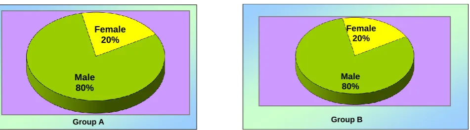
stroke patients with more than six months and less than one year after stroke were selected for the study. Thirty subjects were selected randomly out of forty stroke subjects referred to the physiotherapy department. The samples divided into two groups with fifteen samples in each group. The samples in each group were twelve males and three females for the study. All subjects were collected informed consent before starting the training program in the study.
Inclusion criteria: Patients diagnosed stroke within
six months of duration and are able to walk independently without human support. The subjects were with age group between 45 to 65 years. Subjects those who accepted for the study were evaluation by the assessment Performa before selection of the subjects. Single attack stroke subjects with duration of more than six months and less than one year six months. Subjects in grade 2 and above in modified Ashworth spasticity scale and subjects with the muscle power of 3 and above on manual muscle testing were selected for the study.
Exclusion criteria: Subjects with perceptual and
cognitive deficits were excluded from the study. Subjects with any lower limb joint pathologies like arthritis, limb length discrepancies were excluded from the study. Patients with visual impairments and Visio-spatial disorders also excluded from the study.
Materials used: Exercise mats, Treadmill, Static
bicycle were the materials used for the study. Stop watch used to conform the treatment duration.
Measurement tools: Modified Ashworth scale used
for spasticity measurement. Berg balance scale and Timed get up and go test used to find out the outcome of the study.
Method of collection of data: Subjects diagnosed
with stroke satisfying inclusion criteria with disturbance balance were selected for study. The sample comprising of thirty subjects, the subjects were randomly divided into two groups. (Table 1), Gender and age group of subjects in the study has presented in Fig.1 and Fig. 2.
Table1: Basic characteristics with respect to gender distribution.
Group Male Female Balance Exercise program
A 12 3 Conventional physiotherapy with deprived vision
Figure1: Basic characteristics with respect to gender distribution
Figure2: Basic characteristics with respect to age distribution
PROCEDURE:
Thirty subjects selected on the basis of inclusion and exclusion criteria for the study and randomly divided into two groups. Group-A with fifteen subjects were administered conventional therapy along with deprived vision. Group-B with fifteen subjects was administered conventional therapy along with free vision.
Demographic data and type of stroke, location and side of lesion according to the computed tomographic scan performed at diagnosis. Balance was assessed using berg balance scale it allocates fourteen points to balance. The subjects of both the groups were examined about the purpose and the procedure of the study. A base line reading of berg balance scale and get p and go test was taken before starting the intervention program. Later at the end of fourth week repeated the measurements of same variables. The period of intervention administered to all the subjects of both the groups was sixty minutes daily for five days a week for four consecutive weeks. Experimental approach for Group A
The rehabilitation program was same for the vision deprived group and the free vision group, except that the eyes of the vision-deprived group were blinded with a mask throughout the sessions.
The intervention started with ten minutes of spasticity inhibition followed by fifty minutes devoted to improving balance. This included thirty minutes of exercise performed in following positions during the first week, in the supine or prone position [bridging in spine (Fig. 3), transfer and crawling]; the exercises performed in sitting position (mobility exercises, strengthening and balance training)during the second week; during the third week all fours or kneeling position (mobility exercises, strengthening and balance training) and during the fourth week in upright position (mobility exercises, strengthening and balance training) performed the exercises. Each session included ten minutes of balance training on a treadmill and ended with ten minutes of walking on a foam rubber track with obstacles.
Figure 3: Bridging exercise with masked vision Male
80% Female
20%
Group A
Male 80%
Female 20%
Group B
0 10 20 30 40 50 60 70
Group A Group B
A
ge
i
n
y
ea
Conventional therapy to both Group A and Group B Conventional therapy program included spasticity management, Mobility exercises, Strengthening Exercises and Balance Exercises.
Spasticity management: Slow sustained stretching
with a hold time of thirty counts for every single muscle groups exhibiting spasticity for five repetitions .sustained stretch appeared to be effective. Where distal muscles were found to be more involved than proximal group of muscles, spasticity management preceded all other intervention.
Mobility exercises: Patients were performed active, active assisted or passive as required pertaining to the individual joint ranges for ten repetitions, spinal mobility exercise were also induced apart from the exercises for joints of upper extremity and lower extremity. The program was started with five minutes of cycling on a static bicycle.
Strengthening Exercises: Resistance training was
typically accomplished for the weaker group of muscles pertaining to their grades from manual muscle testing, they were induced with resisted exercises for all individual muscle groups with the help of optimal resistance weigh cuffs throughout the range of motion commands were given for completing the range and positioning of the extremities.
Balance Exercises: Balance training was given
through various positions in mat, exercises for the positions of lying and sitting progressing to balance training in standing with activities of rolling, turning, bridging, progression within the posture to weight shifting from one side to the other, bridging with single limb support, weight shifts in sitting, to quadruped position and progression with-in the posture on bridging, later balance exercises in standing with a minimum of ten repetition.
Statistical analysis: Wilcoxon’s signed rank test and
paired t-test used to find out the significance of balance between pre and post treatment sessions of both group in the study.
RESULTS:
The parametric outcomes based upon the Berg balance scale and the get up and go have been studied in both group; the results of the study were presented on two sections. Section-I was Intra Group analysis and section-II Inter group analysis.
Section I. Intra group analysis 1. Berg balance scale:
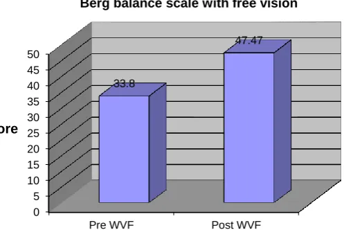
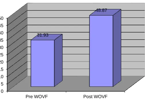
Intra group analysis was done using Wilcoxon’s signed rank test. The data present in table2 shows the P- value for Group –A and Group-B is 0.000(P<0.05), statistically significant at 5% level for both group. The study outcome of balance has improved after intervention in group A and group B.
Berg balance scale Mean rank Sum of ranks Critical value P-value
Group-A 8 0-120.0 30 0.000*
Group-B 8 0-120.0 30 0.000*
Table 2: Intra Group analysis Berg balance scale
Figure 4: Timed get up and go test with visual feedback
0 5 10 15 20 25 30 35 40 45 50
Pre WVF Post WVF
33.8
47.47
score
Figure 5: Timed get up and go test with visual feedback
2. Timed get up and go test
Intra group analysis was done using Paired-t Test and the study found significant at5% level
Get up and go Pre Post T-value P-value
Group Mean S.D Mean S.D
A 14.933 2.251 11.000 0.926 8.50 0.000**
B 14.867 1.727 11.000 0.926 12.61 0.000**
Table 4: Intra group analysis of Get up and go Test
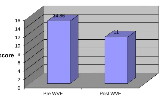
Table 4 shows the pre-treatment mean value of both groups in Timed get up test result as 14.933 with a Standard deviation of ±2.51 and14.867, a Standard deviation of± 1.72the post treatment mean of 11.000 and Standard deviation of±0.926 with the t-value of
8.50 and 12.61 with the P-value as 0.000(P<0.05) respectively for group A and Group B. Here the differences of mean outcome are statistically significant.
Fig.6 Timed get up and go test with visual feedback
0 5 10 15 20 25 30 35 40 45 50
Pre WOVF Post WOVF
31.93
48.87
Score
Berg Scale Without visual feedback
0 2 4 6 8 10 12 14 16
Pre WOVF Post WOVF
14.93
11
score
Fig.7: Timed get up and go test without visual feedback
Section-II Inter group analysis. Berg balance scale:
Inter group analysis is done using the Mann-Whitney test to compare the outcome of balance between
Group A and Group B. Table 5 shows the data’s for inter group analysis using Mann Whitney test with u- value being 289.5 and the p value being 0.0096 The test is significant for P<0.05.
Berg balance scale Mean rank Critical value Sum of ranks U-value P-value
Group-A 8 30 304.5
289.5 0.0096
Group-B 8 30 156.5
Table 5: Inter group analysis of berg balance scale
Fig.8: Mean changes in berg balance with out and with visual feedback
Timed get up and go test:
Inter group analysis of timed get up and go is done using paired t-test .The t-value of 2.63 and the P-value as 0.007(P<0.05) Here the difference of mean outcome with a t value of 2.63 is statistically
significant at 5% level. Hence it can be stated that Visual feedback deprive balance therapy may be more effective over balance therapy with free vision in stroke subjects.
Get up and go test Mean S.D T-value P-value
Group-A 16.93 3.86
2.63 0.007*
Group-B 13.67 2.87
Table 6: Inter group analysis timed get up and go test
0 2 4 6 8 10 12 14 16
Pre WVF Post WVF
14.86
11
score
Timed get up and go test with free vision
0 2 4 6 8 10 12 14 16 18
Berg WOVF Berg WVF
16.93
13.67
Score
DISCUSSION:
In this study the researcher has tried to find out the individual effects of conventional physiotherapy with masked vision and free vision. Parameters named as Berg balance scale with a fourteen phase five point ordinal scale score and the get up and go test with a timed performance scoring. The present study suggests that the vision deprived balance training and the free vision balance training improves the balance parameters significantly (p<0.05) as p=0.000.Intra group analysis with vision masked Balance exercise and conventional physiotherapy was effective in improving the balance in hemeperetic patients following stroke.
Several studied have proved the effect of balance training after stroke by examine the vestibular control of balance following stroke. Visual feedback in retraining balance following acute stroke and concluded that balance training along with visual feedback in addition to regular therapy affords no added benefits when offered in early stages of rehabilitation following stroke.10,11, 12, 13 14, 15
The rehabilitation program includes relaxation exercises; mobilizations exercises, strengthening exercise and treadmill training on mat and on standing have proved effect on reduce spasticity and improve function among stroke subjects.16, 17
Vision overuse may be a compensatory strategy for coping with initial imbalance exacerbated by traditional rehabilitation, hence vision deprived rehabilitation improves balance more effectively than balance with free vision. Masked vision enhances concentration through the somato sensory pathway towards promotion of balance through sensory re-education. This study tries to find out the effects of vision deprived balance therapy in the form of masked vision along with conventional therapy on reestablishing balance in stroke subjects. Conventional therapy brings about improvement in balance, functional mobility and activities of daily living.18
Proprioception and somato sensory pathway has been found to contribute to the vision deprived therapy to a great extent Stroke patients are disabled by the weakness persisting after stroke; deprived visual feedback promotes the use of affected side and prevents the compensatory over use adaptability. The findings of this study are supported by previous studies; hence the alternate hypothesis can be retained.19
Limitations of the study
Small sample size, Skewed effects of stroke and its causes makes the outcomes of balance rehabilitation difficult to be analyzed quantitatively
CONCLUSION:
The results showed that the conventional therapy along with masked vision could bring about significant changes in balance, mobility and function of patients suffering from balance impairment post stroke. The data analysis found the significant difference between the visions deprived balance therapies to that of free vision balance therapy. This study supports the masking of vision to improve balance and functional Mobility in patients with hemiparesis following stroke.
Future recommendation
The study can be replicated with a larger sample size Further studies are required to find the mechanism by which the Vision deprived subjects show better outcomes in terms of balance than those with free vision.
REFERENCES
:
1. Kandel, E.R and Jessell, T M et.al. Principles in neural science. 3rd ed; 1991.
2. Walf.P.a et.al; Secular trends in stroke incidence and mortality The Fleming ham Study. Stroke, 1992; 23:1551-5.
3. Dettmann M.A, Linder M.T, Sepic S.B. Relationship among walking performance, postural stability and functional assessment of the hemi plegic patient. Am j Phys med. 1987; 66 (2): 77-90.
4. Dominic Alien Perennouet.al. Transcutaneous electrical nerve stimulation reduces neglect related postural instability after stroke. Arch.Phys.Med Rehabil. 2001; 82(4): 440-448. 5. Laura Adomatis et.al. An intensive massed
practice approach to retraining balance post-stroke. Gait and Posture. 2005; 22 (2): 154-163. 6. Heller et.al. Postural biofeedback and locomotors
reeducation in stroke patients. Ann Readapt Med Phys. 2005; 48(4): 187-95.
7. Phil Page. Somatosensory training a global approach for balance training. Bodywork and movement therapies; 2006; 10(1): 77-84.
8. Carolyn W et.al, Enduring representational plasticity after somatosensory stimulation. Neuroimage. 2005; 27(4): 872-884.
bio of Learning and Memory; 2005; 84(2): 111-123.
10. Harris-Love M.L et.al. Hemiperetic gait parameters in over ground versus treadmill walking. Neuro Rehab and Neuro Repair. 2001; 15(2): 105-12.
11. Berg et.al. The Balance scale reliability assessment for elderly residents and patients with an acute stroke. Scand J Rehabil Med. 1995; 27(2):27-36.
12. Kathleen M et.al. Reduced ambulatory activity after stroke, the role of balance, gait and cardiovascular fitness. Arch Phys med Rehab. 2005; 86(8): 1552-1556.
13. Marsden J F,D E Playbrol and B L day. The vestibular control of balance after stroke. Journal of Neurology neurosurgery and psychiatry.2005; 76(5): 670-679.
14. Walker C, Bruwer B.J, Culham E.G. Use of visual feedback in retraining balance following acute strok. Phys.ther. 2000; 80(9): 886-895.
15. Richard W. Bohnnon. Patricia A Larkin et.al. Decrease In timed balance test scores with ageing. PhysTher. 1984; 64(1): 1067-1070 16. Bohannon.R and Smith.M;Interrater reliability of
a modified ashworth scale of muscle spasticity; PhysTher. 1987; 67(2): 206-7.
17.
Mizrahi.E and Angel.R. Impairment of voluntary movement by spasticity. Ann Neural.1979; 5(6):594-5.
18. Bonan I.V, F.M Colle, J.P.Guichard et.al, Reliance on visual information after stroke Part-I Balance on dynamic posturography; Arch Phys Med Rehab, 2004; 85(1): 268-273.
19. Isabelle V et.al; Reliance on visual information after stroke Part-II Archive of phys Med Rehabil, 2004; 85(2): 274-278.