Analysis of genetic networks regulating
refractive eye development in collaborative
cross progenitor strain mice reveals new
genes and pathways underlying human
myopia
Tatiana V. Tkatchenko
1, Rupal L. Shah
2, Takayuki Nagasaki
1and Andrei V. Tkatchenko
1,3*Abstract
Background:Population studies suggest that genetic factors play an important role in refractive error
development; however, the precise role of genetic background and the composition of the signaling pathways underlying refractive eye development remain poorly understood.
Methods:Here, we analyzed normal refractive development and susceptibility to form-deprivation myopia in the eight progenitor mouse strains of the Collaborative Cross (CC). We used RNA-seq to analyze gene expression in the retinae of these mice and reconstruct genetic networks and signaling pathways underlying refractive eye
development. We also utilized genome-wide gene-based association analysis to identify mouse genes and pathways associated with myopia in humans.
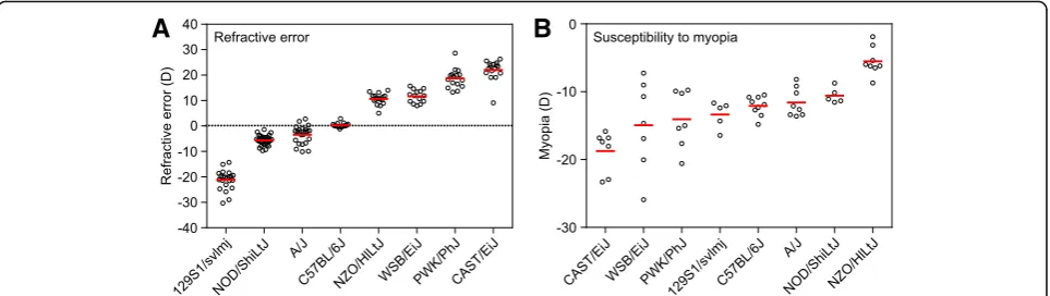
Results:Genetic background strongly influenced both baseline refractive development and susceptibility to environmentally-induced myopia. Baseline refractive errors ranged from −21.2 diopters (D) in 129S1/svlmj mice to + 22.0 D in CAST/EiJ mice and represented a continuous distribution typical of a quantitative genetic trait. The extent of induced form-deprivation myopia ranged from −5.6 D in NZO/HILtJ mice to −20.0 D in CAST/EiJ mice and also followed a continuous distribution. Whole-genome (RNA-seq) gene expression profiling in retinae from CC progenitor strains identified genes whose expression level correlated with either baseline refractive error or susceptibility to myopia. Expression levels of 2,302 genes correlated with the baseline refractive state of the eye, whereas 1,917 genes correlated with susceptibility to induced myopia. Genome-wide gene-based association analysis in the CREAM and UK Biobank human cohorts revealed that 985 of the above genes were associated with myopia in humans, including 847 genes which were implicated in the development of human myopia for the first time. Although the gene sets controlling baseline refractive development and those regulating susceptibility to myopia overlapped, these two processes appeared to be controlled by largely distinct sets of genes.
(Continued on next page)
© The Author(s). 2019Open AccessThis article is distributed under the terms of the Creative Commons Attribution 4.0 International License (http://creativecommons.org/licenses/by/4.0/), which permits unrestricted use, distribution, and reproduction in any medium, provided you give appropriate credit to the original author(s) and the source, provide a link to the Creative Commons license, and indicate if changes were made. The Creative Commons Public Domain Dedication waiver (http://creativecommons.org/publicdomain/zero/1.0/) applies to the data made available in this article, unless otherwise stated.
* Correspondence:[email protected]
1Department of Ophthalmology, Columbia University, New York, NY, USA 3Department of Pathology and Cell Biology, Columbia University, New York,
NY, USA
(Continued from previous page)
Conclusions:Comparison with data for other animal models of myopia revealed that the genes identified in this study comprise a well-defined set of retinal signaling pathways, which are highly conserved across different vertebrate species. These results identify major signaling pathways involved in refractive eye development and provide attractive targets for the development of anti-myopia drugs.
Keywords:Myopia, Refractive eye development, Genetic networks, Signaling pathways, Genetic variation, RNA-seq, Gene-based genome-wide association analysis, Evolutionary conservation of pathways
Background
Myopia is the most common ocular disorder worldwide [1]. The prevalence of myopia in the U.S. has increased from 25% to ~ 48% in the last 40 years [2–4]. The world-wide prevalence of myopia is predicted to increase from the current 25 to 50% in the next three decades [5], while the prevalence already exceeds 80% in several parts of Asia [6,7]. Myopia often leads to serious blind-ing complications such as myopic maculopathy, retinal floaters, chorioretinal atrophy, retinoschisis, retinal tears, retinal detachment, and myopic macular degeneration [8–24]. It also represents a major risk factor for a num-ber of other serious ocular pathologies such as cataract and glaucoma [9, 10, 25–27]. Because of the increasing prevalence, myopia is rapidly becoming one of the lead-ing causes of vision loss in several parts of the world, and World Health Organization designated myopia as one of five priority health conditions [1,8–10,28].
Development of myopia is controlled by both environ-mental and genetic factors [29–32]. Although environ-mental factors, such as reading and nearwork, play a very important role in the development of myopia [33–36], genetic studies suggest that the impact of environmental factors on refractive development is determined by genetic variation in“myopia-susceptibility genes”[37]. The role of genetic background in refractive eye development is also supported by animal studies, which revealed that the ex-tent of myopia experimentally induced in animal models is strongly influenced by genetic background [38–41]. Analysis of the size of ocular components in different strains of mice suggested a significant role of genetic back-ground in the regulation of refractive eye development [42, 43]. Wong and Brown [44] also described significant differences in visual detection, pattern discrimination and visual acuity among different strains of mice. The contri-bution of genetic factors to myopia has been estimated to be as high as 70–80% [45–50], and human genetic map-ping studies have identified over 270 chromosomal loci linked to myopia [51–54]. These loci implicate genes in-volved in multiple cellular and biological processes related to extracellular matrix organization, eye morphogenesis, retinal signaling, and visual perception [53, 54]. Gene ex-pression profiling studies also showed that development of myopia is accompanied by changes in gene expression
in the retina, choroid, and sclera [39, 55–62]. Moreover, these studies suggested that similar biological processes underlie refractive development in animal models and humans [63].
Although these studies revealed important roles of genetic variation and gene expression in the develop-ment of refractive errors, the relationship between gen-etic background, gene expression and development of refractive errors remains unexplored. Here, we systemat-ically analyzed the role of genetic background in the regulation of retinal gene expression and signaling path-ways underlying refractive eye development in eight in-bred strains of mice and their association with human myopia using genome-wide gene expression profiling (RNA-seq) and gene-based genome-wide association analysis in the CREAM and UK Biobank human cohorts. We found that both the baseline refractive state of the eye and susceptibility to myopia are inherited as quanti-tative traits, demonstrating strong dependence on gen-etic background. Furthermore, genetic background strongly influenced expression of genes in the retina and modulated a well-defined set of signaling pathways highly conserved in chickens, mice, monkeys, and humans. Our data suggest that refractive eye develop-ment is regulated by hundreds to thousands of genes across ocular tissues and point to high evolutionary con-servation of signaling pathways underlying refractive de-velopment across vertebrate species.
Methods
Ethics approval and consent to participate
Mice were obtained from the Jackson Laboratory (Bar Harbor, ME) and were maintained as an in-house breed-ing colony. Food and water were provided ad libitum. All procedures adhered to the Association for Research in Vision and Ophthalmology (ARVO) statement on the use of animals in ophthalmic and vision research and were approved by the Columbia University Institutional Animal Care and Use Committee. Animals were anes-thetized via intraperitoneal injection of ketamine (90 mg/kg) and xylazine (10 mg/kg) and were euthanized using CO2followed by cervical dislocation.
and conducted according to the Declaration of Helsinki. All CREAM participants provided written informed con-sent. The UK Biobank received ethical approval from the National Health Service National Research Ethics Service (reference 11/NW/0382).
Analysis of refractive state of the eyes in mice
To examine the effect of genetic background on the fractive state of the eye in mice, we analyzed baseline re-fractive errors in the eight strains of mice comprising Collaborative Cross (129S1/svlmj, A/J, C57BL/6J, CAST/ EiJ, NOD/ShiLtJ, NZO/HlLtJ, PWK/PhJ, and WSB/EiJ mice). The refractive state of both left and right eyes was determined on alert animals at P40 using an auto-mated eccentric infrared photorefractor as previously described [64, 65]. The animal to be refracted was immobilized using a restraining platform, and each eye was refracted along the optical axis in dim room light (< 1 lx), 20–30 min. after instilling 1% tropicamide ophthal-mic solution (Alcon Laboratories) to ensure mydriasis and cycloplegia. Five independent measurement series (~ 300–600 measurements each) were taken for each eye. The measurements were automatically acquired by the photorefractor every 16 msec. Each successful meas-urement series (i.e., Purkinje image in the center of the pupil and stable refractive error for at least 5 s.) was marked by a green LED flash, which was registered by the photorefractor software. Sixty individual measurements from each series, immediately preceding the green LED flash, were combined, and a total of 300 measurements (60 measurements × 5 series = 300 measurements) were collected for each eye. Data for the left and right eyes were combine (600 measurements total) to calculate mean re-fractive error and standard deviation for each animal.
Analysis of form-deprivation myopia in mice
To examine the effect of genetic background on suscep-tibility to environmentally induced myopia in mice, we analyzed the extent of myopia induced by the diffuser-imposed retinal image degradation (visual form deprivation) in the eight strains of mice comprising Col-laborative Cross (129S1/svlmj, A/J, C57BL/6J, CAST/EiJ, NOD/ShiLtJ, NZO/HlLtJ, PWK/PhJ, and WSB/EiJ mice). Visual input was degraded in one of the eyes by applying plastic diffusers, and refractive development of the treated eye was compared to that of the contralateral eye, which was not treated with a diffuser, as previously described [66,67]. Diffusers represented low-pass optical filters, which degraded the image projected onto the ret-ina by removing high spatial frequency details. Frosted hemispherical plastic diffusers were hand-made from zero power rigid contact lenses made from OP3 plastic (diameter = 7.0 mm, base curve = 7.0 mm; Lens.com). Lenses were frosted using a fine sandpaper and inserted into a 3D-printed plastic frames (Proto Labs). On the first day of the experiment (P24), animals were anesthe-tized via intraperitoneal injection of ketamine and xyla-zine, and frames with diffusers were attached to the skin surrounding the right eye with six stitches using size 5– 0 ETHILON™microsurgical sutures (Ethicon) and rein-forced with Vetbond™ glue (3 M Animal Care Products) (the left eye served as a control). Toenails were covered with adhesive tape to prevent mice from removing the diffusers. Animals recovered on a warming pad and were then housed under low-intensity constant light in trans-parent plastic cages for the duration of the experiment as previously described [66, 67]. Following 21 days of visual form deprivation (from P24 through P45), dif-fusers were removed and refractive status of both treated and control eyes was assessed using an automated
A
B
eccentric infrared photorefractor as previously described [64,65]. The interocular difference in refraction between the treated and contralateral control eye served as an in-dication of the extent of induced myopia.
RNA extraction and RNA-seq
Animals were euthanized following an IACUC-ap-proved protocol. Eyes were enucleated, the retinae were dissected from the enucleated eyes and the choroid/RPE removed. The retinae were washed in RNAlater (Thermo Fisher Scientific) for 5 min., fro-zen in liquid nitrogen, and stored at −80 °C until processed for this study. To isolate RNA, tissue
samples were homogenized at 4 °C in a lysis buffer using Bead Ruptor 24 tissue homogenizer (Omni). Total RNA was extracted from each tissue sample using miRNAeasy mini kit (QIAGEN) following the manufac-turer’s protocol. The integrity of RNA was confirmed by analyzing 260/280 nm ratios (Ratio260/280= 2.11–2.13) on
a Nanodrop (Thermo Scientific) and the RNA Integrity Number (RIN = 9.0–10.0) using Agilent Bioanalyzer. Illu-mina sequencing libraries were constructed from 1μg of total RNA using the TruSeq Stranded Total RNA LT kit with the Ribo-Zero Gold ribosomal RNA depletion mod-ule (Illumina). Each library contained a specific index (bar-code) and were pooled at equal concentrations using the
A
C
B
randomized complete block (RCB) experimental design before sequencing on Illumina HiSeq 2500 sequencing system. The number of libraries per multiplexed sample was adjusted to ensure sequencing depth of ~ 70 million reads per library (paired-end, 2 × 100 nucleotides). The ac-tual sequencing depth was 76,773,554 ± 7,832,271 with read quality score 34.5 ± 0.4.
Post-sequencing RNA-seq data validation and analysis The FASTQ raw data files generated by the Illumina se-quencing system were imported into Partek Flow software
package (version 7.0.18.1210, Partek), libraries were sepa-rated based on their barcodes, adapters were trimmed and remaining sequences were subjected to pre-alignment qual-ity control using Partek Flow pre-alignment QA/QC mod-ule. After the assessment of various quality metrics, bases with the quality score < 34 were removed (≤5 bases) from each end. Sequencing reads were then mapped to the mouse reference genome Genome Reference Consortium Mouse Build 38 (GRCm38/mm10, NCBI) using the STAR aligner (version 2.5.2b) resulting in 95.0 ± 0.4% mapped reads per li-brary, which covered 35.4 ± 1.0% of the genome. Aligned
reads were quantified to transcriptome using Partek E/M annotation model and the NCBI’s RefSeq Transcripts 80 an-notation file to determine read counts per gene/genomic re-gion. The generated read counts were normalized by the total read count and subjected to the analysis of variance
(ANOVA) to detect genes whose expression correlates with either refractive error or susceptibility to myopia. Differen-tially expressed transcripts were identified using a P-value threshold of 0.05 adjusted for genome-wide statistical signifi-cance using Storey’s q-value algorithm [68]. To identify sets
A
B
C
of genes with coordinate expression, differentially expressed transcripts were clustered using Partek Flow hierarchical clustering module using average linkage for the cluster dis-tance metric and Euclidean disdis-tance metric to determine the distance between data points. Each RNA-seq sample was analyzed as a biological replicate, thus, resulting in three biological replicates per strain.
Gene ontology analysis and identification of canonical signaling pathways
To identify biological functions (gene ontology categor-ies), which were significantly affected by the genes whose expression correlated with either baseline refractive er-rors or susceptibility to myopia, we used the database
for annotation, visualization and integrated discovery (DAVID) version 6.8 [69] and GOplot R package [70]. DAVID uses a powerful gene-enrichment algorithm and DAVID Gene Concept database to identify biological functions (gene ontology categories) affected by differen-tial genes, while GOplot integrates gene ontology infor-mation with gene expression inforinfor-mation and predicts the effect of gene expression changes on biological pro-cesses. DAVID uses a modified Fisher’s exact test (EASE score) with aP-value threshold of 0.05 to estimate statis-tical significance of enrichment for specific gene ontol-ogy categories. IPA Pathways Activity Analysis module (QIAGEN) was used to identify canonical pathways effected by the genes involved in baseline refractive eye
development or regulating susceptibility to myopia, and predict the effect of gene expression changes in different strains on specific pathways. The activation z-score was employed in the IPA Pathways Activity Analysis module to predict activation or suppression of the canonical path-ways. The z-score algorithm is designed to reduce the chance that random data will generate significant predic-tions. The z-score provides an estimate of statistical quan-tity of change for each pathway found to be statistically significantly affected by the changes in gene expression. The significance values for the canonical pathways were calculated by the right-tailed Fisher’s exact test. The sig-nificance indicates the probability of association of mole-cules from a dataset with the canonical pathway by random chance alone. Pathways Activity Analysis module determines if canonical pathways, including functional end-points, are activated or suppressed based on the gene expression data in a dataset. Once statistically significant canonical pathways were identified, we subjected the data-sets to the Core Functional Analysis in IPA to compare the pathways and identify key similarities and differences in the canonical pathways underlying baseline refractive development and susceptibility to myopia.
Identification of candidate genes for human myopia within known myopia QTLs
To identify candidate genes for myopia in the QTLs pre-viously found to be linked to human myopia, we com-pared the genes that we found to be involved in refractive eye development in mice with a list of genes located within human myopia QTLs. We first compiled a list of all SNPs or markers exhibiting statistically sig-nificant association with myopia in the human linkage or genome-wide association studies (GWAS) using the
Online Mendelian Inheritance in Man (OMIM) (McKu-sick-Nathans Institute of Genetic Medicine, Johns Hop-kins University) and NHGRI-EBI GWAS Catalog [71] databases. The LDlink’s LDmatrix tool (National Cancer Institute) was used to identify SNPs in linkage disequi-librium and identify overlapping chromosomal loci. We then used UCSC Table Browser to extract all genes lo-cated within critical chromosomal regions identified by the human linkage studies or within 200 kb (±200 kb) of the SNPs found by GWAS. The list of genes located within human QTLs was compared with the list of genes that we found to be associated with either baseline re-fractive errors or susceptibility to myopia in mice using Partek Genomics Suite (Partek). The statistical signifi-cance of the overlaps was estimated using probabilities associated with the hypergeometric distribution using Bioconductor software package GeneOverlap version 1.14.0 and associated functions.
Identification of genes associated with refractive error in the human population using gene-based genome-wide association analysis
To identify genes associated with the development of re-fractive errors in humans among the genes whose expres-sion correlated with refractive eye development in mice, human homologs of candidate mouse genes were exam-ined for association with refractive error in the inter-national GWAS study of refractive error carried out by the Consortium for Refractive Error and Myopia (CREAM) [54] and the UK Biobank Eye and Vision con-sortium sample [72] using the Multi-marker Analysis of GenoMic Annotation (MAGMA) [73]. Human homologs of candidate mouse genes were obtained from Ensembl BioMart and were mapped according to gene definitions in the NCBI Entrez Gene database. Genes were defined according to NCBI build 37 (hg19/GRCh37) coordinates with 200 kb flanking regions appended to the transcription start/stop sites. LD patterns were estimated by MAGMA using the 1000 Genomes Phase 1, version 3 European an-cestry reference panel. As summary statistics were used as input, MAGMA gene-based analysis was performed using the default“snp-wise = mean”model.
The CREAM sample included 148,485 individuals of European ancestry from 28 cohorts and 11,935 in-dividuals of Asian ancestry from eight studies. All participants included in this analysis from CREAM were 25 years of age or older.
UK Biobank is a large prospective study following the health and wellbeing of approximately 500,000 UK residents aged between 40 and 69 years-old at the baseline recruit-ment visit (during the period 2006–2010). One hundred thirty thousand five hundred twenty-one participants had non-cycloplegic autorefraction performed for at least one eye using the Tomey RC 5000 autorefractor-keratometer
A
B
(Tomey Corp., Nagoya, Japan), with up to ten mea-surements taken for each eye. After the exclusion of unreliable readings, 130,459 participants had mea-sures for refractive astigmatism and spherical equiva-lent refractive error.
Participants of the UK Biobank and CREAM studies with conditions that might alter refraction, such as cataract gery, laser refractive procedures, retinal detachment sur-gery, keratoconus, or ocular or systemic syndromes were excluded from the analyses. Refractive error was (See figure on previous page.)
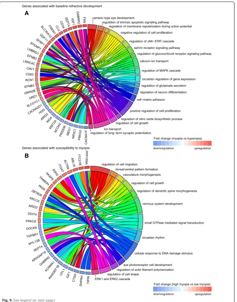
Fig. 9Top biological processes associated with genes linked to refractive eye development in mice and localized in known human myopia QTLs.
aChord diagram showing key genes (left semicircle) and top biological processes (right semicircle) associated with genes correlated with baseline refractive development in mice and localized in known human QTLs linked to myopia.bChord diagram showing key genes (left semicircle) and top biological processes (right semicircle) for genes correlated with susceptibility to myopia in mice and localized in known human QTLs linked to myopia. Colored bars underneath gene names show up- or down-regulation of corresponding genes in either myopic mice versus hyperopic mice (A), or mice with high susceptibility to myopia versus mice with low susceptibility to myopia (B)
A
B
represented by measurements of refraction and analyzed as spherical equivalent (SphE = spherical refractive error + 1/ 2-cylinder refractive error).
Results
Genetic background modulates baseline refractive eye development and susceptibility to myopia in mice Genetic background was shown to influence the size of ocular components in mice. To investigate whether
genetic differences between mice would have an im-pact on the baseline refractive state of the eye and susceptibility to myopia, we analyzed baseline refract-ive development and susceptibility to form-deprivation myopia in eight inbred strains of mice, which served as founder strains for the Collaborative Cross (CC) [74], i.e., 129S1/svlmj, A/J, C57BL/6 J, CAST/EiJ, NOD/ShiLtJ, NZO/HlLtJ, PWK/PhJ, and WSB/EiJ mice.
A
B
Table 1Evolutionary conservation of retinal signaling pathways involved in refractive eye development
Chicken Mouse Marmoset Human Canonical signaling pathways
● ● 4-hydroxyproline degradation I
● ● ● Actin cytoskeleton signaling
● ● ● Aldosterone signaling in epithelial cells
● ● Amyloid processing
● ● ● Amyotrophic lateral sclerosis signaling
● ● ● Androgen receptor signaling
● ● Antiproliferative role of somatostatin receptor 2 signaling
● ● ● ● Calcium signaling
● ● Choline biosynthesis III
● ● Chondroitin sulfate biosynthesis
● ● ● ● Circadian rhythm signaling
● ● ● Clathrin-mediated endocytosis signaling
● ● ● CREB signaling in neurons
● ● ● ● CXCR4 signaling
● ● Diphthamide biosynthesis
● ● Dopamine receptor signaling
● ● ● ● Dopamine-DARPP32 feedback signaling
● ● ● EIF2 signaling
● ● ● eNOS signaling
● ● ● Ephrin B signaling
● ● ● ● Ephrin receptor signaling
● ● ● ● Epithelial adherens junction signaling
● ● ERK/MAPK signaling
● ● ● ● Estrogen receptor signaling
● ● ● GABA receptor signaling
● ● ● Gap junction signaling
● ● ● ● Glucocorticoid receptor signaling
● ● ● ● Glutamate degradation/Glutamate receptor signaling
● ● ● Glutathione biosynthesis
● ● ● Glutathione redox reactions I
● ● ● Glutathione-mediated detoxification
● ● Glycoaminoglycan-protein linkage region biosynthesis
● ● ● Gαq signaling
● ● HIPPO signaling
● ● ● HMGB1 signaling
● ● ● ● Huntington’s disease signaling
● ● ● IGF-1 signaling
● ● ● ILK signaling
● ● ● Insulin receptor signaling
● ● ● Integrin signaling
● ● ● L-cysteine degradation I
● ● ● ● L-cysteine degradation III
● ● Mismatch repair in eukaryotes
We first measured baseline refractive errors in all eight strains at P40, i.e., when baseline refractions reach a plateau in mice [64]. We found that C57BL/6J mice were emmetropic on average (+ 0.3 ± 0.9 D). CAST/EiJ, NZO/ HlLtJ, PWK/PhJ, and WSB/EiJ mice exhibited various degrees of hyperopia ranging from + 10.6 ± 2.2 D to + 22 ± 4.0 D, whereas A/J, NOD/ShiLtJ, and 129S1/svlmj mice developed various degrees of myopia ranging from −3.5 ± 3.6 D to−21.2 ± 3.9 D (Fig.1a, Additional file1: Table S1). The differences between the strains were sta-tistically significant as revealed by ANOVA (F (7, 145) = 429.76, P< 0.00001). More importantly, the distribution
of refractive errors in the CC mice was continuous, sug-gesting that refractive state of the eye in mice is inher-ited as a quantitative trait.
We then analyzed susceptibility to myopia in the same strains of mice by evaluating the extent of induced form-deprivation myopia (Fig. 1b, Additional file 1: Table S2). Susceptibility to form-deprivation myopia was estimated by applying a diffuser to one eye at P24 and comparing the extent of induced myopia in the form-de-prived eye versus the contralateral control eye after 21 days of treatment. We found large differences in suscepti-bility to induced myopia between the strains (Fig. 1b,
Chicken Mouse Marmoset Human Canonical signaling pathways
● ● NF-κB signaling
● ● ● ● nNOS signaling
● ● ● NRF2-mediated oxidative stress response
● ● ● Oncostatin M signaling
● ● ● Phospholipase C signaling
● ● ● Phototransduction pathway
● ● ● ● PI3K/AKT signaling
● ● ● PPAR signaling
● ● ● ● PPARα/RXRαactivation
● ● ● Production of nitric oxide and reactive oxygen species
● ● ● ● Protein kinase A signaling
● ● ● ● Protein ubiquitination pathway
● ● ● PTEN signaling
● ● Purine nucleotides de novo biosynthesis II
● ● ● Purine nucleotides degradation II (aerobic)
● ● RAN signaling
● ● ● RAR activation
● ● ● Regulation of eIF4 and p70S6K signaling
● ● ● Regulation of the epithelial-mesenchymal transition pathway
● ● ● Relaxin signaling
● ● ● ● RhoGDI signaling
● ● Semaphorin signaling in neurons
● ● ● ● Signaling by Rho family GTPases
● ● Sumoylation pathway
● ● Synaptic long-term depression
● ● ● ● Synaptic long-term potentiation
● ● ● TGF-βsignaling
● ● Tight junction signaling
● ● ● tRNA splicing
● ● ● Wnt/Ca + pathway
● ● Xenobiotic metabolism signaling
● ● ● α-adrenergic signaling
Additional file1: Table S2), which ranged from−5.5 ± 2.1 D in NZO/HlLtJ mice to−18.7 ± 3.1 D in CAST/EiJ mice. Other strains occupied intermediate positions between NZO/HlLtJ and CAST/EiJ mice and differences between the strains in the extent of induced myopia were statisti-cally significant as revealed by ANOVA (F (7, 48) = 9.8, P< 0.00001). The distribution of induced refractive errors was continuous, similar to baseline refractive errors, sug-gesting that susceptibility to myopia was also inherited as a quantitative trait in mice. Spearman’s rank-order correl-ation analysis showed that there was no statistically signifi-cant correlation between baseline refractive errors and the extent of induced myopia (rs=−0.60, P= 0.12). Collect-ively, these data suggest that genetic background plays an important role in baseline refractive development and sus-ceptibility to environmentally induced myopia in mice and that both baseline refractive error and susceptibility to myopia are inherited as quantitative traits.
Large number of genes are involved in regulation of baseline refractive error in mice via multiple retinal biological processes and signaling pathways
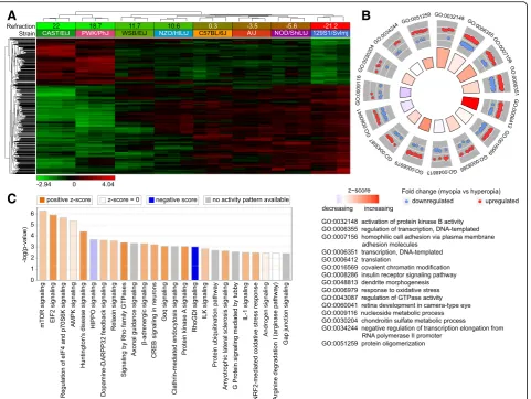
To identify retinal genes influencing baseline refract-ive eye development in mice, we used RNA-seq to analyze gene expression in the retina of eight CC strains at P28 (an age when refractive development is progressing towards its stable plateau) (Fig. 2). We found that expression of 2,302 retinal genes strongly correlated with the baseline refractive state (Fig. 2a, Additional file 2: Table S3). Genes were organized in two distinct clusters. Expression of 793 genes com-prising the first cluster was positively correlated with hyperopia, i.e., expression was increased in the strains with positive refractive errors and decreased in the strains with negative refractive errors. Conversely, ex-pression of 1,509 genes comprising the second cluster was positively correlated with myopia, i.e., expression of these genes was increased in the mouse strains with negative refractive errors and decreased in the strains with positive refractive errors. We observed a clear transition from the “hyperopic” gene expression pattern in CAST/EiJ and PWK/PhJ mice with highly hyperopic refractive errors to the “myopic” gene ex-pression pattern in 129S1/svlmj mice with highly my-opic refractive errors. Other strains occupied intermediate positions between these two extremes and exhibited transitional patterns of gene expression, which correlated with average baseline refractive er-rors in these strains.
Gene ontology analysis revealed that the 2,302 genes whose expression correlated with baseline refractive error were associated with 116 biological processes, 77 cellular components, and 69 molecular functions in the retina (Additional file2: Table S4). Biological processes
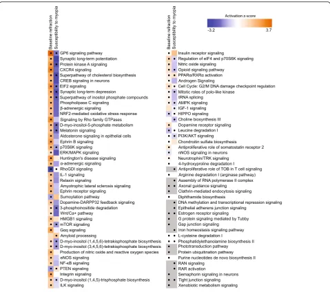
involved in the regulation of baseline refractive devel-opment ranged from regulation of neurogenesis and neuron migration to regulation of DNA methylation, visual perception, and synaptic vesicle endocytosis. Figure 2b shows the top 15 biological processes asso-ciated with these genes, including regulation of pro-tein kinase B, regulation of transcription and translation, covalent chromatin modification, insulin receptor signaling, dendrite morphogenesis, and re-sponse to oxidative stress, among others. Genes underlying baseline refractive development were also associated with multiple canonical signaling pathways in the retina (Fig. 2c, Additional file 2: Table S5). Negative refractive errors were associated with activa-tion of mTOR, EIF2, AMPK, β-adrenergic, and dopa-mine-DARPP32 feedback signaling pathways and suppression of HIPPO and RhoGDI signaling path-ways, among others. Taken together, these data sug-gest that refractive eye development is regulated by a large number of genes and pathways. In summary, the development of hyperopic and myopic refractive errors was associated with specific patterns of gene expression, and the activation or suppression of many retinal signaling pathways.
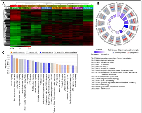
Large number of genes are involved in regulation of susceptibility to myopia in mice via multiple retinal biological processes and signaling pathways
processes, 61 cellular components, and 41 molecular functions were associated with the 1,917 genes corre-lated with susceptibility to myopia (Additional file 3: Table S7). Figure 3b shows the top 15 biological pro-cesses involved in the regulation of susceptibility to my-opia, including regulation of signal transduction, cell-cell adhesion, transcription, translation, protein transport, and lysosome organization, among others. Analysis of the canonical pathways influenced by the genes corre-lated with susceptibility to myopia revealed that in-creased susceptibility to myopia was associated with suppression of mTOR signaling, EIF2 signaling, protein kinase A signaling, D-myo-inositol-5-phosphate metab-olism, cholesterol and choline biosynthesis, as well as with activation of amyloid processing, HIPPO signaling, PTEN signaling, and PPARα/RXRα signaling pathways (Fig. 3c, Additional file 3: Table S8). Collectively, these data implicate an elaborate retinal genetic network and multiple signaling pathways in the regulation of suscepti-bility to myopia in mice.
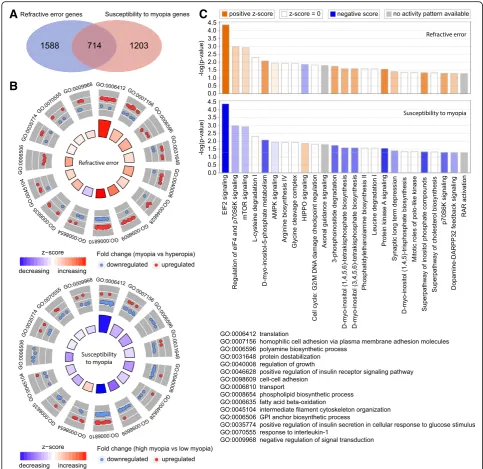
Baseline refractive eye development and susceptibility to myopia in mice are regulated via overlapping but largely distinct retinal genetic networks
To estimate the relative contribution of genes whose ex-pression level correlated with baseline refractive error versus susceptibility to myopia, we analyzed the overlap between the two gene sets (Fig. 4). We found that 714 genes were correlated with both baseline refractive de-velopment and susceptibility to myopia (Fig. 4a, Add-itional file4: Table S9). Gene ontology analysis revealed that these 714 genes were associated with 24 biological processes, 24 cellular components, and 14 molecular functions (Additional file4: Table S10). Figure4b shows the top 15 biological processes for the overlapping genes. Interestingly, the majority of these biological processes were suppressed in animals with high susceptibility to myopia (lower panel) and were activated in animals with negative baseline refractive errors (upper panel). A simi-lar trend was observed when we compared canonical signaling pathways affected by the overlapping genes (Fig. 4c, Additional file 4: Table S11). Signaling path-ways, which were activated in mice with highly negative baseline refractive errors, were suppressed in mice with high susceptibility to myopia, and vice versa. The top pathways associated with both baseline refractive devel-opment and susceptibility to myopia were EIF2 signal-ing, protein kinase A signalsignal-ing, regulation of eIF4 and p70S6K signaling, mTOR pathway, HIPPO pathway, and axonal guidance signaling. Figure5shows a summary of all signaling pathways correlated with baseline refractive eye development and susceptibility to myopia. In addition to the pathways listed above, a number of other
development and susceptibility to myopia, including GP6 signaling pathway, melatonin signaling, RhoGDI signaling, PTEN signaling, opioid signaling pathway, PPARα/RXRα activation, PI3K/AKT signaling, estrogen receptor signaling, and tight junction signaling. Con-versely, synaptic long-term potentiation, α-adrenergic and β-adrenergic signaling, androgen and aldosterone signaling, ephrin receptor signaling, relaxin signaling, dopamine-DARPP32 feedback signaling, dopamine re-ceptor signaling, eNOS and nNOS signaling, somato-statin receptor 2 signaling, neurotrophin/TRK signaling, protein ubiquitination pathway, gap junction signaling, phototransduction pathway, and several other pathways were associated with baseline refractive development but not susceptibility to myopia. The amyloid processing, IGF-1 signaling, DNA methylation and transcriptional repression signaling, epithelial adherens junction signal-ing, iron homeostasis signaling pathway, RAR activation, RAN signaling, and several other pathways were associ-ated with susceptibility to myopia but not baseline re-fractive error. Thus, these data suggest that baseline refractive development and susceptibility to myopia are regulated by genetic networks with considerable overlap; however, the two genetic networks have substantial unique components, which may independently regulate either baseline refractive development or susceptibility to environmentally induced myopia.
Many genes regulating baseline refractive eye development or susceptibility to myopia in mice are localized within chromosomal loci linked to human myopia
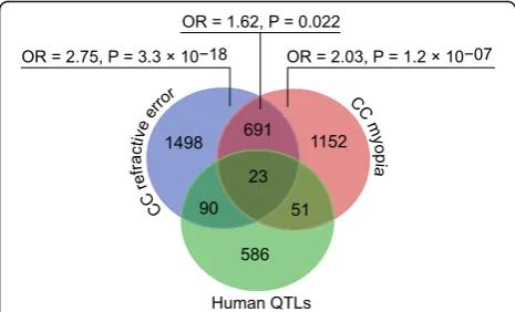
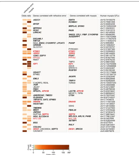
human QTLs and genes associated with both baseline re-fractive development and susceptibility to myopia in mice was weaker (OR = 1.62,P= 0.022); however, overall, these data suggest a functional association between genes asso-ciated with refractive eye development in mice and genes causing myopia in humans. GeneOverlap analysis revealed that a total of 164 mouse genes were located within 109 human QTLs, producing 1.5 candidate genes per QTL (Figs.7and8, Additional file7: Table S16).
We next analyzed biological processes linked to the genes associated with refractive development in mice and local-ized within human myopia QTLs (Fig.9, Additional file8: Tables S17 and S18). Surprisingly, we found that although several biological processes, such as cell growth and prolif-eration, circadian regulation of gene expression, and regula-tion of neuron differentiaregula-tion, were implicated in both baseline refractive development and regulation of suscepti-bility to myopia, many biological processes underlying base-line refractive development and susceptibility to myopia appeared to be different. Our data on the genes associated with baseline refractive development in mice and localized in human QTLs implicate several unique processes, includ-ing camera-type eye development, regulation of ion/cal-cium transport, regulation of membrane polarization during action potential and long-term synaptic potenti-ation, nitric oxide signaling, ephrin receptor signaling, glucocorticoid receptor signaling, regulation of glutamate secretion, and regulation of JAK-STAT and MAPK signaling cascades (Fig.9a, Additional file8: Table S17). Conversely, the genes associated with susceptibility to myopia in mice and localized in human QTLs highlighted several different processes, including regulation of cell shape and cell migra-tion, dorsal/ventral pattern formamigra-tion, vasculature morpho-genesis, photoreceptor cell function and development, cellular response to DNA damage, and small-GTPase-me-diated signal transduction (Fig.9b, Additional file8: Table S18). Collectively, these data suggest that there is a signifi-cant functional overlap between genes we found to be cor-related with refractive eye development in mice and genes causing myopia in humans. Moreover, our data suggest that development of refractive errors in humans is associated with genes that, in mice, regulate both baseline refractive development and susceptibility to refractive changes in-duced by the visual environment.
Gene-based genome-wide association analysis of mouse genes identifies novel gene candidates for human myopia To build on the significant overlap observed above be-tween genes associated with refractive eye development in mice and human myopia QTLs, we examined whether the mouse genes whose expression level correlated with refractive error or myopia susceptibility were enriched for genetic variants associated with refractive error in humans. Human genes enriched for variants associated
with refractive error were identified with MAGMA (Multi-marker Analysis of GenoMic Annotation), using single-marker summary statistics from a genome-wide association study (GWAS) for refractive error and age-of-onset-of-spectacle-wear reported by the CREAM Consortium (N= 160,420 participants; [54]) or a GWAS for refractive error by the UK Biobank Eye & Vision Consortium (N= 88,005 participants; [72]) as input. Flanking regions of 200 kb upstream and downstream of transcription start and stop sites were included, in order to capture regulatory variants influencing the expression of nearby genes.
Of the 2,302 genes associated with baseline refractive development in mice for which there were human ho-mologs, 277 genes in the CREAM dataset were associ-ated with refractive error in humans (FDR< 0.05), with 86 genes surviving Bonferroni correction (PBonferroni< 0.05) (Additional file9: Table S19). Sixty-seven of these genes (including 43 genes withPBonferroni< 0.05) were lo-calized within previously identified human QTLs (Figs.7
and 8), while 210 of the 277 genes (including 43 genes with PBonferroni< 0.05) have not previously been impli-cated in the development of refractive errors in the hu-man population (Additional file9: Table S19). When the above analysis was repeated using the (independent) UK Biobank dataset, 560 genes were associated with refract-ive error (FDR< 0.05), including 156 genes with gen-ome-wide significance (PBonferroni< 0.05) (Additional file
9: Table S20). Four hundred eighty-eight of the five hun-dred sixty genes (including 104 genes with PBonferroni< 0.05) have not previously been implicated in the devel-opment of refractive errors in humans (Additional file9: Table S20). Importantly, 190 genes associated with base-line refractive development in mice were replicated in both the CREAM and UK Biobank cohorts, including 69 genes which achieved genome-wide significance (P Bonfer-roni< 0.05) (Additional file10: Table S23).
opia QTLs (Figs.7and 8), whereas 416 of the 465 genes (including 84 genes withPBonferroni< 0.05) were not previ-ously known to be associated with the development of re-fractive errors in the human population (Additional file9: Table S22). One hundred and fifty-two genes involved in the regulation of susceptibility to myopia in mice were found to be linked to the development of refractive errors in both CREAM and UK Biobank cohorts, including 53 genes which reached genome-wide significance (P Bonfer-roni< 0.05) in both samples (Additional file10: Table S24).
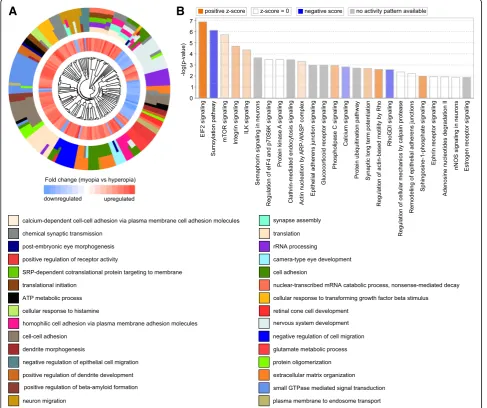
Analysis of the biological processes associated with the above genes linked to either baseline refractive develop-ment or regulation of susceptibility to myopia in humans (Figs. 10a and 11a, Additional file 10: Tables S25 and S26) revealed processes primarily related to calcium-medi-ated cell adhesion, synapse assembly, synaptic transmis-sion, protein translation, small GTPase-mediated signal transduction, and GABA receptor signaling. Several ca-nonical signaling pathways were also identified, including EIF2 and mTOR signaling, eIF4 and p70S6K signaling, epithelial adherence junction signaling, sumoylation path-way, and regulation of cellular mechanics by calpain pro-tease, among others (Figs.10b and11b, Additional file10: Tables S27 and S28).
However, there were also substantial differences be-tween the two sets of human genes identified using ei-ther the mouse baseline refractive error or the mouse myopia susceptibility gene sets. The mouse baseline re-fractive error-derived human gene set was associated with nervous system development, post-embryonic cam-era-type eye development, retinal cone development, neuron migration, dendrite morphogenesis, regulation of glutamate metabolism, extracellular matrix organization, and beta-amyloid formation (Fig. 10a, Additional file 10: Table S25). This gene set was associated with several ca-nonical pathways distinct from those identified using the mouse susceptibility to myopia-derived human gene set. These pathways included integrin signaling, semaphorin signaling in neurons, glucocorticoid receptor signaling, phospholipase C signaling, synaptic long term potenti-ation, ephrin receptor signaling, nNOS signaling in neu-rons, and estrogen receptor signaling (Figs.10b and11b, Additional file10: Tables S27 and S28).
The mouse susceptibility to myopia-derived human gene set, on the other hand, was associated with bio-logical processes related to developmental growth, neuron fate commitment, regulation of mesenchymal cell proliferation, potassium ion transmembrane trans-port, protein transtrans-port, response to estradiol and choles-terol, as well as cellular response to hypoxia (Fig. 11a, Additional file 10: Table S26). Analysis of canonical pathways suggested that pathways associated with
TGF-β signaling, PPARα/RXRα activation, PTEN signaling,
STAT3 signaling, regulation of stem cell pluripotency, VEGF and IGF-1 signaling, NRF2-mediated oxidative stress response, and PI3K/AKT signaling, among others, were involved in the regulation of susceptibility to my-opia (Fig.11b, Additional file10: Table S28).
Discussion
Human population studies and studies in animal models strongly suggest that both environmental and genetic factors play important roles in refractive eye develop-ment. Numerous linkage and genome-wide association studies in humans identified over 270 chromosomal loci linked to the development of myopia in humans [75]; however, very little is known about how genetic variation causing myopia affects gene expression and how changes in gene expression associated with differences in genetic backgrounds affect refractive eye development and sus-ceptibility to myopia.
Our data suggest that differences in genetic back-grounds play a very important role in refractive eye de-velopment and regulation of susceptibility to myopia. Moreover, we found that variations in genetic back-ground produce a continuous distribution of refractive errors and susceptibilities to myopia in a mouse popula-tion, characteristic of quantitative traits. Genetic varia-tions in different mouse strains also produce unique patterns of gene expression, which strongly correlate with either baseline refractive errors or susceptibility to induced myopia. Surprisingly, we found that the baseline refractive development and susceptibility to myopia are controlled by largely distinct sets of genes (Fig.4a, Add-itional file 11: Tables S29 and S30, Additional file 12: Figure S1), which suggests that signaling pathways that regulate the trajectory of refractive eye development to-wards emmetropia, myopia, or hyperopia might be dif-ferent from the pathways that modulate the impact of optical defocus and other environmental factors on re-fractive development. Nevertheless, expression of 714 genes correlated with both baseline refractive errors and susceptibility to myopia, suggesting that at least some genes control both the pathways regulating the trajec-tory of refractive development and the impact of visual input on it.
RhoGDI signaling, PTEN, and HIPPO signaling path-ways, which we found to be activated in animals with high susceptibility to myopia and suppressed in animals with negative baseline refractive errors. The observation that the same pathways were effected in the opposite di-rections in animals with high susceptibility to myopia and animals with highly negative baseline refractive er-rors may be explained by the role of optical defocus in the development of myopia. Considering that mice are housed in small cages and the exposure to distant vision is limited, animals with hyperopic refractive errors would be exposed to high levels of hyperopic optical de-focus (which was shown to cause myopia) compared to the animals with negative refractive errors; thus explain-ing why signalexplain-ing pathways are effected in the same dir-ection in animals with high susceptibility to myopia and animals with hyperopic baseline refractive errors.
Remarkably, we also found that many genes whose ex-pression correlated with either baseline refractive errors or susceptibility to myopia in mice were localized in the known human QTLs linked to myopia. The majority of these genes (90 genes) were exclusively involved in base-line refractive eye development, 51 genes were linked to the regulation of susceptibility to myopia, and 23 genes affected both baseline refractive development and sus-ceptibility to myopia.
Gene-based genome-wide association analysis of the genes we found in mice against CREAM and UK Bio-bank human samples revealed that 647 genes whose ex-pression correlated with baseline refractive errors in mice were also associated with refractive errors in humans, including 173 genes that withstood Bonferroni correction. Using gene-based analysis, we also found that 536 genes whose expression correlated with susceptibil-ity to myopia were associated with refractive errors in humans, including 138 genes which exhibited genome-wide significance (PBonferroni< 0.05). One hundred and ninety-eight of these genes were involved in both base-line refractive development and regulation of susceptibil-ity to myopia, including 34 genes which withstood Bonferroni correction. Although many of these genes were previously implicated in the development of human myopia, 572 genes (92 genes with PBonferroni< 0.05) whose expression correlated with baseline refractive de-velopment and 486 genes (79 genes with PBonferroni< 0.05) whose expression correlated with susceptibility to myopia were linked to human myopia for the first time, including 211 genes (54 genes with PBonferroni< 0.05) whose expression correlated with both baseline refract-ive development and susceptibility to myopia.
Genes that we found to be involved in refractive error development in mice and humans affect a multitude of biological functions in the retina; how-ever, many biological functions underlying refractive
development appear to be highly conserved across species. These include camera-type eye development and post-embryonic eye morphogenesis, ephrin recep-tor signaling, glucocorticoid receprecep-tor signaling, regula-tion of circadian rhythms and circadian regularegula-tion of gene expression, glutamate signaling, regulation of neurogenesis and dendrite morphogenesis, regulation of nitric oxide biosynthesis, regulation of long-term synaptic potentiation, synapse assembly and chemical synapse transmission, calcium-dependent signaling, regulation of translation, small GTPase mediated sig-nal transduction, photoreceptor function and develop-ment, cell-cell adhesion, regulation of beta-amyloid formation, regulation of mesenchymal cell prolifera-tion, cellular response to hypoxia. We also found that many retinal signaling pathways involved in refractive development in mice are also subjected to genetic variation causing myopia in humans (Additional file 10: Tables S27 and S28).
the level of photoreceptors in refractive eye development [84]. A GTP-binding protein GNL2, involved in baseline refractive development, was demonstrated to play an im-portant role in retinal neurogenesis [85]. Finally, AGRN gene, found by this study to be involved in baseline re-fractive development, was shown to interact with EGR1 (previously implicated in refractive eye development) and regulate synaptic physiology in the retina [86,87].
Although many pathways that we found to under-lie refractive development in mice and humans in this study are novel, many of them are conserved across vertebrate species (Table 1). Seven out of 47 pathways found to be involved in optical defocus re-sponse in chickens by Riddell et al. [62] were also found to be involved in refractive eye development in this study (Additional file 2: Table S5, Additional file 3: Table S8, Additional file 10: Tables S27 and S28). We replicated 12 out of 20 canonical pathways identified by Stone et al. [78] in chickens with lens-induced myopia (Additional file 2: Table S5, Additional file 3: Table S8, Additional file 10: Tables S27 and S28). We also found that 35 out of 75 path-ways that we recently found to be involved in the development of hyperopia and myopia in marmosets [76] were also among the pathways which we found to be involved in refractive eye development in this study (Additional file 2: Table S5, Additional file 3: Table S8, Additional file 10: Tables S27 and S28). An important role of the amyloid signaling pathway, which we found to be involved in the regulation of susceptibility to myopia (Additional file 3: Table S8), is in agreement with our recent finding that a com-ponent of the amyloid signaling pathway APLP2 reg-ulates susceptibility to myopia in mice and humans [37]. Our data also suggest that the phototransduction pathway is involved in refractive eye development (Add-itional file2: Table S5), in agreement with a recent GWAS study [54] and recent marmoset data [76].
Conclusions
We have identified 2,302 genes which are involved in baseline refractive eye development and 1,917 genes which regulate susceptibility to myopia in mice. Our data suggest that at least 985 of these genes are sub-jected to genetic variation in the human population and are involved in refractive error development in humans. Eight hundred forty-seven of these genes were impli-cated in the development of human myopia for the first time (Additional file 9: Tables S19-S22). A large number of common genes and canonical pathways that we found to be involved in the regulation of refractive eye devel-opment in mice, chickens and humans suggests strong evolutionary conservation of the signaling pathways
the number of genes involved in the regulation of re-fractive eye development may be as high as 3,505 in the retina alone, suggesting that refractive eye development may be regulated by hundreds to thousands of genes across ocular tissues. Interestingly, in spite of significant overlap between genes that control baseline refractive development and genes regulating susceptibility to my-opia, these two processes appear to be controlled by largely distinct sets of genes. The genes that we found to be involved in refractive eye development control, how-ever, a well-defined set of retinal signaling pathways that we begin to consistently find to be involved in refractive eye development across different species. This provides a solid framework for future studies and for the develop-ment of anti-myopia drugs.
Additional files
Additional file 1Table S1.Baseline refractive errors in Collaborative Cross mice measured at P40 (diopters).Table S2.Form-deprivation myopia in Collaborative Cross mice (deprived eye versus control eye, diopters). (XLSX 14 kb)
Additional file 2Table S3.List of genes whose expression correlates with baseline refractive error in Collaborative Cross mice.Table S4.Gene ontology categories significantly associated with genes whose expression correlates with baseline refractive error in Collaborative Cross mice (BP, biological process; CC, cellular component; MF, molecular function).
Table S5.Canonical signaling pathways affected by genes whose expression correlates with baseline refractive error in Collaborative Cross mice. (XLSX 192 kb)
Additional file 3Table S6.List of genes whose expression correlates with susceptibility to myopia in Collaborative Cross mice.Table S7.Gene ontology categories significantly associated with genes whose expression correlates with susceptibility to myopia in Collaborative Cross mice (BP, biological process; CC, cellular component; MF, molecular function).
Table S8.Canonical signaling pathways affected by genes whose expression correlates with susceptibility to myopia in Collaborative Cross mice. (XLSX 158 kb)
Additional file 4Table S9.List of genes whose expression correlates with both baseline refractive error and susceptibility to myopia in Collaborative Cross mice.Table S10.Gene ontology categories significantly associated with genes whose expression correlates with both baseline refractive error and susceptibility to myopia in Collaborative Cross mice (BP, biological process; CC, cellular component; MF, molecular function).Table S11.Canonical signaling pathways affected by genes whose expression correlates with both baseline refractive error and susceptibility to myopia in Collaborative Cross mice. (XLSX 110 kb)
Additional file 5Table S12.List of human myopia QTLs and candidate genes located within 200 kb of the lead SNP (or within critical region). (XLSX 28 kb)
Additional file 6Table S13.List of genes localized within human myopia QTLs and whose expression correlates with baseline refractive error in mice.Table S14.List of genes localized within human myopia QTLs and whose expression correlates with susceptibility to myopia in mice.Table S15.List of genes localized within human myopia QTLs and whose expression correlates with both baseline refractive error and susceptibility to myopia in mice. (XLSX 28 kb)
Additional file 8Table S17.Gene ontology categories significantly associated with genes localized within human myopia QTLs and whose expression correlates with baseline refractive error in mice (BP, biological process; CC, cellular component; MF, molecular function).Table S18.
Gene ontology categories significantly associated with genes localized within human myopia QTLs and whose expression correlates with susceptibility to myopia in mice (BP, biological process; CC, cellular component; MF, molecular function). (XLSX 15 kb)
Additional file 9Table S19.List of mouse genes displaying significant association with refractive error in CREAM cohort and whose expression correlates with baseline refractive error in mice (Blue identifies PBonferroni< 0.05 in both cohorts).Table S20.List of mouse genes
displaying significant association with refractive error in UK Biobank cohort and whose expression correlates with baseline refractive error in mice (Blue identifies PBonferroni< 0.05 in both cohorts).Table S21.List of
mouse genes displaying significant association with refractive error in CREAM cohort and whose expression correlates with susceptibility to myopia in mice (Blue identifies PBonferroni< 0.05 in both cohorts).Table
S22.List of mouse genes displaying significant association with refractive error in UK Biobank cohort and whose expression correlates with susceptibility to myopia in mice (Blue identifies PBonferroni< 0.05 in both
cohorts). (XLSX 172 kb)
Additional file 10Table S23.List of mouse genes displaying significant association with refractive error in both UK Biobank and CREAM cohorts, and whose expression correlates with baseline refractive error in mice (Blue identifies PBonferroni< 0.05 in both cohorts).Table S24.List of
mouse genes displaying significant association with refractive error in both UK Biobank and CREAM cohorts, and whose expression correlates with susceptibility to myopia in mice (Blue identifies PBonferroni< 0.05 in
both cohorts).Table S25.Biological processes significantly associated with genes linked to refractive error in UK Biobank and CREAM cohorts, and whose expression correlates with baseline refractive error in mice (BP, biological process).Table S26.Biological processes significantly associated with genes linked to refractive error in UK Biobank and CREAM cohorts, and whose expression correlates with susceptibility to myopia in mice (BP, biological process).Table S27.Canonical signaling pathways significantly associated with genes linked to refractive error in UK Biobank and CREAM cohorts, and whose expression correlates with baseline refractive error in mice.Table S28.Canonical signaling pathways significantly associated with genes linked to refractive error in UK Biobank and CREAM cohorts, and whose expression correlates with susceptibility to myopia in mice. (XLSX 91 kb)
Additional file 11Table S29.List of genes whose expression correlates with baseline refractive error in Collaborative Cross mice (molecular function).Table S30.List of genes whose expression correlates with susceptibility to myopia in Collaborative Cross mice (molecular function). (XLSX 163 kb)
Additional file 12Figure S1.Venn diagrams showing overlaps between different functional classes of genes underlying baseline refractive eye development and genes regulating susceptibility to myopia in Collaborative Cross progenitor strain mice. (EPS 1034 kb)
Abbreviations
AGRN:Agrin; AKT: Protein kinase B; AMPK: AMP-activated protein kinase; ANOVA: Analysis of variance; CC: Collaborative Cross; CDK5: Cyclin-dependent kinase 5; CREAM: Consortium for Refractive Error and Myopia; CREB: cAMP response element binding protein; DARPP32: Dopamine- and cAMP-regulated phosphoprotein 32 kDa; DAVID: Database for annotation, visualization and integrated discovery; EFEMP1: EGF containing fibulin-like extracellular matrix protein 1; EGR1: Early growth response 1; EIF2: Eukaryotic initiation factor 2; eIF4: Eukaryotic initiation factor 4; eNOS: Endothelial nitric oxide synthase; EPHA10: Ephrin receptor A10; GABA: Gamma-aminobutyric acid; GNL2: Guanine nucleotide binding protein-like 2; GP6: Glycoprotein VI; GWAS: Genome-wide association study; IGF-1: Insulin-like growth factor 1; MAGMA: Multi-marker Analysis of GenoMic Annotation; mTOR: Mammalian target of rapamycin; nNOS: Neuronal nitric oxide synthase; NRF2: Nuclear factor erythroid 2-related factor 2; PI3K: Phosphatidylinositol 3-kinase; PPARα: Peroxisome proliferator-activated receptor alpha; PTEN: Phosphatase and tensin homolog; QTL: Quantitative trait locus; RAN: Ras-related nuclear
protein; RAR: Retinoic acid receptor; RhoGDI: Rho GDP-dissociation inhibitor; RIN: RNA Integrity Number; RNA-seq: Massive parallel RNA sequencing; RPE: Retinal pigment epithelium; RXRα: Retinoid X receptor alpha;
SNP: Single-nucleotide polymorphism; STAT3: Signal transducer and activator of transcription 3; TGF-β: Transforming growth factor beta; TRK: Tyrosine receptor kinase; TTC21B: Tetratricopeptide repeat domain 21B; VEGF: Vascular endothelial growth factor; Wnt: Wingless-related protein
Acknowledgements
We are grateful to the CREAM Consortium, the UK Biobank Eye and Vision Consortium, and all the individuals who took part in the CREAM and UK Biobank studies, the personnel who recruited them, interviewers, computer and laboratory technicians, clerical workers, research scientists, volunteers, managers, receptionists and nurses. This research has been conducted using the CREAM datasets and the UK Biobank Resource (application #17351). Special thanks to Jeremy Guggenheim who provided valuable feedback on an early version of the manuscript.
Authors’contributions
TVT and AVT conceptualized the study, analyzed refractive development in mice, performed RNA-seq, and analyzed the data. RLS performed gene-based genome-wide association analysis. TN contributed to the analysis of mouse data. AVT supervised the entire study, analyzed and validated data, and wrote the original draft of the manuscript. All authors read, edited, and approved the final version of the manuscript.
Funding
This work was supported by the National Institutes of Health grants R01EY023839 (AVT), P30EY019007 (Core Support for Vision Research received by the Department of Ophthalmology, Columbia University), and Research to Prevent Blindness (Unrestricted funds received by the Department of Ophthalmology, Columbia University). The funders had no role in study design, data collection and analysis, decision to publish, or preparation of the manuscript.
Availability of data and materials
All data generated or analyzed during this study are included in this article and its supplementary information files.
Ethics approval and consent to participate
Mice were obtained from the Jackson Laboratory (Bar Harbor, ME) and were maintained as an in-house breeding colony. All procedures adhered to the Association for Research in Vision and Ophthalmology (ARVO) statement on the use of animals in ophthalmic and vision research and were approved by the Columbia University Institutional Animal Care and Use Committee. Animals were anesthetized via intraperitoneal injection of ketamine (90 mg/ kg) and xylazine (10 mg/kg) and were euthanized using CO2followed by
cer-vical dislocation.
All human studies were approved by the relevant institutional review boards and/or medical ethics committees and conducted according to the Declaration of Helsinki. All CREAM participants provided written informed consent. The UK Biobank received ethical approval from the National Health Service National Research Ethics Service (reference 11/NW/0382).
Consent for publication
Not applicable.
Competing interests
AVT is a named inventor on two US patent applications related to the development of a pharmacogenomics pipeline for anti-myopia drug development. The remaining authors declare that they have no competing interests.
Author details
1
Department of Ophthalmology, Columbia University, New York, NY, USA. 2School of Optometry & Vision Sciences, Cardiff University, Cardiff, UK. 3Department of Pathology and Cell Biology, Columbia University, New York,
References
1. Pararajasegaram R. VISION 2020-the right to sight: from strategies to action. Am J Ophthalmol. 1999;128(3):359–60.
2. Kempen JH, Mitchell P, Lee KE, Tielsch JM, Broman AT, Taylor HR, et al. The prevalence of refractive errors among adults in the United States, Western Europe, and Australia. Arch Ophthalmol. 2004;122(4):495–505.
3. Javitt JC, Chiang YP. The socioeconomic aspects of laser refractive surgery. Arch Ophthalmol. 1994;112(12):1526–30.
4. Vitale S, Sperduto RD, Ferris FL 3rd. Increased prevalence of myopia in the United States between 1971-1972 and 1999-2004. Arch Ophthalmol. 2009; 127(12):1632–9.
5. Holden BA, Fricke TR, Wilson DA, Jong M, Naidoo KS, Sankaridurg P, et al. Global prevalence of myopia and high myopia and temporal trends from 2000 through 2050. Ophthalmology. 2016;123(5):1036–42.
6. Lin LL, Shih YF, Hsiao CK, Chen CJ. Prevalence of myopia in Taiwanese schoolchildren: 1983 to 2000. Ann Acad Med Singap. 2004;33(1):27–33. 7. Lam CS, Goldschmidt E, Edwards MH. Prevalence of myopia in local and
international schools in Hong Kong. Optom Vis Sci. 2004;81(5):317–22. 8. Alexander LJ. Primary care of the posterior segment. 2nd ed. Appleton &
Lange: Connecticut; 1994.
9. Saw SM, Gazzard G, Shih-Yen EC, Chua WH. Myopia and associated pathological complications. Ophthalmic Physiol Opt. 2005;25(5):381–91. 10. Flitcroft DI. The complex interactions of retinal, optical and environmental
factors in myopia aetiology. Prog Retin Eye Res. 2012;31(6):622–60. 11. Hurst J, Johnson D, Law C, Schweitzer K, Sharma S. Value of subjective
visual reduction in patients with acute-onset floaters and/or flashes. Can J Ophthalmol. 2015;50(4):265–8.
12. Shunmugam M, Ang GS, Lois N. Giant retinal tears. Surv Ophthalmol. 2014; 59(2):192–216.
13. Rumelt S, Sarrazin L, Averbukh E, Halpert M, Hemo I. Paediatric vs adult retinal detachment. Eye (Lond). 2007;21(12):1473–8.
14. Brasil OF, Brasil MV, Japiassu RM, Biancardi AL, Souza DD, Oliveira RC, et al. Fundus changes evaluation in degenerative myopia. Arq Bras Oftalmol. 2006;69(2):203–6.
15. Ndiaye PA, Koffane RR, Wade A, Ndiaye CS, Gomez JC, Ndiaye MR. Frequency of retinal changes in myopia in a black population. J Fr Ophtalmol. 2001;24(9):927–9.
16. Bonnet M. Myopia and rhegmatogenous retinal detachment. Rev Prat. 1993; 43(14):1779–83.
17. Pierro L, Camesasca FI, Mischi M, Brancato R. Peripheral retinal changes and axial myopia. Retina. 1992;12(1):12–7.
18. Grossniklaus HE, Green WR. Pathologic findings in pathologic myopia. Retina. 1992;12(2):127–33.
19. Burton TC. The influence of refractive error and lattice degeneration on the incidence of retinal detachment. Trans Am Ophthalmol Soc. 1989;87:143–55 discussion 55-7.
20. Pruett RC, Weiter JJ, Goldstein RB. Myopic cracks, angioid streaks, and traumatic tears in Bruch's membrane. Am J Ophthalmol. 1987;103(4):537–43. 21. Chaine G, Sebag J, Coscas G. The induction of retinal detachment. Trans
Ophthalmol Soc U K. 1983;103(Pt 4):480–5.
22. Menezo JL, Suarez-Reynolds R, Frances J, Vila E. Shape, number and localization of retinal tears in myopic over 8D, aphakic and traumatic cases of retinal detachment. An experience report. Ophthalmologica. 1977;175(1):10–8.
23. Kanski JJ. Giant retinal tears. Am J Ophthalmol. 1975;79(5):846–52. 24. Verhoeven VJ, Wong KT, Buitendijk GH, Hofman A, Vingerling JR, Klaver CC.
Visual consequences of refractive errors in the general population. Ophthalmology. 2015;122(1):101–9.
25. Qiu M, Wang SY, Singh K, Lin SC. Association between myopia and glaucoma in the United States population. Invest Ophthalmol Vis Sci. 2013;54(1):830–5. 26. Praveen MR, Vasavada AR, Jani UD, Trivedi RH, Choudhary PK. Prevalence of
cataract type in relation to axial length in subjects with high myopia and emmetropia in an Indian population. Am J Ophthalmol. 2008;145(1):176–81. 27. Loyo-Berrios NI, Blustein JN. Primary-open glaucoma and myopia: a
narrative review. WMJ. 2007;106(2):85–9 95.
28. Pizzarello L, Abiose A, Ffytche T, Duerksen R, Thulasiraj R, Taylor H, et al. VISION 2020: the right to sight: a global initiative to eliminate avoidable blindness. Arch Ophthalmol. 2004;122(4):615–20.
–
30. Young TL. Molecular genetics of human myopia: an update. Optom Vis Sci. 2009;86(1):E8–E22.
31. Baird PN, Schache M, Dirani M. The GEnes in myopia (GEM) study in understanding the aetiology of refractive errors. Prog Retin Eye Res. 2010; 29(6):520–42.
32. Wojciechowski R. Nature and nurture: the complex genetics of myopia and refractive error. Clin Genet. 2011;79(4):301–20.
33. Parssinen O, Lyyra AL. Myopia and myopic progression among schoolchildren: a three-year follow-up study. Invest Ophthalmol Vis Sci. 1993;34(9):2794–802.
34. Goss DA. Nearwork and myopia. Lancet. 2000;356(9240):1456–7.
35. Hepsen IF, Evereklioglu C, Bayramlar H. The effect of reading and near-work on the development of myopia in emmetropic boys: a prospective, controlled, three-year follow-up study. Vis Res. 2001;41(19):2511–20. 36. Saw SM, Chua WH, Hong CY, Wu HM, Chan WY, Chia KS, et al. Nearwork in
early-onset myopia. Invest Ophthalmol Vis Sci. 2002;43(2):332–9. 37. Tkatchenko AV, Tkatchenko TV, Guggenheim JA, Verhoeven VJ, Hysi PG,
Wojciechowski R, et al. APLP2 regulates refractive error and myopia development in mice and humans. PLoS Genet. 2015;11(8):e1005432. 38. Troilo D, Li T, Glasser A, Howland HC. Differences in eye growth and the
response to visual deprivation in different strains of chicken. Vis Res. 1995; 35(9):1211–6.
39. Tkatchenko AV, Walsh PA, Tkatchenko TV, Gustincich S, Raviola E. Form deprivation modulates retinal neurogenesis in primate experimental myopia. Proc Natl Acad Sci U S A. 2006;103(12):4681–6.
40. Schaeffel F, Burkhardt E, Howland HC, Williams RW. Measurement of refractive state and deprivation myopia in two strains of mice. Optom Vis Sci. 2004;81(2):99–110.
41. Chen YP, Hocking PM, Wang L, Povazay B, Prashar A, To CH, et al. Selective breeding for susceptibility to myopia reveals a gene-environment interaction. Invest Ophthalmol Vis Sci. 2011;52(7):4003–11.
42. Zhou G, Williams RW. Mouse models for the analysis of myopia: an analysis of variation in eye size of adult mice. Optom Vis Sci. 1999;76(6):408–18. 43. Puk O, Dalke C, Favor J, de Angelis MH, Graw J. Variations of eye size
parameters among different strains of mice. Mamm Genome. 2006;17(8):851–7. 44. Wong AA, Brown RE. Visual detection, pattern discrimination and visual
acuity in 14 strains of mice. Genes Brain Behav. 2006;5(5):389–403. 45. Peet JA, Cotch MF, Wojciechowski R, Bailey-Wilson JE, Stambolian D.
Heritability and familial aggregation of refractive error in the old order Amish. Invest Ophthalmol Vis Sci. 2007;48(9):4002–6.
46. Lyhne N, Sjolie AK, Kyvik KO, Green A. The importance of genes and environment for ocular refraction and its determiners: a population based study among 20-45 year old twins. Br J Ophthalmol. 2001;85(12):1470–6. 47. Hammond CJ, Snieder H, Gilbert CE, Spector TD. Genes and environment in
refractive error: the twin eye study. Invest Ophthalmol Vis Sci. 2001;42(6):1232–6.
48. Teikari JM, Kaprio J, Koskenvuo MK, Vannas A. Heritability estimate for refractive errors--a population-based sample of adult twins. Genet Epidemiol. 1988;5(3):171–81.
49. Dirani M, Chamberlain M, Shekar SN, Islam AF, Garoufalis P, Chen CY, et al. Heritability of refractive error and ocular biometrics: the genes in myopia (GEM) twin study. Invest Ophthalmol Vis Sci. 2006;47(11):4756–61. 50. Lopes MC, Andrew T, Carbonaro F, Spector TD, Hammond CJ. Estimating
heritability and shared environmental effects for refractive error in twin and family studies. Invest Ophthalmol Vis Sci. 2009;50(1):126–31.
51. Verhoeven VJ, Hysi PG, Wojciechowski R, Fan Q, Guggenheim JA, Hohn R, et al. Genome-wide meta-analyses of multiancestry cohorts identify multiple new susceptibility loci for refractive error and myopia. Nat Genet. 2013;45(3):314–8.
52. Kiefer AK, Tung JY, Do CB, Hinds DA, Mountain JL, Francke U, et al. Genome-wide analysis points to roles for extracellular matrix remodeling, the visual cycle, and neuronal development in myopia. PLoS Genet. 2013; 9(2):e1003299.
53. Flitcroft DI, Loughman J, Wildsoet CF, Williams C, Guggenheim JA, for the CC. Novel myopia genes and pathways identified from syndromic forms of myopia. Invest Ophthalmol Vis Sci. 2018;59(1):338–48.