R E V I E W
Open Access
Transcranial direct current stimulation for
promoting motor function in cerebral palsy:
a review
Melanie K. Fleming
1*, Tim Theologis
2, Rachel Buckingham
2and Heidi Johansen-Berg
1Abstract
Transcranial direct current stimulation (tDCS) has the potential to improve motor function in a range of neurological conditions, including Cerebral Palsy (CP). Although there have been many studies assessing tDCS in adult stroke, the literature regarding the efficacy of tDCS in CP is more limited. This review therefore focuses on the neurophysiological and clinical findings in children and adolescents with CP. Initial studies applying anodal tDCS to promote lower limb function are promising, with improvements in gait, mobility and balance reported. However, the results of upper limb studies are mixed and more research is needed. Studies investigating neurophysiological changes or predictors of response are also lacking. Large-scale longitudinal studies are needed for the lower limb to ascertain whether the initial pilot results translate into clinically meaningful improvements. Future studies of the upper limb should focus on determining the optimal stimulation parameters and consider tailoring stimulation to the individual based on the (re)organisation of their motor system.
Keywords:Cerebral palsy, Motor function, Transcranial direct current stimulation, Brain stimulation, Upper limb, Lower limb
Introduction
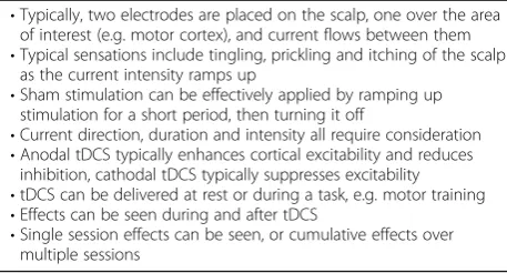
Transcranial direct current stimulation (tDCS), a form of non-invasive brain stimulation, has received considerable interest as a neuromodulatory technique with the poten-tial to enhance cortical plasticity and improve motor func-tion in a range of neurological condifunc-tions. Low intensity, direct, constant current is applied to the scalp (Fig.1), typ-ically over the primary motor cortex (M1), and cortical excitability and inhibition is altered depending on the stimulation parameters [1,2] (Table1).
One of the neurological conditions which may benefit from the neuromodulatory effects of tDCS is Cerebral Palsy (CP), whereby motor function and development are affected by an insult to the developing brain [3]. Since functional deficits limit independence and quality of life long term, the potential to utilise tDCS as an ad-junct to physical therapy for enhancing motor function
is an attractive concept. However, although there has been considerable investigation of the effectiveness of tDCS for adult stroke, the findings cannot be assumed to translate directly into children and adolescents with CP, due to differences in brain size, maturity, anatomy and reorganisation. The application of tDCS in this population appears to be safe [4] and safety guidelines have been developed [5]. This narrative review therefore focuses on the neurophysiological and clinical findings with use of tDCS in children and adolescents (6–21 years) with CP.
Information sources
References for this review were identified, by MF, through searches of PubMed for articles published up to July 2018. Combinations of the terms “cerebral palsy”, “tdcs”, “brain stimulation”, “child stroke” and “pediatric stroke” were used. Additionally, articles were identified through article reference lists. The final reference list was selected, by MF, on the basis of topic relevance. * Correspondence:[email protected]
1Wellcome Centre for Integrative Neuroimaging, FMRIB, Nuffield Department
of Clinical Neurosciences, University of Oxford, John Radcliffe Hospital, Oxford OX3 9DU, UK
Full list of author information is available at the end of the article
Neurophysiological findings
Assessment of change in cortical activity or excitability is important in order to understand the mechanism of action of tDCS. Additionally, differences in neurophysio-logical outcomes may potentially be of use to explain variability in clinical outcome, while variations in neuro-physiological measures at baseline may be able to predict who will benefit from tDCS. Currently, there are very few studies in CP which have reported using brain im-aging or neurophysiological measures alongside tDCS.
TDCS is known to alter cortical excitability, intra-cortical inhibition, and intra-cortical plasticity [1, 2, 6, 7] and these neuromodulatory effects are thought to underlie the behavioural or clinical efficacy of tDCS. Transcranial Magnetic Stimulation (TMS) is com-monly used to assess changes in cortical excitability or intracortical inhibition following a single session of tDCS in adult stroke [8, 9]. However, to our know-ledge, there are no published studies of this type in CP. One study [10] reported an increase in motor evoked potential (MEP) amplitude elicited by TMS following 10 days of anodal tDCS (1 mA, 20 min) tar-geting the lower limb. MEPs were elicited at 110% rest motor threshold (RMT) from the abductor muscle of the thumb and the quadriceps muscle of the lower limb at rest. Each hemisphere was stimulated separ-ately, but the results do not separate the findings from
each muscle or hemisphere. Therefore, although an-odal tDCS appeared to increase cortical excitability, as hypothesised, it is unclear as to how specific the changes are to the targeted region or the time-scale over which these changes occurred.
Changes in brain metabolites following tDCS can be assessed using Magnetic Resonance Spectroscopy (MRS) [2, 11, 12]. This can provide insights into alterations in measures of neuronal health or changes in levels of cor-tical inhibitory or excitatory neurotransmitters. Auvi-chayapat et al. [13] attempted to assess changes in brain metabolites following tDCS using MRS in children with CP. Anodal tDCS (20 min, 1 mA) was delivered for 5 consecutive days to the left M1 in children 8–12 years old with spastic CP affecting their right upper limb. They reported a significant increase in concentrations of N-acetylaspartate (NAA), Choline and Myoinositol in the left basal ganglia and an increase in the ratio of Glx (a combination of glutamate and glutamine) to Creatine in the left M1. Although there was no sham control group, the authors speculated that the tDCS-induced in-crease in activity of the M1 leads to an inin-crease in the concentration of NAA, Choline and Myoinositol in the basal ganglia. There was a negative correlation between the ratio of Glx:Creatine in the M1 and the spasticity (Tardieu scale score) of the right upper limb (shoulder flexors, shoulder external rotators, elbow flexors and elbow pronators) following tDCS. However, the authors did not report whether this relationship existed at base-line or whether the change in metabolite ratios corre-lated with change in spasticity. There was also no indication of the quality of the MRS data, which is typic-ally an important consideration in MRS studies. High quality MRS data may be difficult to obtain in this popu-lation, especially in regions such as the basal ganglia.
Upper limb function
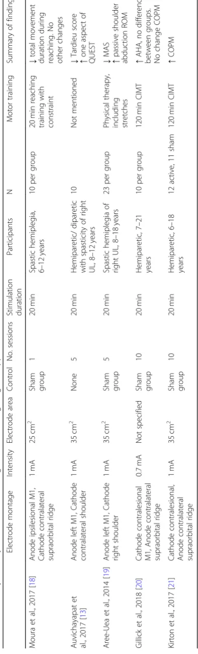
Research on the effect of tDCS on upper limb function in CP is limited to date (Table2). Similar to adult stroke [14] the studies that have been published have utilised the
Fig. 1Diagrammatic representation of tDCS.aAnodal stimulation applied over the motor cortex contralateral to the trained limb.b
Cathodal stimulation applied over the motor cortex ipsilateral to the trained limb, based on the interhemispheric imbalance model
Table 1Fundamentals of tDCS
•Typically, two electrodes are placed on the scalp, one over the area of interest (e.g. motor cortex), and current flows between them
•Typical sensations include tingling, prickling and itching of the scalp as the current intensity ramps up
•Sham stimulation can be effectively applied by ramping up stimulation for a short period, then turning it off
•Current direction, duration and intensity all require consideration
•Anodal tDCS typically enhances cortical excitability and reduces inhibition, cathodal tDCS typically suppresses excitability
•tDCS can be delivered at rest or during a task, e.g. motor training
•Effects can be seen during and after tDCS
“interhemispheric imbalance model”as rationale. The in-terhemispheric imbalance model proposes that there are abnormal levels of interhemispheric inhibition from the contralesional to ipsilesional M1, resulting in a reduction in activity of the ipsilesional M1 during movement of the affected limb and an increase in activity of the contrale-sional M1 [15–17]. Therefore, this model provides ration-ale for applying anodal tDCS to the ipsilesional M1 to increase excitability, or cathodal tDCS to the contrale-sional M1 in an attempt to decrease excitability and thereby upregulate the ispilesional M1 through a reduc-tion in interhemispheric inhibireduc-tion from the contralesional hemisphere.
A single session study [18] delivered 20 min of 1 mA anodal tDCS (or sham) to the ipsilesional M1 of chil-dren with spastic hemiplegia, alongside 20 min of motor training of the affected arm with constraint of the other arm. Using motion analysis, a significant re-duction in total movement duration during reaching movements with the affected hand was observed for the tDCS group compared with sham. Although this ini-tially seems promising, there were numerous compari-sons made, and none of the other change values (e.g. smoothness, velocity or accuracy parameters) showed significant between-group differences.
Two studies have delivered multiple sessions of anodal tDCS in CP [13,19]. Auvichayapat et al. [13] delivered 5 days of tDCS to the left M1. Although there was a mix-ture of hemiparetic and diparetic participants, all had spasticity of their right upper limb. However, there is no mention as to whether there was any motor training alongside the tDCS. Although the authors reported an improvement in spasticity (Tardieu scale) and one aspect of the Quality of Upper Extremity Skills Test (QUEST), there was no sham group for comparison. A randomised, double-blinded study [19] aimed to assess changes in spasticity with 5 consecutive days of anodal tDCS (20 min, 1 mA) to the left M1 of children with spastic hemi-plegia affecting the right arm. In addition to the tDCS, participants engaged in “routine physical therapy”, in-cluding passive and active stretching, therapeutic posi-tioning and aerobic exercise. There were improvements in spasticity of the shoulder, elbow, wrist and fingers and an improvement in shoulder abduction passive range of movement for the active tDCS group only. However, there were no active motion function measures assessed. Two double-blind randomised trials [20,21] have com-bined 20 min of cathodal tDCS of the contralesional M1 with motor training, including constraint induced move-ment therapy (CIMT), over 10 sessions in children with hemiparetic CP. Both active and sham groups demon-strated a significant increase in the Assisting Hand Assess-ment (AHA), which measures bimanual function during novel play or functional tasks, but there was no difference
between groups. Kirton et al. [21] did find greater improvement in self-reported performance (using the Canadian Occupational Performance Measure (COPM)) for the active tDCS group, and a higher proportion of par-ticipants achieved a clinically significant improvement on this measure compared to the sham group. However, the COPM did not show between-group differences in the study by Gillick et al. [20], indicating that more research is needed with both objective and subjective measures.
The intensity of the current for cathodal tDCS may be an issue in the studies so far. Contrary to effects in adults [1], in a study with healthy children (11–16 years) [22], corticospinal excitability was found to increase, ra-ther than decrease, following 1 mA cathodal tDCS. If the intensity of stimulation was lowered to 0.5 mA then the hypothesised decrease in MEP amplitude for cathodal tDCS was evident. Moliadze et al. therefore speculated that 0.5 mA cathodal stimulation in children may pro-duce similar effects as 1 mA in adults. The situation is different from anodal stimulation: 0.5 mA anodal stimu-lation was found to be ineffective at increasing MEP amplitude in children whereas 1 mA anodal stimulation did lead to a significant increase [22], consistent with ef-fects of anodal tDCS in adults [1].
Therefore, there is currently no indication that tDCS provides additional benefit for active motor function over motor training or CIMT alone in children and young people with CP, but spasticity appears to improve with anodal tDCS.
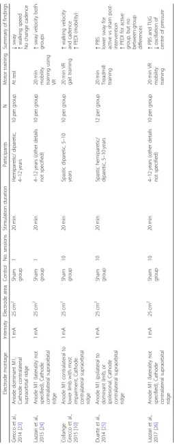
Lower limb function
increase in sway velocity observed immediately could represent a deterioration in balance due to fatigue for both groups following the mobility training. If this is the case, then it would appear that tDCS was not effective at ameliorating this fatigue effect.
Three studies have assessed multiple sessions of anodal tDCS for promoting lower limb function [10, 25, 26]. Duarte et al. [25] delivered 10 sessions of anodal tDCS (1 mA, 20 min), in combination with treadmill training in chil-dren with spastic CP. There was a mixture of hemiparetic and diparetic participants and the anode was placed over the motor cortex ipsilateral to the dominant limb (thereby stimulating the more-affected hemisphere). Interpretation is complicated as the authors report within group changes and between group score comparisons separately, rather than using a mixed analysis of variance or change scores. Nonetheless, within-group comparisons demonstrated an improvement in the Pediatric Balance Scale (PBS) for the active tDCS group only, and between-group comparisons showed that the active group had a higher PBS score and lower sway than the sham group when assessed following the intervention and at the 1 month follow-up. Similarly, there was an improvement for the active group on the mobility subsection of the Pediatric Evaluation Disability Inventory (PEDI), which is a subjective assessment of func-tional performance in activities of daily living. However, the scores did not differ between groups.
Collange Grecco et al. [10] used virtual reality for gait training in combination with 10 sessions of anodal tDCS (1 mA, 20 min) in children with spastic diparetic CP. The virtual reality training involved walking around a simulated race track at varying speeds (Xbox 360 with Kinect movement sensor (Microsoft Corporation, Red-mond, WA)). Participants were asked which lower limb they found had most difficulty during gait and the anode was placed over the contralateral motor cortex. Their primary outcome measure was gait kinematics, using motion analysis. There was a greater improve-ment in walking velocity and cadence for the tDCS group compared to sham, but not for any of the other gait variables assessed. Mobility, assessed using the PEDI, also improved for the active tDCS group, but not for sham. Similarly promising results were found by Lazzari et al. [26], who combined anodal tDCS (1 mA, 20 min) with 20 min of mobility training using virtual reality over 10 sessions. The virtual reality training in-volved a game that simulates stationary walking requir-ing complete flexion of the hip, knee and ankle, and weight transfer from one limb to the other (Xbox 360 with Kinect movement sensor (Microsoft Corporation, Redmond, WA)). They demonstrated a significantly greater improvement in the PBS and the Timed Up and Go (TUG) for the active tDCS group compared with sham. There was also a greater improvement in static
balance, assessed as the oscillation of the centre of pressure. However, variability within groups was high and there was no investigation of variables to account for variability.
Predictors of response
Data on the predictors of response to tDCS are currently lacking in this population. The only study to attempt to analyse potential predictors [27] did so by combining 3 studies that delivered anodal tDCS alongside gait train-ing (for a total of 56 participants) in children with spas-tic hemiparespas-tic or diparespas-tic CP. The authors reported that two predictors were significantly associated with the responsiveness to the intervention; MEP presence during initial evaluation (indicating preservation of the corti-cospinal tract) and location of the injury (cortical or sub-cortical). However, it is unclear whether this is specific to the modulatory effects of the tDCS per se or simply an indicator of who has the potential to improve motor function, as is the case for adult stroke survivors [28].
For the upper limb, it is currently unclear whether anodal or cathodal tDCS should be applied in unilateral CP. In-deed, this decision may depend on the extent to which the activity of each M1 is required for motor function, based on the degree to which the ipsilesional M1 and corticosp-inal tract are damaged. Although in some instances, over-activity of the contralesional hemisphere may be mal-adaptive [29] and benefit from downregulation, in other cases the motor system may be reorganised towards ipsilat-eral control [30, 31]. If the control of the paretic hand is through fast-conducting ipsilateral projections from the contralesional M1, then attempting to enhance ipsilesional M1 excitability with anodal tDCS may be futile. Equally, de-creasing excitability of the contralesional M1 with cathodal tDCS might be detrimental, as is seen for people with severe upper limb impairment after adult stroke [32]. It is difficult to determine whether someone with CP relies on ipsilateral control from the contralesional hemisphere based on clinical presentation alone, as children with ipsilateral projections can show a useful grasp, or no movement at all [30]. Therefore, measures, such as Diffusion Tensor Im-aging (DTI) to assess fractional anisotropy of the corticosp-inal tract, or TMS to assess corticospcorticosp-inal tract integrity through the presence or absence of MEPs, may be neces-sary for informing choices with regard to electrode place-ment. We therefore propose that future studies attempt to optimise tDCS delivery, based on knowledge of the (re)or-ganisation of the individual’s motor system.
Conclusions
Application of tDCS for enhancing lower limb function in young people with CP appears effective, although large-scale longitudinal studies are required to confirm the ini-tially promising findings. Further single-session and longi-tudinal studies are required to determine the efficacy of tDCS for the upper limb and to elucidate mechanisms of action and predictors of response in this population.
Abbreviations
AHA:Assisting Hand Assessment; CIMT: Constraint induced movement therapy; COPM: Canadian Occupational Performance Measure; CP: Cerebral Palsy; DTI: Diffusion Tensor Imaging; M1: Primary motor cortex; MEP: Motor evoked potential; MRS: Magnetic Resonance Spectroscopy; NAA: N-acetylaspartate; PBS: Pediatric Balance Scale; PEDI: Pediatric Evaluation Disability Inventory; QUEST: Quality of Upper Extremity Skills Test; RMT: Rest motor threshold; tDCS: Transcranial direct current stimulation; TMS: Transcranial magnetic stimulation; TUG: Timed Up and Go
Acknowledgements
Not applicable.
Funding
H.J.B. holds a Wellcome Principal Research Fellowship (Grant 110027/Z/15/Z). The Wellcome Centre for Integrative Neuroimaging is supported by core funding from the Wellcome Trust (Grant 203139/Z/16/Z). The funders had no role in the writing of the manuscript.
Availability of data and materials
Not applicable.
Authors’contributions
MKF reviewed the articles and was the major contributor to the article. HJB, TT and RB contributed to the basic concept of the paper and criticially revised the draft paper. All authors read and approved the manuscript.
Ethics approval and consent to participate
Not applicable.
Consent for publication
Not applicable.
Competing interests
The authors declare that they have no competing interests.
Publisher’s Note
Springer Nature remains neutral with regard to jurisdictional claims in published maps and institutional affiliations.
Author details
1
Wellcome Centre for Integrative Neuroimaging, FMRIB, Nuffield Department of Clinical Neurosciences, University of Oxford, John Radcliffe Hospital, Oxford OX3 9DU, UK.2Nuffield Orthopaedic Centre, Oxford University Hospitals NHS Foundation Trust, Oxford, UK.
Received: 30 August 2018 Accepted: 14 December 2018
References
1. Nitsche M, Paulus W. Excitability changes induced in the human motor cortex by weak transcranial direct current stimulation. J Physiol. 2000;527(Pt 3):633–9 Available from:http://www.ncbi.nlm.nih.gov/pubmed/10990547. 2. Stagg CJ, Best JG, Stephenson MC, O’Shea J, Wylezinska M, Kincses ZT, et al.
Polarity-sensitive modulation of cortical neurotransmitters by transcranial stimulation. J Neurosci. 2009;29:5202–6 Available from:http://www.jneurosci. org/cgi/doi/10.1523/JNEUROSCI.4432-08.2009.
3. Rosenbaum P, Paneth N, Leviton A, Goldstein M, Bax M, Damiano D, et al. A report : The definition and classification of cerebral palsy April 2006. Dev
Med child Neurol Suppl. 2007;109:8–14 Available from:https://www.ncbi. nlm.nih.gov/pubmed/17370477.
4. Krishnan C, Santos L, Peterson MD, Ehinger M. Safety of noninvasive brain stimulation in children and adolescents. Brain Stimul. 2016;8:76–87 Available from:https://www.ncbi.nlm.nih.gov/pubmed/25499471.
5. Gillick BT, Gordon AM, Feyma T, Krach LE, Carmel J, Rich TL, et al. Non-invasive brain stimulation in children with unilateral cerebral palsy: a protocol and risk mitigation guide. Front Pediatr. 2018;6:1–9 Available from: http://journal.frontiersin.org/article/10.3389/fped.2018.00056/full.
6. Kidgell DJ, Daly RM, Young K, Lum J, Tooley G, Jaberzadeh S, et al. Different current intensities of anodal transcranial direct current stimulation do not differentially modulate motor cortex plasticity. Neural Plast. 2013;2013:13–5 Available from:https://www.ncbi.nlm.nih.gov/pubmed/23577272. 7. Huang YZ, Lu MK, Antal A, Classen J, Nitsche M, Ziemann U, et al. Plasticity
induced by non-invasive transcranial brain stimulation: a position paper. Clin Neurophysiol. 2017;128:2318–29.https://doi.org/10.1016/j.clinph.2017. 09.007International Federation of Clinical Neurophysiology.
8. Bastani A, Jaberzadeh S. Does anodal transcranial direct current stimulation enhance excitability of the motor cortex and motor function in healthy individuals and subjects with stroke: a systematic review and meta-analysis. Clin Neurophysiol. 2012;123:644–57.https://doi.org/10.1016/j.clinph.2011.08. 029International Federation of Clinical Neurophysiology.
9. Fleming MK, Pavlou M, Newham DJ, Sztriha L, Teo JT. Non-invasive brain stimulation for the lower limb after stroke: what do we know so far and what should we be doing next? Disabil Rehabil. 2017;39:714–20 Available from:https://www.ncbi.nlm.nih.gov/pubmed/27013330.
10. Collange Grecco LA, De Almeida Carvalho Duarte N, Mendonça ME, Galli M, Fregni F, Oliveira CS. Effects of anodal transcranial direct current stimulation combined with virtual reality for improving gait in children with spastic diparetic cerebral palsy: a pilot, randomized, controlled, double-blind, clinical trial. Clin Rehabil. 2015;29:1212–23 Available from:https://www.ncbi. nlm.nih.gov/pubmed/25604912.
11. Bachtiar V, Johnstone A, Berrington A, Lemke C, Johansen-Berg H, Emir U, et al. Modulating regional motor cortical excitability with non-invasive brain stimulation results in neurochemical changes in bilateral motor cortices. J Neurosci. 2018;38:2853–17 Available from:http://www.jneurosci.org/lookup/ doi/10.1523/JNEUROSCI.2853-17.2018.
12. Bachtiar V, Near J, Johansen-Berg H, Stagg CJ. Modulation of GABA and resting state functional connectivity by transcranial direct current stimulation. Elife. 2015;4:1–9 Available from:https://www.ncbi.nlm.nih.gov/ pubmed/26381352.
13. Auvichayapat P, Aree-uea B, Auvichayapat N, Phuttharak W, Janyacharoen T, Tunkamnerdthai O, et al. Transient changes in brain metabolites after transcranial direct current stimulation in spastic cerebral palsy: a pilot study. Front Neurol. 2017;8:1–9 Available from:http://journal.frontiersin.org/article/ 10.3389/fneur.2017.00366/full.
14. Ward NS, Cohen LG. Mechanisms underlying recovery of motor function after stroke. Arch Neurol. 2004;61:1844–8 Available from:https://www.ncbi. nlm.nih.gov/pubmed/15596603.
15. Murase N, Duque J, Mazzocchio R, Cohen LG. Influence of interhemispheric interactions on motor function in chronic stroke. Ann Neurol. 2004;55:400–9. 16. Takeuchi N, Tada T, Toshima M, Ikoma K. Correlation of motor function with transcallosal and intracortical inhibition after stroke. J Rehabil Med. 2010;42: 962–6.
17. Takeuchi N, Izumi S-I. Noninvasive brain stimulation for motor recovery after stroke: mechanisms and future views. Stroke Res Treat. 2012;2012:1–10 Available from:http://www.hindawi.com/journals/srt/2012/584727/. 18. Moura RCF, Santos C, Collange Grecco L, Albertini G, Cimolin V, Galli M, et
al. Effects of a single session of transcranial direct current stimulation on upper limb movements in children with cerebral palsy: a randomized, sham-controlled study. Dev Neurorehabil. 2017;20:368–75 Available from: https://www.ncbi.nlm.nih.gov/pubmed/28632467.
19. Aree-Uea B, Auvichayapat N, Janyacharoen T, Siritaratiwat W, Amatachaya A, Prasertnoo J, et al. Reduction of spasticity in cerebral palsy by anodal transcranial direct current stimulation. J Med Assoc Thail. 2014;97(9):954-62. 20. Gillick B, Rich T, Nemanich S, Chen CY, Menk J, Mueller B, et al. Transcranial direct current stimulation and constraint-induced therapy in cerebral palsy: A randomized, blinded, sham-controlled clinical trial. Eur J Paediatr Neurol. 2018;22:358–68.https://doi.org/10.1016/j.ejpn.2018.02.001Elsevier Ltd. 21. Kirton A, Ciechanski P, Zewdie E, Andersen J, Nettel-Aguirre A, Carlson H,
stroke and hemiparesis. Neurology. 2017;88:259–67 Available from:http:// cochranelibrary-wiley.com/o/cochrane/clcentral/articles/733/CN-01297733/ frame.html.
22. Moliadze V, Schmanke T, Andreas S, Lyzhko E, Freitag CM, Siniatchkin M. Stimulation intensities of transcranial direct current stimulation have to be adjusted in children and adolescents. Clin Neurophysiol. 2015;126:1392–9. https://doi.org/10.1016/j.clinph.2014.10.142International Federation of Clinical Neurophysiology.
23. Grecco LAC, Duarte NAC, Zanon N, Galli M, Fregni F, Oliveira CS. Effect of a single session of transcranial direct-current stimulation on balance and spatiotemporal gait variables in children with cerebral palsy: a randomized sham-controlled study. Brazilian J Phys Ther. 2014;18:419–27 Available from: https://www.ncbi.nlm.nih.gov/pubmed/25372004.
24. Lazzari RD, Politti F, Santos CA, Dumont AJL, Rezende FL, Grecco LAC, et al. Effect of a single session of transcranial direct-current stimulation combined with virtual reality training on the balance of children with cerebral palsy: a randomized, controlled, double-blind trial. J Phys Ther Sci. 2015;27:763–8 Available from:https://www.jstage.jst.go.jp/article/jpts/ 27/3/27_jpts-2014-603/_article.
25. De Almeida Carvalho Duarte N, Grecco LAC, Galli M, Fregni F, Santos Oliveira C. Effect of transcranial direct-current stimulation combined with treadmill training on balance and functional performance in children with cerebral palsy: A double-blind randomized controlled trial. PLoS One. 2014;9 Available from:https://www.ncbi.nlm.nih.gov/pubmed/25171216. 26. Lazzari RD, Politti F, Belina SF, Collange Grecco LA, Santos CA, Dumont AJL,
et al. Effect of transcranial direct current stimulation combined with virtual reality training on balance in children with cerebral palsy: a randomized, controlled, double-blind, clinical trial. J Mot Behav. 2017;49:329–36 Available from:https://www.ncbi.nlm.nih.gov/pubmed/27644454.
27. Grecco LAC, Oliveira CS, Galli M, Cosmo C, de Duarte N AC, Zanon N, et al. Spared primary motor cortex and the presence of MEP in cerebral palsy dictate the responsiveness to tDCS during Gait Training. Front Hum Neurosci. 2016;10:1–11 Available from:http://journal.frontiersin.org/Article/ 10.3389/fnhum.2016.00361/abstract.
28. Stinear CM, Barber PA, Smale PR, Coxon JP, Fleming MK, Byblow WD. Functional potential in chronic stroke patients depends on corticospinal tract integrity. Brain. 2007;130:170–80 Available from:https://www.ncbi.nlm. nih.gov/pubmed/17148468.
29. Kirton A. Advancing non-invasive neuromodulation clinical trials in children: lessons from perinatal stroke. Eur J Paediatr Neurol. 2017;21:75–103.https:// doi.org/10.1016/j.ejpn.2016.07.002Elsevier Ltd.
30. Staudt M. Reorganization after pre- and perinatal brain lesions. J Anat. 2010; 217:469–74 Available from:https://www.ncbi.nlm.nih.gov/pubmed/20649910. 31. Mackey A, Stinear C, Stott S, Byblow WD. Upper limb function and cortical organization in youth with unilateral cerebral palsy. Front Neurol. 2014;5 JUL:1–9 Available from:https://www.ncbi.nlm.nih.gov/pubmed/25071705. 32. Bradnam LV, Stinear CM, Barber PA, Byblow WD. Contralesional