R E S E A R C H
Open Access
Modulation of event-related desynchronization
during kinematic and kinetic hand movements
Kosei Nakayashiki
1†, Midori Saeki
1, Yohei Takata
2, Yoshikatsu Hayashi
3and Toshiyuki Kondo
1*†Abstract
Background: Event-related desynchronization/synchronization (ERD/ERS) is a relative power decrease/increase of electroencephalogram (EEG) in a specific frequency band during physical motor execution and mental motor imagery, thus it is widely used for the brain-computer interface (BCI) purpose. However what the ERD really reflects and its frequency band specific role have not been agreed and are under investigation. Understanding the underlying mechanism which causes a significant ERD would be crucial to improve the reliability of the ERD-based BCI. We systematically investigated the relationship between conditions of actual repetitive hand movements and resulting ERD.
Methods: Eleven healthy young participants were asked to close/open their right hand repetitively at three different speeds (Hold, 1/3 Hz, and 1 Hz) and four distinct motor loads (0, 2, 10, and 15 kgf). In each condition, participants repeated 20 experimental trials, each of which consisted of rest (8–10 s), preparation (1 s) and task (6 s) periods. Under the Hold condition, participants were instructed to keep clenching their hand (i.e., isometric contraction) during the task period. Throughout the experiment, EEG signals were recorded from left and right motor areas for offline data analysis. We obtained time courses of EEG power spectrum to discuss the modulation of mu and beta-ERD/ERS due to the task conditions.
Results: We confirmed salient mu-ERD (8–13 Hz) and slightly weak beta-ERD (14–30 Hz) on both hemispheres during repetitive hand grasping movements. According to a 3×4 ANOVA (speed×motor load), both mu and beta-ERD during the task period were significantly weakened under the Hold condition, whereas no significant difference in the kinetics levels and interaction effect was observed.
Conclusions: This study investigates the effect of changes in kinematics and kinetics on resulting ERD during repetitive hand grasping movements. The experimental results suggest that the strength of ERD may reflect the time differentiation of hand postures in motor planning process or the variation of proprioception resulting from hand movements, rather than the motor command generated in the down stream, which recruits a group of motor neurons.
Keywords: BCI, EEG, ERD, Grasping
Background
In recent years many countries are faced with aged soci-ety. As growth of the elderly population continues, the number of stroke patients with motor paralysis increases [1]. Brain-Computer Interfaces (BCIs) have been sug-gested as one of effective neurorehabilitation means for
*Correspondence: [email protected]
†Equal contributors
1Department of Computer and Information Sciences, Tokyo University of Agriculture and Technology, 2-24-16 Naka-cho, Koganei, 184-8588, Tokyo, Japan
Full list of author information is available at the end of the article
the stroke patients, because it can be closing the impaired sensorimotor loop by compensating somatosensory feed-back on their motor attempt [2-4]. The experience of the BCI neurorehabilitation should be important for promot-ing brain neuroplasticity to recover from motor paralysis. A significant factor for a successful BCI neuroreha-bilitation is reliable detection of human motor intent. Neurophysiological studies have demonstrated that not only neuronal spike recordings but also local field poten-tial (LPF) in cortical [5] and sub-cortical [6,7] areas can be used to decode the movements. In particular, event-related power changes in the neural oscillatory activities
have been studied in relation of sensorimotor processes. For example, LFP in the subthalamic nucleus (STN) dur-ing voluntary grip with different motor efforts has been investigated [7]; they reported that power suppression in the beta band (13–30 Hz) and power increase in the theta/alpha (4–12 Hz), the gamma (55–90 Hz) and higher frequency bands were observed during motor execution, and the power changes correlated with effort levels. Engel et al. (2010) reported that the beta band activity is atten-uated by voluntary movements, but is increased during steady contractions; thus they suggested that the beta band oscillations may reflect maintenance of status quo in both sensory and motor circuits [6].
The event-related and frequency-band specific power decrease/increase are known as event-related desynchro-nization (ERD) and synchrodesynchro-nization (ERS). For the practi-cal use of BCI, attempts to decode the oscillatory activities associated with human sensorimotor processes in non-invasive manner have been intensively investigated [8-13]. Those research are mainly focused on the ERD/ERS in alpha and beta bands. The salient brain oscillations in alpha band (8–13 Hz) over the sensorimotor area is known as mu-rhythm, which is desynchronized during motor planning, execution and imagery of hand/finger move-ments [8,12,13], even when one observes the movement by others [11], though it does not coincide with the results of LPF in STN [7]. Whereas the sensorimotor beta power (14–30 Hz) is totally consistent with invasive studies in the above [6,7], it is attenuated by a voluntary execution and imagery of hand/foot movements, and even passive movements [14], but it prominently increases after move-ment offset (known as beta rebound) and during steady contractions [13,15].
Because the ERD can be observed in both alpha and beta bands during voluntary motor execution and imagery, it is expected to be an intuitive and self-paced (i.e., asyn-chronous) BCI [16,17]. However it is suggested that the ability of voluntary ERD generation varies with individual and it is difficult for most novice BCI users [10]; thus it is generally agreed that sufficient neurofeedback training is necessary to utilize this type of BCI [18,19].
Understanding the conditions (and underlying brain mechanism) which causes a significant ERD would be cru-cial to improve the reliability of the ERD-based BCI. In this context, Cassim et al. (2000) investigated the relation-ship between movement durations under brief/sustained wrist extensions and resulting ERD; they reported there was no difference in pre and post movement periods [20]. Jeon et al. (2011) reported consistent movement duration effect in their motor imagery study [21]. Yuan et al. (2010) reported that mu and beta-ERD are correlated with the speed of repetitive hand grasping movements in both actual execution and motor imagery conditions [12]. Stancak et al. (1997) reported that post movement
mu-ERD and beta-ERS under the heaviest external load condition showed longer duration, concluding that the ERD/ERS is influenced by external load opposing fin-ger movements [22]. In contrast, Chakarov et al. (2009) reported that EEG and EMG spectral power did not show any significant difference among the three force condi-tions, but the beta range EEG-EMG coherence increases as the load increases [15].
Even though several works investigated the effect of kinematics and kinetics on resulting ERD, the sensorimo-tor processes the ERD really reflects and its frequency band specific role have not been agreed and are under investigation. In the paper, we systematically investigated the effects of kinematics (speeds) and kinetics (motor loads) during repetitive hand grasping movements on resulting mu and beta-ERD to be clear some controversial points in the previous literature. Accordingly we discuss about brain function in terms of human sensorimotor execution process.
Methods
Participants
Eleven healthy young participants aged 19–23 years (mean age, 21.1 years) took part in the following exper-iment. All were right handed and had no record of any neurological disorders. The recruitment of the partici-pants and the experimental procedure were approved by the ethics committee of the Tokyo University of Agricul-ture and Technology. All the participants were informed the aim and the procedure of experiments and provided written informed consent prior to participate a series of trials under supervision.
Experimental environment
During the experiment, the participants wore an EEG cap with electrodes; they were seated in a comfortable high-back chair and placed their right arm on the armrest so that a group of muscles of their upper limb is relaxed against the gravity. An LCD monitor was located in front of them and they could see a visual cue on the display.
Experimental design
Figure 1Kinematics pattern of the instructed hand grasping movement.Participants were instructed to relax during the rest and preparation periods, whereas they were asked to grasp their right hand at three distinct speeds (i.e., Hold, Slow, and Fast) and four different grasping loads (0, 2, 10, and 15 kgf) during the task period.
conducted in a fixed order for all participants, namely starting from no load and three different speed conditions (Hold, Slow, and Fast in this order), then the grasping load condition was changed to be one rank heavier.
In each experimental condition, the participants repeated 20 experimental trials, each of which consisted of rest, preparation, and task periods, respectively. To avoid anticipatory response, a random time duration from 8 to 10 s was set in the rest period. Immediately after the rest period, a visual cue (a colored filled circle) was displayed on the LCD monitor to notify the participants of the preparation period (1 s). Participants were instructed
to move their hand paced by the visual cue which was periodically moving up and down in vertical direction indicating closing/opening hand during the task period of 6 s. Figure 1 represents the instructed hand kinematics during an experimental trail. Note that participants had to keep clenching their hand (4 s) under the Hold condition.
EEG recording
To focus on the oscillatory activities in primary motor area, we recorded EEG signals from eight active dry electrodes (g.SAHARA electrode, g.tec, Vienna, Austria) placed around the C3 (left hemisphere) and C4 (right
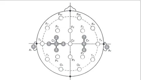
hemisphere) of the international 10–20 system. These areas are well known that 1) mainly reflecting contralat-eral hand movement/imagery and 2) activated bilatcontralat-erally in the case of actual hand movements [12]. The active dry electrodes is a latest technology, and its availability is validated in [23] and [24]. The configuration of eight electrodes are shown in Figure 2. Five electrodes were located in the cross-configuration with C3 at the cen-ter, whereas three electrodes were located in line C4 at the center and other two in anterior and posterior loca-tion. The distance between them was kept at 35-mm in each configuration. The reference and ground electrodes were placed at A1 and A2 (i.e., left and right mastoids), respectively.
The EEG signal was sampled at 512 Hz, preampli-fied in a specific electrode box (g.SAHARAbox, g.tec)
and amplified using a digital multi-telemeter system (WEB5000, NIHON KOHDEN, Tokyo, Japan). The EEG data were band-passed from 0.3 to 100 Hz in the amplifier. The analog signal was converted into digital data by an AD converter board (LPC-321416, Interface, Japan), and stored in a personal computer (Windows 7, Core i5-760, 2.8 GHz).
Signal processing
In offline analysis EEG data were re-treated by bipolar spatial derivation between C3 (channel 1) and other near-est neighbor electrodes (channels 2–5), and were identi-fied as Ch1–2, Ch1–3, Ch1–4, and Ch1–5; in the same manner, the signals by bipolar derivation with respect to C4 (channel 6) were identified as Ch6–7 and Ch6–8 (see Figure 2).
To calculate ERD as a function of time, the time win-dow of 1 s (i.e., 512 samples) was employed to perform the short time Fourier transform (STFT), and the time win-dow was shifted by 1/16 s, we thus obtained instantaneous power spectrum (Pn) at every time window. The ERD was defined as a percentage of power decrease in a specific fre-quency band relative to the baseline period (i.e., the rest period). We calculated the relative power (RP) using the instantaneous power spectrum (Pn):
Prest =
1
|Trest|
n∈Trest Pn,
Ptask =
1
|Ttask|
n∈Ttask Pn,
RP(n)=Pn−Prest Prest ×
100
RP=Ptask−Prest Prest ×
100
wherePrestandPtaskare the mean power spectra during the rest period (Trest) and the task period (Ttask), respec-tively.RPwas averaged across trials within a participant. To obtain salient mu and beta-ERD individually, we evalu-ated the most significant frequency bin (3 Hz band width) and derivation channel (for both C3 and C4) at every experimental condition. The frequency range searching for the salient mu and beta-ERD were selected from 8 to 13 Hz and from 14 to 30 Hz, respectively.
Results
Time course of ERD/ERS
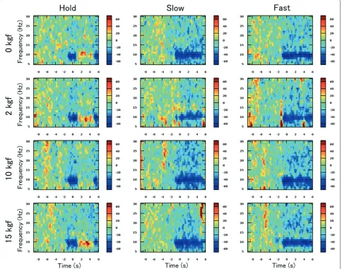
Figure 3 illustrates the time courses of the percentage change in relative power (i.e.RP(n)) on C3 of a typical par-ticipant (subject G) under each speed and grasping load condition. In the figure, significant mu-ERD (i.e., 8–13 Hz) and slightly weak beta-ERD (14–30 Hz) right after the visual cue onset (-1 s in the horizontal time scale) can be observed.
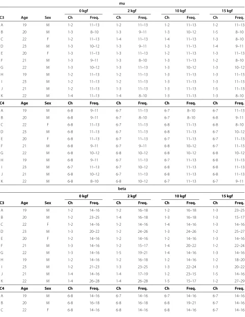
Table 1 Participants-specific bipolar channels (C3/C4) and frequency bands used for the statistical evaluation of mu and beta-ERD
mu
0 kgf 2 kgf 10 kgf 15 kgf
C3 Age Sex Ch Freq. Ch Freq. Ch Freq. Ch Freq.
A 19 M 1-2 11–13 1-2 11–13 1-2 11–13 1-2 11–13
B 20 M 1-3 8–10 1-3 9–11 1-3 10–12 1-5 8–10
C 22 F 1-2 11–13 1-4 11–13 1-4 11–13 1-3 8–10
D 23 M 1-3 10–12 1-3 9–11 1-3 11–13 1-4 9–11
E 20 F 1-3 11–13 1-3 11–13 1-2 11–13 1-3 11–13
F 21 M 1-3 9–11 1-3 8–10 1-3 11–13 1-2 8–10
G 22 M 1-3 10–12 1-3 11–13 1-3 10–12 1-3 10–12
H 19 M 1-2 11–13 1-2 11–13 1-3 11–13 1-3 11–13
I 23 M 1-2 11–13 1-2 11–13 1-3 11–13 1-3 11–13
J 21 M 1-2 11–13 1-3 11–13 1-3 11–13 1-5 11–13
K 22 M 1-4 11–13 1-4 8–10 1-3 11–13 1-3 8–10
C4 Age Sex Ch Freq. Ch Freq. Ch Freq. Ch Freq.
A 19 M 6-8 9–11 6-7 11–13 6-7 8–10 6-7 11–13
B 20 M 6-8 9–11 6-7 8–10 6-7 8–10 6-8 9–11
C 22 F 6-8 11–13 6-7 11–13 6-8 11–13 6-8 8–10
D 23 M 6-8 11–13 6-7 11–13 6-8 11–13 6-7 10–12
E 20 F 6-8 11–13 6-7 11–13 6-7 11–13 6-7 11–13
F 21 M 6-8 9–11 6-7 9–11 6-8 10–12 6-7 11–13
G 22 M 6-8 10–12 6-8 10–12 6-8 10–12 6-8 10–12
H 19 M 6-8 9–11 6-7 11–13 6-7 11–13 6-8 11–13
I 23 M 6-7 11–13 6-7 10–12 6-8 11–13 6-8 11–13
J 21 M 6-8 10–12 6-7 11–13 6-8 11–13 6-8 11–13
K 22 M 6-8 8–10 6-8 10–12 6-7 11–13 6-7 9–11
beta
0 kgf 2 kgf 10 kgf 15 kgf
C3 Age Sex Ch Freq. Ch Freq. Ch Freq. Ch Freq.
A 19 M 1-2 14–16 1-2 16–18 1-2 16–18 1-3 23–25
B 20 M 1-2 23–25 1-4 16–18 1-3 16–18 1-3 15–17
C 22 F 1-2 14–16 1-2 14–16 1-4 14–16 1-3 14–16
D 23 M 1-3 20–22 1-2 24–26 1-3 24–26 1-2 25–27
E 20 F 1-2 14–16 1-2 14–16 1-2 14–16 1-3 14–16
F 21 M 1-3 14–16 1-2 15–17 1-4 20–22 1-2 22–24
G 22 M 1-3 14–16 1-5 19–21 1-4 14–16 1-3 14–16
H 19 M 1-2 14–16 1-2 16–18 1-2 14–16 1-2 18–20
I 23 M 1-2 21–23 1-3 23–25 1-3 22–24 1-3 20–22
J 21 M 1-4 14–16 1-4 17–19 1-2 23–15 1-5 14–16
K 22 M 1-4 26–28 1-4 26–28 1-5 15–17 1-2 27–29
C4 Age Sex Ch Freq. Ch Freq. Ch Freq. Ch Freq.
A 19 M 6-8 14–16 6-7 14–16 6-7 14–16 6-7 14–16
B 20 M 6-8 16–18 6-8 16–18 6-8 19–21 6-7 14–16
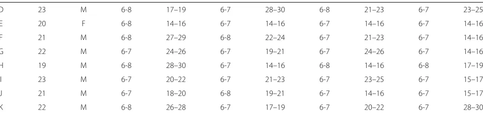
Table 1 Participants-specific bipolar channels (C3/C4) and frequency bands used for the statistical evaluation of mu and beta-ERD(continued)
D 23 M 6-8 17–19 6-7 28–30 6-8 21–23 6-7 23–25
E 20 F 6-8 14–16 6-7 14–16 6-7 14–16 6-7 14–16
F 21 M 6-8 27–29 6-8 22–24 6-7 21–23 6-7 14–16
G 22 M 6-7 24–26 6-7 19–21 6-7 24–26 6-7 14–16
H 19 M 6-8 28–30 6-7 14–16 6-8 14–16 6-8 17–19
I 23 M 6-7 20–22 6-7 21–23 6-7 23–25 6-7 15–17
J 21 M 6-7 18–20 6-8 19–21 6-7 14–16 6-7 15–17
K 22 M 6-8 26–28 6-7 17–19 6-7 20–22 6-7 28–30
Channel 1 represents the C3, and channels 2, 3, 4 and 5 indicate the anterior, posterior, right and left of C3. Channel 6 represents the C4, and channels 7 and 8 indicate the anterior and posterior of C4.
Moreover in the Hold condition, the mu and beta-ERD was changed into mu-ERS immediately after hand closing, regardless of the motor load (i.e., isometric contraction) conditions. This tendency was commonly observed in all the other participants.
Effect of kinematics and kinetics of hand movement on resulting ERD
Figure 4 demonstrates the mean and the standard error of the relative power (RP) in C3/C4 and mu/beta frequency bands across all the participants (n=11) under the dif-ferent grasping speeds and loads condition. Participant-specific bipolar channels (C3/C4) and frequency ranges (mu/beta bands) used for the statistical evaluation are listed in Table 1.
A 3 ×4 repeated-measures ANOVA (speed × motor load) was applied to each hemisphere (C3/C4) and fre-quency band (mu/beta). Under the case of C3 and mu-rhythm, a significant main effect in grasping speed (F(2, 120)=11.214,p<0.001) whereas neither the effect of motor load (F(3, 120)=0.250,p=0.862) nor interac-tion effect (F(6, 120) =0.259,p= 0.955) was confirmed. More importantly, Tukey’s HSD for multiple comparisons exhibits a significant difference between Hold and the other speed conditions (Hold–Slow: p < 0.001, Hold– Fast:p<0.001). In all the cases, tendency of the statistical analyses were identical as shown in the figure.
Discussion
In the present study, we investigated the modula-tion of event-related and frequency-band specific power decrease/increase (i.e., mu and beta ERD/ERS) elicited by actual hand grasping movements under various kinemat-ics (three different velocity patterns) and kinetkinemat-ics (four distinct motor loads) conditions. Our results demon-strated that (1) both time-averaged mu and beta-ERD levels during actual movement period were significantly weakened under the Hold condition and (2) there was no significant difference among different kinetics conditions.
Note that the hand grasping movements under the Hold condition included isometric contraction in the middle phase of the task period (i.e., 1–5 s) whereas the move-ments in the other phases and speed conditions were isotonic contraction. This implies that the time differenti-ation in kinematics (change of hand posture) is correlated with the strength of mu/beta-ERD but maintenance of the current sensorimotor state (i.e., keeping the hand pos-ture in the Hold condition) was related to the mu/beta rebound. These results seem to be consistent with (Engel and Fries, 2010) [6]. Moreover our results suggested that the modulation of the ERD/ERS may not depend on the muscle activities to resist the motor loads.
different. The task used in our study is also hand grasp-ing; thus we suggest that the ERD might be insensitive to the change in kinetics. To our knowledge as the clos-est piece of work to our results and indication, Chakarov et al. (2009) reported that EEG and EMG spectral power did not show any significant difference among the three force conditions, though the beta range EEG-EMG coher-ence increases as the load increases [15].
Even though the above works appears to be contro-versial, the modulation of ERD/ERS may be dependent on several factors such as 1) experimental paradigm, for example single execution or repetitive motion 2) fre-quency range 3) how the average is performed over task duration and 4) brain regions; invasive or non-invasive. Our experimental protocol allows us to systematically dis-cuss the effects of kinematics and kinetics on resulting ERD, and clarify some controversial points in previous lit-erature in the systematic experimental conditions in terms of 1) isometric and isotonic contraction condition, 2) time averaging of ERD/ERS over task period, and 3) kinematics patterns (speed) of grasping motion.
Human sensorimotor process consists of sub-processes; motor intention, planning of motion trajectory, motor command generation, and receiving sensory feedback. Our findings showed that the ERD might not reflect the strength of motor load. However motor commands should be continuously generated to maintain the hand grasp-ing (isometric contraction). Thus we consider that the motor command generation process have little effect on the resulting mu and beta ERD. In contrast, changes in hand posture were found out to be correlated with the ERD strength. We suggest that the strength of mu and beta-ERD may reflect the time differentiation of hand postures in our motor planning process or the varia-tion in proprioceptive sensavaria-tion resulting from the hand movements rather than the motor command generated in the down stream, which recruits a group of motor neurons.
Abbreviations
BCI: Brain-computer interface; EEG: Electroencephalogram; ERD: Event-related desynchronization.
Competing interests
The authors declare that they have no competing interests.
Authors’ contributions
KN and TK supervised the study, designed the experiment, developed the experimental system, signal processing and statistical analysis. TK and YH contributed to discussion of the obtained results. KN, TK, and YH wrote the manuscript. MS, YT, and YH provided fruitful comments for the experimental procedure and analysis. All the authors have read and approved the final manuscript.
Acknowledgements
This research was partially supported by JSPS KAKENHI, Grant-in-Aid for Scientific Research (B) (#23300216). We thank to Mr. Norito Takiuchi (MIYUKI GIKEN Co., Ltd, Japan) who supported the EEG measurement using active dry electrodes. We are also grateful to all participants in the experiment.
Author details
1Department of Computer and Information Sciences, Tokyo University of
Agriculture and Technology, 2-24-16 Naka-cho, Koganei, 184-8588, Tokyo, Japan.2Hamamatsu Photonics K.K. Branch 5000, Hirakuchi, Hamamatsu, Shizuoka, Japan.3Brain Embodiment Laboratory, School of Systems
Engineering, University of Reading, RG6 6AY, Whiteknights, Reading, UK.
Received: 7 February 2014 Accepted: 22 May 2014 Published: 30 May 2014
References
1. McCall M, McEwen S, Colantonio a, Streiner D, Dawson DR:Modified constraint-induced movement therapy for elderly clients with subacute stroke.Am J Occup Therapy2011,65(4):409–418.
2. Takahashi M, Takeda K, Otaka Y, Osu R, Hanakawa T, Gouko M, Ito K:Event related desynchronization-modulated functional electrical stimulation system for stroke rehabilitation: a feasibility study.
J NeuroEng Rehabil2012,9(1):56.
3. Shindo K, Kawashima K, Ushiba J, Ota N, Ito M, Ota T, Kimura A, Liu M: Effects of neurofeedback training with an electroencephalogram-based brain-computer interface for hand paralysis in patients with chronic stroke: a preliminary case series study.J Rehabil Med2011, 43(10):951–957.
4. Hayashi Y, Nagai K, Ito K, Nasuto SJ, Loureiro RCV, Harwin WS:A feasible study of EEG-driven assistive robotic system for stroke
rehabilitation.InProceedings of the 4th IEEE RAS and EMBS International Conference on Biomedical Robotics and Biomechatronics. Rome, Italy; 2012:1733–1739.
5. Pistohl T, Schmidt TSB, Ball T, Schulze-Bonhage A, Aertsen A, Mehring C: Grasp detection from human ECoG during natural reach-to-grasp movements.PloS One2013,8(1):54658.
6. Engel AK, Fries P:Beta-band oscillations–signalling the status quo?
Curr Opinion Neurobiol2010,20(2):156–165.
7. Tan H, Pogosyan A, Anzak A, Ashkan K, Bogdanovic M, Green AL, Aziz T, Foltynie T, Limousin P, Zrinzo L, Brown P:Complementary roles of different oscillatory activities in the subthalamic nucleus in coding motor effort in Parkinsonism.Exp Neurol2013,248:187–195. 8. Pfurtscheller G, Lopes da Silva, FH:Event-related EEG/MEG
synchronization and desynchronization: basic principles.Clin Neurophysiol1999,110(11):1842–1857.
9. Wolpaw JR, Birbaumer N, Mcfarland DJ, Pfurtscheller G, Vaughan TM: Brain-computer interfaces for communication and control.Clin Neurophysiol2002,113(6):767–791.
10. Leeb R, Lee F, Keinrath C, Scherer R, Bischof H, Pfurtscheller G: Brain-computer communication: motivation, aim and impact of exploring a virtual apartment.IEEE Trans Neural Syst Rehabil Eng2007, 15(4):473–482.
11. Neuper C, Scherer R, Wriessnegger S, Pfurtscheller G:Clinical Neurophysiology Motor imagery and action observation:
Modulation of sensorimotor brain rhythms during mental control of a brain-computer interface.Clin Neurophysiol2009,120(2):239–247. 12. Yuan H, Perdoni C, He B:Relationship between speed and EEG activity
during imagined and executed hand movements.J Neural Eng2010, 7(2):26001.
13. Kilavik BRE, Zaepffel M, Brovelli A, MacKay WA, Riehle A:The ups and downs ofβoscillations in sensorimotor cortex.Exp Neurol2013, 245:15–26.
14. Alegre M, Labarga A, Gurtubay IG, Iriarte J, Malanda A, Artieda J:Beta electroencephalograph changes during passive movements: sensory afferences contribute to beta event-related desynchronization in humans.Neurosci Lett2002,331(1):29–32. 15. Chakarov V, Omlor W, Huethe F:Beta-range EEG-EMG coherence with
isometric compensation for increasing modulated low-level forces.
J Neurophysiol2009,102:1115–1120.
16. Ron-Angevin R, Velasco-Alvarez F, Sancha-Ros S, da Silva-Sauer L:A two-class self-paced BCI to control a robot in four directions.In
Proceedings of the 12th IEEE International Conference on Rehabilitation Robotics. Zurich, Switzerland; 2011:977–982.
environments: a case study with a tetraplegic.Comput Intell Neurosci
2007,2007:79642.
18. Hashimoto Y, Ushiba J, Kimura A, Liu M, Tomita Y:Change in brain activity through virtual reality-based brain-machine communication in a chronic tetraplegic subject with muscular dystrophy.BMC Neurosci2010,11:117.
19. Saeki M, Takata Y, Kondo T:Effect of visual stimuli on training of spontaneous ERD.InProceedings of the 16th International Graphonomics Society Conference. Nara, Japan; 2013:90–93.
20. Cassim F, Szurhaj W, Sediri H, Devos D, Bourriez J-L, Poirot I, Derambure P, Defebvre L, Guieu J-D:Brief and sustained movements: differences in event-related (de) synchronization (ERD/ERS) patterns.Clin Neurophysiol2000,111:2032–2039.
21. Jeon Y, Nam CS, Kim Y-J, Cheol M:Event-related (De) synchronization (ERD/ERS) during motor imagery tasks: implications for
brainecomputer interfaces.Int J Ind Ergon2011,41(5):428–436. 22. Stancák a, Riml a, Pfurtscheller G:The effects of external load on
movement-related changes of the sensorimotor EEG rhythms.
Electroencephalogr Clin Neurophysiol1997,102(6):495–504.
23. Saab J, Battes B, Grosse-Wentrup M:Simultaneous EEG recordings with dry and wet electrodes in motor-imagery.InProceedings of the 5th International Brain-Computer Interface Conference. Graz, Austria; 2011:312–315.
24. Guger C, Krausz G, Edlinger G:Brain-computer interface control with dry EEG electrodes.InProceedings of the 5th International
Brain-Computer Interface Conference. Graz, Austria; 2011:316–319. 25. Cramer SC, Weisskoff RM, Schaechter JD, Nelles G, Foley M, Finklestein SP,
Rosen BR:Motor cortex activation is related to force of squeezing.
Hum Brain Mapping2002,16(4):197–205.
26. Keisker B, Hepp-Reymond M-C, Blickenstorfer A, Meyer M, Kollias SS: Differential force scaling of fine-graded power grip force in the sensorimotor network.Hum Brain Mapping2009,30(8):2453–2465. doi:10.1186/1743-0003-11-90
Cite this article as:Nakayashikiet al.:Modulation of event-related desynchronization during kinematic and kinetic hand movements.Journal of NeuroEngineering and Rehabilitation201411:90.
Submit your next manuscript to BioMed Central and take full advantage of:
• Convenient online submission
• Thorough peer review
• No space constraints or color figure charges
• Immediate publication on acceptance
• Inclusion in PubMed, CAS, Scopus and Google Scholar
• Research which is freely available for redistribution