R E V I E W
Open Access
Regenerative abilities of mesenchymal
stem cells through mitochondrial transfer
Swati Paliwal
1, Rituparna Chaudhuri
1, Anurag Agrawal
2*and Sujata Mohanty
1*Abstract
The past decade has witnessed an upsurge in studies demonstrating mitochondrial transfer as one of the emerging mechanisms through which mesenchymal stem cells (MSCs) can regenerate and repair damaged cells or tissues. It has been found to play a critical role in healing several diseases related to brain injury, cardiac myopathies, muscle sepsis, lung disorders and acute respiratory disorders. Several studies have shown that various mechanisms are involved in mitochondrial transfer that includes tunnel tube formation, micro vesicle formation, gap junctions, cell fusion and others modes of transfer. Few studies have investigated the mechanisms that contribute to mitochondrial transfer, primarily comprising of signaling pathways involved in tunnel tube formation that facilitates tunnel tube formation for movement of mitochondria from one cell to another. Various stress signals such as release of damaged mitochondria, mtDNA and mitochondrial products along with elevated reactive oxygen species levels trigger the transfer of mitochondria from MSCs to recipient cells. However, extensive cell signaling pathways that lead to mitochondrial transfer from healthy cells are still under investigation and the changes that contribute to restoration of mitochondrial bioenergetics in recipient cells remain largely elusive. In this review, we have discussed the phenomenon of mitochondrial transfer from MSCs to neighboring stressed cells, and how this aids in cellular repair and regeneration of different organs such as lung, heart, eye, brain and kidney. The potential scope of mitochondrial transfer in providing novel therapeutic strategies for treatment of various pathophysiological conditions has also been discussed.
Keywords:Mesenchymal stem cells, Mitochondrial transfer mechanism, Regenerative medicine
Background
Recent advancements in regenerative medicine have capitalized on using stem cells for cellular repair and regeneration. Mesenchymal stem cells (MSCs) have emerged as promising candidates to treat various dis-eases owing to their unique characteristic properties such as self-renewal, immuno-suppressive potential and ability to trans-differentiate. They are easily avail-able from various tissue sources such as bone marrow, adipose tissue, dental, umbilical cord etc. with low risk of rejection and several clinical trials have provided evidences of their beneficial roles. Interestingly, apart from through engraftment, MSCs are also being shown to exert their actions through paracrine mechanisms, thus repairing cellular damage [1–3]. These paracrine effects
through which MSCs mediate repair can be through release of several immuno-modulatory factors, microvesi-cles, microRNAs, exosomes and mitochondrial transfer [4]. Mitochondrial dysfunction is associated with a major-ity of degenerative diseases like ischemic heart diseases, lung disorders, stroke, brain injury and several degenera-tive diseases including cardiomyopathy, Parkinson’s and Alzheimer’s [5–7]. Thus, mitochondrial transfer alone has appeared as a great therapeutic strategy as it can restore the bioenergetic needs of damaged cells. The transfer of healthy mitochondria appears to be an effective reparative strategy to rejuvenate several kinds of damaged cells such as epithelial cells, endothelial cells, cardiomyocytes and cells of other origins as well. In this review article, we will be discussing the role of mitochondrial transfer through MSCs and how it aids in repair and regeneration of in-jured and damaged cells under stress.
* Correspondence:[email protected];[email protected] 2
Molecular Immunogenetics Laboratory and Centre of Excellence for Translational Research in Asthma & Lung disease, CSIR-Institute of Genomics and Integrative Biology, Delhi 110007, India
1Stem Cell Facility, DBT Centre of Excellence for Stem Cell Research, All India Institute of Medical Sciences, New Delhi 110029, India
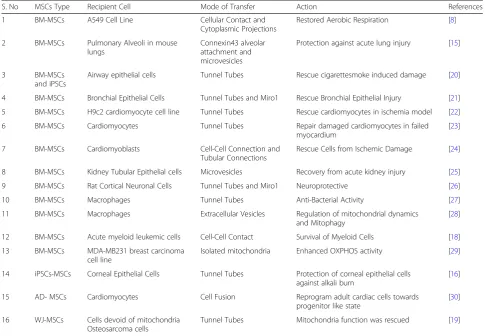
stored when co-cultured with MSCs through mitochondrial transfer. This was demonstrated by presence of DsRed2 tagged mitochondria in MSCs and observed by time-lapse photo microscopy. It was also confirmed that uptake of mitochondria was facilitated by active and not passive transfer [8]. Dysfunctional mitochondria can lead to cellular damage and apoptosis, thus a rescue mechan-ism wherein healthy mitochondria can be transferred has shown tremendous potential. Other cells such as fibroblast and somatic cells have also shown the ability to transfer mitochondria. Mitochondrial donation by MSCs is a faster and more economical physiological process in the cell to replace dysfunctional mitochondria in diseased cells/ tissue, as compared to the mitochondrial biogenesis and thus, appears as an efficient means to attenuate a disease conditions. During the time that Spees et al. published their work, many research groups had simultaneously acknowl-edged and identified the formation of tunnel tube forma-tion for cell-cell communicaforma-tions [9–12]. Rustom et al.,had demonstrated de novo formation of multiple tunnel tubes leading to complex networks between cells and facilitating transfer of membrane vesicles and organelles between cells [9]. These tunnel tubes comprise of F-actin based connec-tions between distant cells and exist in diverse morpholo-gies carrying multiple cargos and signals between cells [13]. These studies were followed by a plethora of studies that provided evidence for mitochondrial transfer be-tween MSCs and damaged cells of varied origins [14– 18]. The discovery of remarkable mitochondrial transfer ability of MSCs to cells with dysfunctional mitochon-dria paved way for numerous studies [14]. MSCs from different tissue sources like bone marrow, adipose, and Wharton’s jelly have now been shown to transfer mito-chondria to various damaged cells, like osteosarcoma cells, that aid in the restoration of their respiratory ac-tivities [19]. Table1provides a list of studies performed using different sources of stem cells in different condi-tions, described in detail in later sections of the review.
Mitochondrial bioenergetics status in MSCs
Mitochondria play important roles in oxidative phos-phorylation, ATP generation, and cellular apoptosis. At
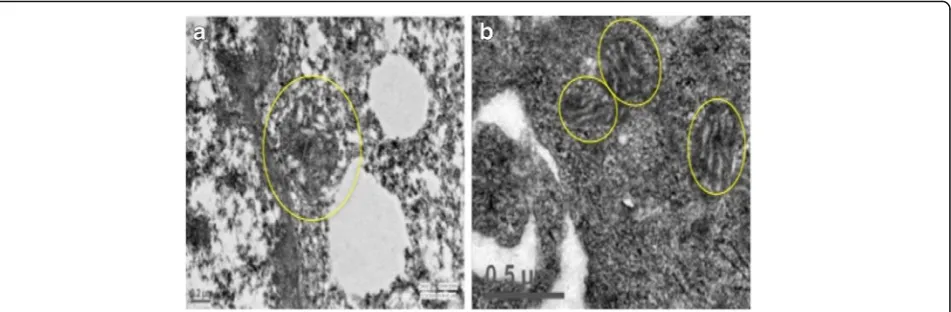
cells [20,26,32]. In Fig.1, we have shown mitochondrial status in transmission electron images of human BM-MSCs and human choriocarcinoma cell line JEG-3, to demonstrate a striking difference in the mitochondria of two cells with low and high-energy demands respectively (unpublished data). The mitochondria in MSCs are spher-ical in shape with underdeveloped cristae, cytoplasmic localization and less in number, owing to lower energy demand (Fig.1a). In contrast, cell lines, here JEG-3, have higher energy and thus, branched, elongated mitochondria with well-organized and localized in peri-nuclear region (Fig. 1b). These naive mitochondria are transferred from MSCs to cells with dysfunctional mitochondria to with high oxidative energy demands thereby modulating their cellular metabolism. Mitochondria transfer appears to be a lucrative strategy for regenerative processes, as mitochondrial biosynthesis usually take longer than can be afforded by damaged cells in crisis. Thus, mitochon-drial transfer is an efficient method for regeneration of injured cells. Although complete mechanisms of mito-chondria transfer have not been elucidated yet, but cer-tain studies suggests that mitochondria transfer from MSCs modulate cellular metabolism of recipient cells. Up regulation of mitochondrial respiration and ATP
levels along with reduction in oxidative damage has been observed in several studies wherein MSCs have been co-cultured with injured cells or tissues [16,33–35]. It is plaus-ible that mitochondria transferred from MSCs modulate cellular bioenergetics by regulating mtDNA replication, maintaining its copy number and regulating other mito-chondrial dynamics and cellular processing required for maintenance of mitochondrial homeostasis in cell.
Signals that trigger mitochondrial transfer from mesenchymal stem cells
The signals that trigger transfer of mitochondria from MSCs have intrigued scientists for a long time. Researchers have postulated theories suggesting that oxidative stress of dysfunctional mitochondria of recipient cell sends en-vironment cues to the MSCs [15, 34]. The local micro-environment of an injured cell has been shown to release physiological cues that trigger transfer of mitochondria. It is compelling to note that bidirectional mitochondrial transfer occurs between MSCs and their microenviron-ment. Few studies have illustrated that stress signals re-leased by recipient cells include damaged mitochondria, released mtDNA and mitochondrial products that are characterized as damage associated molecular patterns Table 1Mitochondrial transfer from Different Tissue Specific MSCs to Recipient Cells of Different Origins
S. No MSCs Type Recipient Cell Mode of Transfer Action References
1 BM-MSCs A549 Cell Line Cellular Contact and Cytoplasmic Projections
Restored Aerobic Respiration [8]
2 BM-MSCs Pulmonary Alveoli in mouse lungs
Connexin43 alveolar attachment and microvesicles
Protection against acute lung injury [15]
3 BM-MSCs
and iPSCs
Airway epithelial cells Tunnel Tubes Rescue cigarettesmoke induced damage [20]
4 BM-MSCs Bronchial Epithelial Cells Tunnel Tubes and Miro1 Rescue Bronchial Epithelial Injury [21]
5 BM-MSCs H9c2 cardiomyocyte cell line Tunnel Tubes Rescue cardiomyocytes in ischemia model [22]
6 BM-MSCs Cardiomyocytes Tunnel Tubes Repair damaged cardiomyocytes in failed myocardium
[23]
7 BM-MSCs Cardiomyoblasts Cell-Cell Connection and Tubular Connections
Rescue Cells from Ischemic Damage [24]
8 BM-MSCs Kidney Tubular Epithelial cells Microvesicles Recovery from acute kidney injury [25]
9 BM-MSCs Rat Cortical Neuronal Cells Tunnel Tubes and Miro1 Neuroprotective [26]
10 BM-MSCs Macrophages Tunnel Tubes Anti-Bacterial Activity [27]
11 BM-MSCs Macrophages Extracellular Vesicles Regulation of mitochondrial dynamics and Mitophagy
[28]
12 BM-MSCs Acute myeloid leukemic cells Cell-Cell Contact Survival of Myeloid Cells [18]
13 BM-MSCs MDA-MB231 breast carcinoma cell line
Isolated mitochondria Enhanced OXPHOS activity [29]
14 iPSCs-MSCs Corneal Epithelial Cells Tunnel Tubes Protection of corneal epithelial cells against alkali burn
[16]
15 AD- MSCs Cardiomyocytes Cell Fusion Reprogram adult cardiac cells towards progenitor like state
[30]
16 WJ-MSCs Cells devoid of mitochondria Osteosarcoma cells
(DAMPS) [36–39]. Interestingly, mtDNA released by injured cells are engulfed by MSCs that subsequently triggers the cytoprotective function of MSCs and en-hanced mitochondrial biogenesis through retrograde signaling, thereby preparing MSCs for mitochondrial donation [40]. In addition, reactive oxygen species re-leased by cells under oxidative stress and inflammation status have also been postulated to trigger donation of mitochondria [15,34,41,42]. Elevated levels of reactive oxygen species have been found in cells with high en-ergy demands such as in cancer cells, and these usually have been seen to receive mitochondria from surrounding cells to meet its energy demands [43,44]. In acute myeloid leukemic cells, NADPH oxidase-2 derived superoxide has been reported to stimulate mitochondrial transfer from BM-MSCs through tunnel tube formation [45]. As dis-cussed previously, that co-culturing MSCs with cells under oxidative stress reversed the oxidative stress state by de-creasing mitochondrial ROS and activities of electron trans-port chain complexes [34]. Thus, it is possible that under retrograde signaling is triggered by ROS, Ca2+, AMP/ATP and NAD+/NADH ratio in cells under oxidative stress. This further stimulates transfer of mitochondria from MSCs to recipient cells. In another study it was shown that calcium dependent mechanism of CD38/cyclic ADP ribose signaling mediated transfer of mitochondria from neurons to astro-cytes for recycling and disposal of damaged mitochondria. This was seen to contribute to neuroprotective and neuro-recovery mechanism after stroke [46]. As CD38, catalyzes the synthesis of calcium messenger cyclic ADP-ribose in mitochondria membranes, its up regulation further increased mitochondria donation in a calcium dependent manner [47]. Another interesting study has shown that loss of cytochrome c in stressed cells, characterized by caspase 3 activation also triggered mitochondrial donation to rescue
UV-damaged PC12 cells by MSCs in the early stages of apoptosis [48].
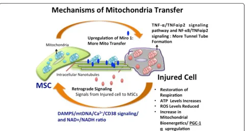
In addition, under stress, MSCs are programmed to secrete depolarized mitochondria for their own protec-tion through transcellular mitophagy [49]. In an in vivo model of myocardial infarction, mitochondria released from damaged cells activated anti-apoptotic signals in MSCs. Foreign mitochondria from other cells were shown to be engulfed and degraded in MSCs, leading to induction of cytoprotective enzyme HO-1, and stimulation of mito-chondrial biogenesis. This triggered enhanced mitochon-drial donation from MSCs to these cells [40]. Phinney et al., have suggested that mitochondrial donation by MSCs is a protective mechanism adopted by MSCs rather than being an altruistic strategy [49]. Simultaneously, mitochondrial DNA that evades autophagy stimulates inflammatory re-sponses mediated by Toll-like Receptor (TLR) 9 and other inflammatory responses, and eventually leads to cellular apoptosis [49–51]. Mitochondria proteins like TFAM and DAMPs including N-formyl peptides, mtDNA, cardiolipin or extracellular ATP are also known to participate in in-flammatory responses and trigger transfer of mitochondria from donor to acceptor cells [52]. These studies have shown that various signaling molecules participate in mitochondria transfer from MSCs, but the exact mecha-nisms and choice of release of a particular signal molecule in a given situation followed by other downstream signals has not been well elucidated.
Mechanisms of mitochondrial transfer
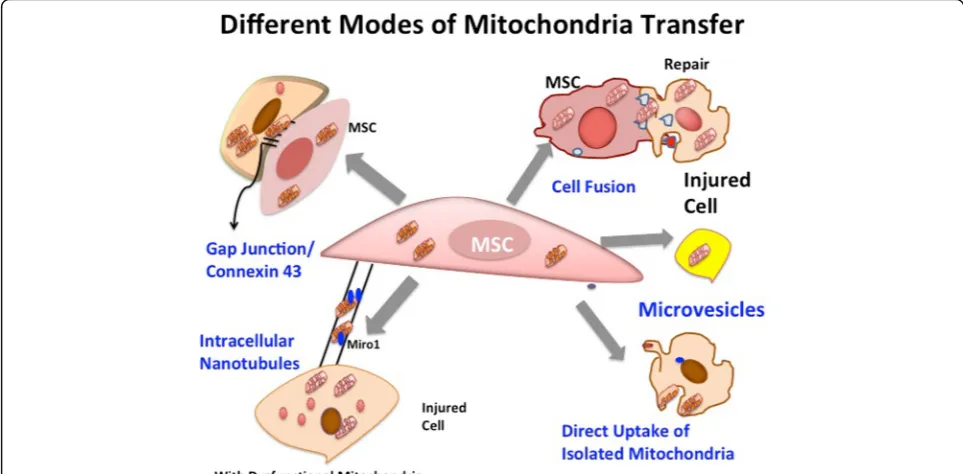
Mitochondrial transfer has been found to be mediated by different modes that include tunnel tube formation, gap junctions, micro vesicles, cell fusion and transfer of isolated mitochondria [15, 34, 42, 53–56]. Figure 2 dia-grammatically illustrates several modes of mitochondrial
transfer from MSCs to recipient cells with damaged or dysfunctional mitochondria and Fig. 3 shows overview of mechanism involved in mitochondria trasnfer. A ma-jority of studies illustrate mitochondrial transfer through formation of nanotubes between MSCs and damaged cells. Motor-adaptor protein complexes regulate mito-chondrial transport and homeostasis, Miro 1 and Miro 2 are two dynamin related Rho-GTPases that interact with other accessory proteins to allow movement of mito-chondria on cytoplasmic tunnel tube extensions that connect two cells. These calcium sensitive proteins bind mitochondria to KLF 5 kinesin motor protein with the help of other accessory proteins like TRAK 1 and TRAK 2, Myo 10 and Myo 19. Together, they form a motor-adaptor complex that contributes to mitochondria trans-port machinery and regulates movement of mitochon-dria on microtubules. The role of Miro1 was shown in nanotube transport in intercellular mitochondrial trans-port in a co-culture system of BM-MSCs and alveolar epithelial cells. MSCs overexpressing Miro1 were shown to perform enhanced rescue potential, leading to greater epithelial cell repair [34]. Down regulation of Miro1 ex-pression and inhibition of tunnel tube formation pre-vented mitochondrial transfer from MSCs to epithelial cells. This study provided supporting evidence that established the significant role of Miro1 in mitochon-drial transfer through tunnel tubes [34].
In an interesting study by Zhang et al., iPSCs show su-perior efficiency than BM-MSCs in mitochondrial trans-fer in rescue of Anthracycline induced cardiomyopathy
due to higher expression of Miro 1 [41]. This was con-firmed by various experiments showing that knockdown of Miro1 with short hairpin RNA (shRNA) that led to remarkable decrease in Miro1 expression and subse-quent reduction in mitochondrial donation. Nonetheless, overexpression of Miro1 enhanced intracellular mito-chondria transfer; thereby providing supporting evidence to confirm that mitochondria transfer is modulated by Miro1 expression. They also investigated tunnel tube for-mation between iPSCs and cardiomyocytes during mito-chondrial transfer. It was shown that no mitochondria transfer occurred when cells were treated with cytochalasin B, that inhibits actin polymerisation and tunnel tube forma-tion. These iPSCs were also found to be more responsive to tumor necrosis factor alpha (TNFα) that induced tunneling nanotube (TNT) formation in cardiomyocytes by elevating TNFαIP2 expression, which was further, regulated by
TNF-α/NF-kappaB/TNFαip2 signaling pathway. A comparative analysis between iPSCs and BM-MSCs showed that iPSCs displayed higher expression of TNFαip2, and more number tunnel tube formation per cell followed by increased mitochondrial transfer. Thus, this study shows that higher expression of both Miro1 and TNFαip2 leads to more mitochondrial transfer and tunnel tube formation and im-parts a cell with higher efficiency to transfer mitochondria as in case of iPSCs as compared to BM-MSCs [41].
Similar results have been observed in another study where MSCs transferred mitochondria to corneal epithelial cells through tunnel tube formation via NF-κB/TNFαip2 signaling. This was confirmed by western blot analysis of
protein showing that enhanced levels of phosphorylated of NF-κB subunit p-IκB along with elevated levels of TNFα and TNFαip2 was markedly increased during tunnel tube formation. It was observed that NF-κB inhibitor SC-514 significantly attenuated tunnel tube formation thus; demon-strating the role of NF-κB mediated signaling mechanism in tunnel tube formation. Furthermore, the role of ROS sig-nal in stimulating of tunnel tube formation was validated by using antioxidant scavenger, N-acetylcysteine (NAC) treatment that reduced ROS levels and concomitantly re-sulted in inhibition of tunnel tube formation and mitochon-dria transfer between MSCs and CECs [16].
Majority of the studies have shown that mitochondrial transfer is mainly mediated through tunnel tube forma-tion and micro vesicles rather than gap juncforma-tions. This could be because most of the studies are carried out in in vitro co-culture system wherein tunnel tube forma-tion is easily observed. However, connexin 43 containing gap junctions channels have been particularly well observed in in vivo acute lung injury mouse model [15]. Sinclair and colleagues have characterized and compared different modes of mitochondrial transfer from different types of MSCs to bronchial epithelial cells [57]. Complete abroga-tion of mitochondrial transfer was observed by blocking tunnel tube formation by cytochalasin B and inhibition of microvesicles by dynasore [57]. Cytoplasmic transfer was found unaffected by Gap26 inhibitor of connexin 43. However, a cocktail of inhibitors exhibited conspicuous attenuation of mitochondrial transfer and at greater extents
suggesting synergy between these modes of mitochondrial transfer [57]. Uptake of mitochondria isolated from stem cells has also been observed in case of MDA-MB231 cell lines and cardiomyocytes [54, 58]. Although, the exact mechanism of mitochondrial uptake is yet un-known but few studies have suggested that internaliza-tion of mitochondria is mediated by actin-dependent cellular mechanisms [53,58,59]. Different modes of trans-fer have been observed under various pathophysiological conditions as illustrated further in this review. However, the basis for choice of mode of mitochondrial transfer under a particular condition is unclear. Though, it is quite possible that the proximity of damaged cells to MSCs and the environmental condition might act as contributing factors regulating the mode of transfer for mitochondrial transfer.
In a study by Spees et al., BM-MSCs were shown to transfer functional mitochondria to rhoφcells devoid of functional mitochondria. Careful examination of these rescued cells excluded cell fusion as one of the main mechanisms of mitochondrial transfer. It was also shown that active formation of TNTs or vesicular transfer of mitochondria, but not passive uptake of mitochondrial fragments, resulted in functional complementation. Islam et al showed that formation of connexin-43 gap junction channels is essential for mitochondrial transfer from BM-MSCs to alveolar epithelial cells, thus increasing epithelial ATP production. BM-MSCs expressing dysfunctional con-nexin 43 could not adhere to alveolar epithelium, and also
could not transfer mitochondria. Cytosolic calcium ions and cellular bioenergetics levels of ATP and glucose are also considered as major players in regulating movement of mitochondria [42]. Further comprehensive studies and elucidations of mitochondrial transfer mechanisms are re-quired to better understand the signaling pathways and molecules involved in this process to employ mitochon-drial transfer to many therapeutic applications.
Mitochondrial transfer in regeneration of diseased tissues
Mitochondria transfer from MSCs has been studied in various disease models that have not only provided evi-dence for its significance in regenerative medicine but also helped to gain insight on mitochondria transfer mecha-nisms. Different preferred mechanism and modes have been observed on the basis of concerned recipient cells and stress condition. In lung cells, renal cells, corneal epithelial cells and brain cortical cells, mitochondria transfer has oc-curred mostly through tunnel tube formation. However, cell fusion and reprogramming of progenitor cells have been observed in cardiac cells. Micro vesicle formation has been observed predominantly in studies where immune-mechanisms are involved as in the case of anti-microbial activity. Nonetheless, remarkable restoration of cellular bio-energetics and reduction in oxidative stress was observed in all the studies that demonstrates that mitochondria transfer from MSCs plays critical role in cellular repair and regener-ation. Also, another therapeutic strategy that targets inhib-ition of tunnel tube formation and mitochondrial transfer in case of tumors has also been discussed.
Lung diseases
Interestingly, mitochondrial transfer has been studied mostly in lung diseases. One of the remarkable works in-cludes study carried out by Islam et al., wherein they found that BM-MSCs transfer mitochondria to pulmon-ary alveoli contributing to protection from acute lung in-jury [15]. This study reported mitochondrial transfer in intact lungs in in vivo mice model treated with lipopoly-saccharide (LPS). It was noted that human or mice BM-MSCs instilled in mice airways were able to transfer mitochondria and repair mitochondrial bioenergetics in lungs. The mitochondrial transfer occurred over a period of 24 h in the lung epithelium and was supported by the expression of connexin 43 that mediated mitochondrial transfer through nanotubes and microvesicles in a cal-cium dependent manner. Mitochondrial transfer led to increased ATP concentrations in the recipient cells suggesting its role in bioenergetics. This study was followed by another remarkable discovery of role of Miro1, a mito-chondrial Rho-GTPase that regulated mitomito-chondrial trans-fer from MSCs to lung epithelial cells through connecting nanotubes [34]. In this study, Ahmad et al., reported that over expression of Miro1 enhanced mitochondrial transfer
whereas its knockdown lead to the loss of rescue efficacy of MSCs. This study was performed in rotenone induced lung injury mouse model and further confirmed in allergen in-duced asthma models. Another study investigated the effect of mitochondrial transfer in rat models exposed to cigarette smoke for 56 days that induced lung damage and manifest-ation of chronic obstructive pulmonary disease [60]. It was reported that BM-MSCs mitigated the damage by transfer-ring mitochondria to lung epithelium. Similar to results of previous studies, they also found significant mitochondrial transfer occurred after 24 h of MSC and BEAS-2B co-culture followed by subsequent elevation in ATP levels through tunnel tube formation. However, further research along these lines is warranted as the reasons for difference are vastly unknown and a comparative analysis of mito-chondrial transfer abilities of stem cells from different sources remain unexplored. Li et al., have also suggested that mitochondrial dysfunction is observed in case of prolonged inflammation and stem cells can transfer mito-chondria to mitigate inflammation indicative of their res-cue potential by promoting anti-inflammatory effects.
Cardiac tissues
have also been devised that further shed light on mito-chondrial transfer and cellular contact between MSCs and cardiomyocytes [70]. These studies have shown that mitochondrial transfer from stem cells can prove to be an effective therapeutic strategy to treat several cardiomyopathies.
Corneal epithelium and renal tubular cells
MSCs were found to transfer functional mitochondria to human corneal epithelial cells, which improved mitochon-drial bioenergetics in recipient cells, studied using extracel-lular flux analyzer. Comparison of ATP production, resting respiration and maximal respiration rates showed that rates of these bioenergetics parameters were found to be lower in rotenone-treated corneal epithelial cells (CECs) as com-pared to normal CECs. All these parameters were improved in CECs co-cultured with MSCs. It was demonstrated that mitochondria were transferred from MSCs to damaged CECs, which also protected them from rotenone-induced cell death and proliferation-inhibition [16]. This study sug-gests that mitochondrial transfer can have huge implica-tions in treating alkali-eye burns and other eye related damages. It can further prove to be an efficient strategy to provide protection to cornea against oxidative stress-induced mitochondrial damage. Interestingly, this study has also suggested a two-way flow of cytoplasmic com-ponents between cells that trigger the response of stem cells to the differentiated cells. Intercellular communica-tion between renal epithelial cells has also been shown through exchange of cytoplasmic materials [71]. Mito-chondrial transfer from MSCs to renal tubular cells has been observed in a co-culture of rat renal tubular cells and human MSCs through intercellular connections [72].
Brain cortical cells
Intercellular communication and mitochondrial transfer from MSCs to rat cortical neurons has been observed in
co-culture in in vitro system [73]. A prominent neuro-protective behavior of MSCs was established through mitochondrial transfer and overexpression of Miro1, followed by subsequent increase in brain derived neuro-trophic factor. Furthermore, intravenous injection of MSCs post ischemia was found to mitigate the patho-logical symptoms of stroke and restoration of neuro-logical activity [73]. The role of mitochondrial transfer in brain cells is also evident from a recent interesting study that has shown transfer of mitochondria from as-trocytes to neurons as a means of neuroglia cellular cross-talk in alleviation of stroke [47]. There is a press-ing need to further investigate the role of mitochondrial transfer from stem cell to brain that can aid in devising better therapeutic strategies.
WJ-MSCs were co-cultured with mitochondrial DNA-depleted cells. Importantly, these cells regained the expres-sion of mtDNA-encoded proteins and showed functional oxygen consumption and respiratory control, as well as the activity of electron transport chain complexes I, II, III and IV. In addition, metabolic shifting and ETC complex V-inhibitor-sensitive ATP production were also recovered. Furthermore, cellular behaviors like attachment-free prolif-eration, aerobic viability and OXPHOS-dependent cellular motility were also recovered after mitochondrial trans-fer by WJMSCs [74]. This study, along with the others cited above, provide strong evidence that MSCs can serve as potential therapeutics to ameliorate diseases with mitochondrial dysfunction, through donation of healthy mitochondria.
Antimicrobial activity of MSCs through mitochondrial transfer
MSCs are known to aid in regeneration through multiple ways that include anti-oxidant property, anti-inflammatory activities of MSCs and differentiation to regenerate dam-aged tissue. MSCs also demonstrate ability to combat
infection by transferring mitochondria to macrophages. This leads to modulation of phagocytic ability of macro-phages through tunnel tube by enhancing their bioenerget-ics evidenced by increased ATP activity. This facilitates more efficient clearing of bacterial cells, thereby influencing innate immune response of these cells serving as an im-portant mechanism in pre-clinical models of acute respira-tory distress syndrome (ARDS) and sepsis [75]. To establish that anti-microbial effect of MSCs is mediated by alveolar macrophage, effect of MSCs was determined in normal E. coliinfected model with animal model treated with intrana-sal Clodronate Lipososmes (CL) that completely abrogated alveolar macrophages (AM). It was found that administra-tion of MSC treatment in AM depleted mice was not able to restore levels of several cytokines involved in anti-inflammatory affects. But, in contrast MSCs could restore levels of cytokines in normal mice suggesting that the anti-microbial activity of MSCs is mediated through macro-phages. Interestingly, macrophage phagocytosis of MSCs or fusion of MSC and macrophage was not observed in monocytes or neutrophils. This suggests that mitochon-dria transfer as the only means for enhanced engulfment by macrophages by both confocal and flow cytometry ana-lysis. It was also observed that non-contact transfer of mito-chondria through microvesicles or exosomes significantly enhanced mitochondria transfer and phagocytosis index of macrophages suggesting it as another critical mechanism for mitochondrial transfer. Another study by Phinney et al., has also shown that mitochondria transfer through micro-vesicles to macrophages and support paracrine and im-mune reaction. However, further investigation to better understand the role of mitochondria in anti-microbial and immune-modulation can provide better insights of multi-level reparative contributions of mitochondria in mainten-ance of cellular health post transfer to recipient cells.
Inhibition of mitochondrial transfer: A potential target for anti-tumor therapies
Mitochondria are the key regulators of cellular bioenerget-ics and metabolism that greatly impacts cancer progression [27]. Many researchers have pointed out mitochondria as a potential target for cancer therapies [7, 76]. In terms of MSCs, many studies have reported transfer of mitochondria through stem cells to tumor cells via tunnel tube formation contributing to their chemo resistance and proliferation [18,43]. A very interesting study by Dong et al., have dem-onstrated that metastatic melanoma cells without mito-chondrial DNA and with defective respiratory function were unable to form tumours. However, upon acquiring in-tact mitochondria along with their mtDNA from their host through horizontal transfer, the cellular respiration is re-stored. This was shown to be critical in tumor generation in mice [77]. It has been found that mitochondrial transfer from bone marrow stem cells renders survival advantage to
acute myeloid leukemic cells thereby making them resistant to chemotherapy [18]. In a recent study, it was shown that inhibition of ICAM-1 an adhesion molecule prevented mitochondria transfer from MSCs to Jurkat Cells treated with chemotherapeutic drug. This prevented tunnel tube formation and stopped transfer of mitochondria from MSCs to Jurkat cells and eventually induced chemotherapy induced cell-death [74]. Thus, inhibition of mitochondrial transfer can serve as a potential therapy target for cancer, such as selective blockage of tunnel tube formation for pre-venting mitochondrial transfer [11,78]. However, in view of the importance of aerobic glycolysis in cancer cells it re-mains possible that altering mitochondrial transfer to can-cer cells may have context dependent effects and this merits further investigation [79].
Challenges and future scope
evident from enhanced mitochondrial transfer from BM-MSCs upon treatment of anti-oxidants such as of N-acetyl-L-cysteine (NAC) and L-ascorbic acid 2-phosphate (AAP) [81]. Such innovative strategies can further be employed for efficient treatments using mitochondrial transfer. Further-more, inhibition of mitochondrial transfer to tumor cells can serve as a novel strategy for cancer treatment [7, 18]. The use of isolated and artificial mitochondria for treat-ment of diseases appears to be a lucrative future therapy [55]. Further investigations are imperative to use artificial mitochondria for treatment of diseases. The optimum dose, packaging, delivery methods and ethical issues for using isolated mitochondria have yet not been studied. It is inter-esting to note that isolated mitochondria may emerge as an off-the shelf therapy in future, as many researchers are now exploring the role of isolated mitochondria in regenerative medicine.
Abbreviations
AAP:ascorbic acid 2-phosphate; AD-MSCs: adipose derived stem cell; AM: alveolar macrophages; ARDS: Acute Respiratory Distress Syndrome; BM-MSCs: Bone marrow mesenchymal stem cell; CL: Clodronate Lipososmes; DAMPS: damage associated molecular patterns; DP-MSCs: Dental Pulp derived mesenchymal stem cell; iPSCs: Induced Pluripotent Stem Cells;
MSCs: Mesenchymal stem cells; WJ-MSCs: Wharton’s Jelly, mesenchymal stemcell
Acknowledgements
We thank Sonali Rawat for contribution to diagrammatic representation of mitochondrial transfer in Fig.2and Fig.3of this review. We thank Manish Kumar for assisting with confocal microscope imaging. We also thank Dr. Swati Midha, Dr. Manu Dalela and Manisha Singh for their suggestions and proofreading this review.
Funding
Funds obtained from Department of Biotechnology, Ministry of Science and Technology, India.
Availability of data and materials
Data and materials related to this work are available upon request.
Authors’contributions
SP and SM have jointly contributed to writing the review. SP, RC, AA and SM have jointly revised the review in its current form. All authors read and approved the final manuscript.
Ethics approval and consent to participate
This study was ethically approved by the Institutional Committee for Stem Cell Research (IC-SCR), All India Institute of Medical Sciences (AIIMS), New Delhi.
References
1. Wei X, et al. Mesenchymal stem cells: a new trend for cell therapy. Acta Pharmacol Sin. 2013;34(6):747–54.
2. Dimarino AM, Caplan AI, Bonfield TL. Mesenchymal stem cells in tissue repair. Front Immunol. 2013;4:201.
3. Patel DM, Shah J, Srivastava AS. Therapeutic potential of mesenchymal stem cells in regenerative medicine. Stem Cells Int. 2013;2013:496218.
4. Liang X, et al. Paracrine mechanisms of mesenchymal stem cell-based therapy: current status and perspectives. Cell Transplant. 2014;23(9):1045–59. 5. Liu CY, Lee CF, Wei YH. Role of reactive oxygen species-elicited apoptosis in
the pathophysiology of mitochondrial and neurodegenerative diseases associated with mitochondrial DNA mutations. J Formos Med Assoc. 2009; 108(8):599–611.
6. Mabalirajan U, et al. Mitochondrial structural changes and dysfunction are associated with experimental allergic asthma. J Immunol. 2008;181(5):3540–8. 7. Loureiro R, et al. Mitochondria in cancer stem cells: a target for therapy.
Recent Pat Endocr Metab Immune Drug Discov. 2013;7(2):102–14. 8. Spees JL, et al. Mitochondrial transfer between cells can rescue aerobic
respiration. Proc Natl Acad Sci U S A. 2006;103(5):1283–8.
9. Rustom A, et al. Nanotubular highways for intercellular organelle transport. Science. 2004;303(5660):1007–10.
10. Onfelt B, et al. Cutting edge: membrane nanotubes connect immune cells. J Immunol. 2004;173(3):1511–3.
11. Bukoreshtliev NV, et al. Selective block of tunneling nanotube (TNT) formation inhibits intercellular organelle transfer between PC12 cells. FEBS Lett. 2009;583(9):1481–8.
12. Gerdes HH, Bukoreshtliev NV, Barroso JF. Tunneling nanotubes: a new route for the exchange of components between animal cells. FEBS Lett. 2007;581(11):2194–201.
13. Abounit S, Zurzolo C. Wiring through tunneling nanotubes–from electrical signals to organelle transfer. J Cell Sci. 2012;125(Pt 5):1089–98.
14. Cho YM, et al. Mesenchymal stem cells transfer mitochondria to the cells with virtually no mitochondrial function but not with pathogenic mtDNA mutations. PLoS One. 2012;7(3):e32778.
15. Islam MN, et al. Mitochondrial transfer from bone-marrow-derived stromal cells to pulmonary alveoli protects against acute lung injury. Nat Med. 2012;18(5):759–65.
16. Jiang D, et al. Mitochondrial transfer of mesenchymal stem cells effectively protects corneal epithelial cells from mitochondrial damage. Cell Death Dis. 2016;7(11):e2467.
17. Figeac F, et al. Nanotubular crosstalk with distressed cardiomyocytes stimulates the paracrine repair function of mesenchymal stem cells. Stem Cells. 2014;32(1):216–30.
18. Moschoi R, et al. Protective mitochondrial transfer from bone marrow stromal cells to acute myeloid leukemic cells during chemotherapy. Blood. 2016;128(2):253–64.
19. Lin HY, et al. Mitochondrial transfer from Wharton's jelly-derived mesenchymal stem cells to mitochondria-defective cells recaptures impaired mitochondrial function. Mitochondrion. 2015;22:31–44. 20. Chen CT, et al. Coordinated changes of mitochondrial biogenesis and
antioxidant enzymes during osteogenic differentiation of human mesenchymal stem cells. Stem Cells. 2008;26(4):960–8.
22. Nagley P, Linnane AW. Biogenesis of mitochondria. XXI. Studies on the nature of the mitochondrial genome in yeast: the degenerative effects of ethidium bromide on mitochondrial genetic information in a respiratory competent strain. J Mol Biol. 1972;66(1):181–93.
23. Youle RJ, Narendra DP. Mechanisms of mitophagy. Nat Rev Mol Cell Biol. 2011;12(1):9–14.
24. Watkins J, Basu S, Bogenhagen DF. A quantitative proteomic analysis of mitochondrial participation in p19 cell neuronal differentiation. J Proteome Res. 2008;7(1):328–38.
25. Ding WX, Yin XM. Mitophagy: mechanisms, pathophysiological roles, and analysis. Biol Chem. 2012;393(7):547–64.
26. Lonergan T, Bavister B, Brenner C. Mitochondria in stem cells. Mitochondrion. 2007;7(5):289–96.
27. Vega-Naredo I, et al. Mitochondrial metabolism directs stemness and differentiation in P19 embryonal carcinoma stem cells. Cell Death Differ. 2014;21(10):1560–74.
28. Nishitai G, et al. Stress induces mitochondria-mediated apoptosis independent of SAPK/JNK activation in embryonic stem cells. J Biol Chem. 2004;279(3):1621–6.
29. Arranz L, Urbano-Ispizua A, Mendez-Ferrer S. Mitochondria underlie different metabolism of hematopoietic stem and progenitor cells. Haematologica. 2013;98(7):993–5.
30. Xu X, et al. Mitochondrial regulation in pluripotent stem cells. Cell Metab. 2013;18(3):325–32.
31. Armstrong L, et al. Human induced pluripotent stem cell lines show stress defense mechanisms and mitochondrial regulation similar to those of human embryonic stem cells. Stem Cells. 2010;28(4):661–73.
32. Bukowiecki R, Adjaye J, Prigione A. Mitochondrial function in pluripotent stem cells and cellular reprogramming. Gerontology. 2014;60(2):174–82. 33. Acquistapace A, et al. Human mesenchymal stem cells reprogram adult cardiomyocytes toward a progenitor-like state through partial cell fusion and mitochondria transfer. Stem Cells. 2011;29(5):812–24.
34. Ahmad T, et al. Miro 1 knockdown in stem cells inhibits mitochondrial donation mediated rescue of bronchial epithelial injury. Biophys J. 2014;104(2):659a.
35. Liu K, et al. Mesenchymal stem cells rescue injured endothelial cells in an in vitro ischemia-reperfusion model via tunneling nanotube like structure-mediated mitochondrial transfer. Microvasc Res. 2014;92:10–8.
36. Galluzzi L, Kepp O, Kroemer G. Mitochondria: master regulators of danger signalling. Nat Rev Mol Cell Biol. 2012;13(12):780–8.
37. Nakahira K, Hisata S, Choi AM. The roles of mitochondrial damage-associated molecular patterns in diseases. Antioxid Redox Signal. 2015;23(17):1329–50. 38. Zhang Q, et al. Circulating mitochondrial DAMPs cause inflammatory
responses to injury. Nature. 2010;464(7285):104–7.
39. Maeda A, Fadeel B. Mitochondria released by cells undergoing TNF-alpha-induced necroptosis act as danger signals. Cell Death Dis. 2014;5:e1312. 40. Mahrouf-Yorgov M, et al. Mesenchymal stem cells sense mitochondria
released from damaged cells as danger signals to activate their rescue properties. Cell Death Differ. 2017;24(7):1224–38.
41. Zhang Y, et al. iPSC-MSCs with High Intrinsic MIRO1 and Sensitivity to TNF-alpha Yield Efficacious Mitochondrial Transfer to Rescue Anthracycline-Induced Cardiomyopathy. Stem Cell Reports. 2016;7(4):749–63.
42. Torralba D, Baixauli F, Sanchez-Madrid F. Mitochondria know no boundaries: mechanisms and functions of intercellular mitochondrial transfer. Front Cell Dev Biol. 2016;4:107.
43. Pasquier J, et al. Preferential transfer of mitochondria from endothelial to cancer cells through tunneling nanotubes modulates chemoresistance. J Transl Med. 2013;11:94.
44. Kaneyuki Y, Yoshino H, Kashiwakura I. Involvement of intracellular reactive oxygen species and mitochondria in the radiosensitivity of human hematopoietic stem cells. J Radiat Res. 2012;53(1):145–50.
45. Marlein CR, et al. NADPH oxidase-2 derived superoxide drives mitochondrial transfer from bone marrow stromal cells to leukemic blasts. Blood. 2017; 130(14):1649–60.
46. Huang PJ, et al. Transferring Xenogenic mitochondria provides neural protection against ischemic stress in ischemic rat brains. Cell Transplant. 2016;25(5):913–27.
47. Hayakawa K, et al. Transfer of mitochondria from astrocytes to neurons after stroke. Nature. 2016;535(7613):551–5.
48. Wang X, Gerdes HH. Transfer of mitochondria via tunneling nanotubes rescues apoptotic PC12 cells. Cell Death Differ. 2015;22(7):1181–91.
49. Phinney DG, et al. Mesenchymal stem cells use extracellular vesicles to outsource mitophagy and shuttle microRNAs. Nat Commun. 2015;6:8472. 50. Oka T, et al. Mitochondrial DNA that escapes from autophagy causes
inflammation and heart failure. Nature. 2012;485(7397):251–5.
51. Boudreau LH, et al. Platelets release mitochondria serving as substrate for bactericidal group IIA-secreted phospholipase A2 to promote inflammation. Blood. 2014;124(14):2173–83.
52. Weinberg SE, Sena LA, Chandel NS. Mitochondria in the regulation of innate and adaptive immunity. Immunity. 2015;42(3):406–17.
53. Koyanagi M, et al. Cell-to-cell connection of endothelial progenitor cells with cardiac myocytes by nanotubes: a novel mechanism for cell fate changes? Circ Res. 2005;96(10):1039–41.
54. Caicedo A, et al. MitoCeption as a new tool to assess the effects of mesenchymal stem/stromal cell mitochondria on cancer cell metabolism and function. Sci Rep. 2015;5:9073.
55. Caicedo A, et al. Artificial mitochondria transfer: current challenges, advances, and future applications. Stem Cells Int. 2017;2017:7610414. 56. Sinha P, et al. Intercellular mitochondrial transfer: bioenergetic crosstalk
between cells. Curr Opin Genet Dev. 2016;38:97–101.
57. Sinclair KA, et al. Characterization of intercellular communication and mitochondrial donation by mesenchymal stromal cells derived from the human lung. Stem Cell Res Ther. 2016;7(1):91.
58. Pacak CA, et al. Actin-dependent mitochondrial internalization in cardiomyocytes: evidence for rescue of mitochondrial function. Biol Open. 2015;4(5):622–6. 59. Kitani T, et al. Internalization of isolated functional mitochondria:
involvement of macropinocytosis. J Cell Mol Med. 2014;18(8):1694–703. 60. Li X, et al. Mitochondrial transfer of induced pluripotent stem cell-derived
mesenchymal stem cells to airway epithelial cells attenuates cigarette smoke-induced damage. Am J Respir Cell Mol Biol. 2014;51(3):455–65. 61. Barbash IM, et al. Systemic delivery of bone marrow-derived mesenchymal
stem cells to the infarcted myocardium: feasibility, cell migration, and body distribution. Circulation. 2003;108(7):863–8.
62. Vallabhaneni KC, Haller H, Dumler I. Vascular smooth muscle cells initiate proliferation of mesenchymal stem cells by mitochondrial transfer via tunneling nanotubes. Stem Cells Dev. 2012;21(17):3104–13.
63. Plotnikov EY, et al. Cell-to-cell cross-talk between mesenchymal stem cells and cardiomyocytes in co-culture. J Cell Mol Med. 2008;12(5A):1622–31. 64. Ikeda Y, et al. Molecular mechanisms mediating mitochondrial dynamics
and mitophagy and their functional roles in the cardiovascular system. J Mol Cell Cardiol. 2015;78:116–22.
65. Masuzawa A, et al. Transplantation of autologously derived mitochondria protects the heart from ischemia-reperfusion injury. Am J Physiol Heart Circ Physiol. 2013;304(7):H966–82.
66. Nair V, et al. Efficacy of stem cell in improvement of left ventricular function in acute myocardial infarction–MI3 trial. Indian J Med Res. 2015;142(2):165–74. 67. Cselenyak A, et al. Mesenchymal stem cells rescue cardiomyoblasts from cell
death in an in vitro ischemia model via direct cell-to-cell connections. BMC Cell Biol. 2010;11:29.
68. McCully JD, et al. Injection of isolated mitochondria during early reperfusion for cardioprotection. Am J Physiol Heart Circ Physiol. 2009;296(1):H94–H105.
69. Han H, et al. Bone marrow-derived mesenchymal stem cells rescue injured H9c2 cells via transferring intact mitochondria through tunneling nanotubes in an in vitro simulated ischemia/reperfusion model. Mol Med Rep. 2016; 13(2):1517–24.
70. Ma Z, et al. Mesenchymal stem cell-cardiomyocyte interactions under defined contact modes on laser-patterned biochips. PLoS One. 2013;8(2):e56554. 71. Domhan S, et al. Intercellular communication by exchange of cytoplasmic
material via tunneling nano-tube like structures in primary human renal epithelial cells. PLoS One. 2011;6(6):e21283.
72. Plotnikov EY, et al. Cytoplasm and organelle transfer between mesenchymal multipotent stromal cells and renal tubular cells in co-culture. Exp Cell Res. 2010;316(15):2447–55.
73. Babenko VA, et al. Improving the post-stroke therapeutic potency of mesenchymal multipotent stromal cells by Cocultivation with cortical neurons: the role of crosstalk between cells. Stem Cells Transl Med. 2015;4(9):1011–20. 74. Yang X, et al. Mesenchymal stem cells derived from Wharton jelly of the
human umbilical cord ameliorate damage to human endometrial stromal cells. Fertil Steril. 2011;96(4):1029–36.
mesenchymal stem cells. Oxidative Med Cell Longev. 2017;2017:8510805.
• We accept pre-submission inquiries
• Our selector tool helps you to find the most relevant journal
• We provide round the clock customer support
• Convenient online submission
• Thorough peer review
• Inclusion in PubMed and all major indexing services
• Maximum visibility for your research
Submit your manuscript at www.biomedcentral.com/submit