Placental Circulation*
ELIZABETH M. RAMSEY, M.D.
Visiting Professor of Obstetrics and Gynecology, University of Virginia School of Medicine, Charlottesville, Virginia
One of the most important developments of recent years in the field of uterine physiology has been the recognition that the endometrial changes occurring during the menstrual cycle and those associated with pregnancy are interlocking, sequen-tial events in an ordered progression from the first day of the cycle to parturition-and not separate phenomena as was formerly believed. No com-ponent of the endometrium illustrates this pro-gression more strikingly than does the vasculature.
Much of the story of uterine vascular pattern and circulatory mechanism is based upon studies in the rhesus monkey, employing in vivo techniques inapplicable to clinical patients. (These studies were carried out in the Department of Embryology, Carnegie Institution of Washington at Baltimore.) Subsequent checking of the monkey findings against their human counterparts, in operative and necropsy specimens, etc., has shown the monkey to be a valid experimental model with reproductive sys-tem anatomy and physiology closely similar to the human (Ramsey and Harris, 1966).
Following the menstrual slough the vasculature regenerates pari passu with the endometrial stroma and glands (Fig. 1). Initially a long capillary net-work forms between the stumps of spiral arteries in the basalis and the epithelial surface. Subsequently, muscular and elastic layers forming around the capil-laries transform them into true arteries. It may be noted parenthetically that this is a more accurate description than the familiar statement that "spiral arteries grow toward the endometrial surface." A rich capillary bed remains in the immediately sub-epithelial layer and connects the arteries with veins which run perpendicularly toward the myometrium.
Although the follicular phase of the cycle is frequently referred to as the "growth phase,"
*
Presented at the 43rd Annual McGuire Lecture Series, December 3, 1971, at the Medical College ofVir-ginia, Richmond.
MCV QUARTERLY 8(1): 61·68, 1972
growth of spiral arteries continues unabated during the corpus luteum phase and even further on, as we will see. Indeed, vascular growth during the lutein phase outstrips stromal growth, so that the increasing length of the arteries must be accomo-dated within the endometrium by ever increasing coiling (Fig. 1 ) .
Fig. 1-Camera lucida drawings of the vascular bed at three
stages of the menstrual cycle in the rhesus monkey. Left. postmenstrual; Center. postovulatory; Right. late secretory.
Myometrium stippled. (Reprinted with permission from Bartelmez. Contrib. Embryo/. 36: 153-182, 1957.)
Fig. 2- Photomicrograph of an early human implantation. (a) trophoblastic lacunae; (b) maternal capillaries. Carnegie Collection 8004, 7th day of pregnancy, section 11-4-4. (Reprinted with permission from Hertig and Rock. Contrib. Embryo/. 31: 65-84, 1945.)
lacunae of the trophoblastic shell. With progressive penetration by trophoblast, the terminal tips of spiral arteries are opened up and maternal arterial blood flows into the shell. This meanwhile has itself been enlarged and transformed by the develop-ment of chorionic villi (Fig. 3). It is around the villi, in the inter-villous space, that the maternal blood now flows and from now on we may speak of a placenta and placental circulation.
Reconstructions of representative uteroplacen-tal arteries, both human and monkey, at comparable stages of gestation (Fig. 4), show that there is very little qualitative change in growth pattern during the first weeks after implantation (Harris and Ram-sey, 1966; Ramsey, 1949). The coiling of the arteries continues and there is just a slight indica-tion of a new process at the arterial tips where trophoblast is beginning to replace normal wall structure. Soon however a change does become manifest. Arterial elongation (as determined by
careful micromeasurements) is continuing, but the thickness of the endometrium is being diminished as the result of trophoblastic erosion combined with pressure of the overlying conceptus. Thus, the previously vertical arterial stems are diverted toward the margins of the implantation site, an increasingly sharp angulation developing. With the continuation of these processes in succeeding weeks, the in-creased coiling of the artery is no longer sufficient to effect its accomodation in the thinned endome-trium, so back and forth and lateral looping is added. A terminal dilatation of the artery devel-ops proximal to its point of entry into the inter-vil1ous space.
RAMSEY: PLACENTAL CIRCULATION
\
Fig. 3-Photomicrograph of a portion of a monkey placenta
in situ. Chorionic plate above; entrance of an endometrial
spiral artery into the intervillous space at the left. Carnegie Collection C-477, 29ih day of pregnancy, section 47b.
and anchorage is increased. The coils are more fully smoothed away in the monkey than in man, probably becaus.e monkey endometrium undergoes the greater stretching.
The terminal dilatations of arteries communi-cating with the interviUous space appear to be the result of the weakening of the vessel wall brought about by replacement of muscle and elastic tissue by trophoblast. Appearing first as an
intraluminal accumulation (Fig. Sa), the tropho-blastic cells gradually invade and replace the vessel wall (Fig. Sb). The invasion is earlier in the monkey and baboon than ih the human, but it is deeper and more extensive in the latter. Human cytotrophoblast penetrates the endometrial stroma as well as entering the arterial lumen and invasion of the wall proceeds from without as well as from within (Fig. 6). The more drastic elimination of normal vascular wall resistance in man doubtless occasions the larger and more persistent terminal dilatations of human uteroplacental arteries. A further result of greater trophoblastic activity in the human is the erosion of arteries all the way to the midendometrium where branches arise from the main spiral stems. These branches then
com-63
municate with the intervillous space which ex-plains why there is a proportionately greater num-ber of arterial entries in humans than in monkeys. Upon occasion the trophoblastic action, in con-trary fashion, may cause occlusion of branches or even main arterial stems.
Venous drainage, at all stages of the reproduc-tive cycle, is a less dynamic affair than arterial inflow. The basic venous pattern in the endome-trium is a grid with dilatations into venous lakes at the junction of vertical and lateral limbs. These relationships continue into pregnancy with certain of the vertical channels increasingly distended as they are required to accommodate the ever in-creasing volume of placental blood. Other channels
are passively obliterated by external
compres-s10n.
On the physiological side there is again con-tinuity between prepregnant and pregnant states. From the standpoint of circulation, this is most
apparent in the persistence of an intrinsic
con-tractile potential in the spiral arteries. This is manifested during the menstrual cycle by isolated
contractions at the myoendometrial junction which
produce ischemia leading to foci of endometrial necrosis and slough (Bartelmez, 19S7), and in pregnancy by intermittency of flow through indi-vidual spiral arteries into the intervillous space (Martin, McGaughey, et al, 1964).
The opposite number to uteroplacental cir-culation is of course fetoplacental circir-culation. Pro-pelled by the vis a tergo bf fetal blood pressure, fetal blood courses through the umbilical arteries
into the subdivisions which run laterally through the chorionic plate. Finally, the vessels dip into the
substance of the placenta and travel through the
arborizations of the fetal villous tree. They
pro-ceed in comparable subdivisions to the terminal villi. There the fetal capillary bed, coming into its closest proximity to maternal blood in the
inter-villous space, forms the ultimate area of
maternal-fetal exchange. Oxygenated blood returns via ves-sels running through the same villous stems to the umbilical vein and thence to the fetal body (Martin
and Ramsey, 1970).
The mechanism of circulation within the pla-centa, first hypothesized upon the basis of
ana-tomical data, has been established by radioangi-ographic studies (Donner, et al, 1963). Especially with cirieradioangiography, it is possible to visualize directly the inflow of arterial blood to the intervillous
space (Fig. 7), its circulation through the space, and
Monkey
_
/
ARTERIES , , - - - -- -- -- ---- - - ,
PERITONEAL SURFACE
MARGIN OF INTERVILLOUS SPACE
3rd WEEK
A
15
c
8!hWEEK PERITONEAL SURFACE
ARCUATE ARTERY 19th. WEEK
Human
16 WEEKS 20 WEEKS
8 WEEKS
c
!
A
J
16
Fr1nk8 Proc~Fig. 4-Diagrammatic representations of the course and configuration of the uteroplacental arteries in the rhesus monkey and man, at comparable stages of gestation. (Reprinted with permission from Harris and Ramsey. Contrib. Embryo/. 38: 43-58, r9~6.)
The propulsive force throughout is the head of maternal blood pressure which drives blood into the intervillous space in discreet, fountainlike "spurts." The incoming blood wafts aside the villi surrounding the orifices of entry, but once the
propulsive force is reduced, in part by the, baffie
action of the villi, the b.lood disperses laterally crowd
-ing the exist-ing content of blood through the
ve-nous orifices in the basal plate into the' uterine
con-RAMSEY: PLACENTAL CIRCULATION
Fig. Sa-Photomicrograph of uteroplacental arteries in the monkey illustrating early accumulation of trophoblast within the lumen of the artery. Carnegie Collection C-477, 29th day of pregnancy. (Reprinted with permission from Wislocki and Streeter. Contrib. Embryo/. 27: 1-66, 1938.) ·
Fig. Sb-Photomicrograph of uteroplacental arteries in monkey illustrating subsequent replacement of the arterial wall without trophoblastic penetration of stroma. Carnegie Collection C-629, S3rd day of pregnancy. (Reprinted with permission from Ramsey. Contrib. Embryo/. 33: 113-147, 1949.)
tractions both inflow and outflow cease, in whole or in part, depending upon the strength of the con-traction (Ramsey, Martin, McGaughey, et al, 1966).
The volume of the placental pool, however, is main-tained. That is to say, the old concept that "contrac-tions squeeze the placenta like a sponge" is incor-rect; rather blood is trapped in the placenta during contractions.
Radioangiography of the fetal side of placental
circulation (Martin, Ramsey, and Donner, 1966)
65
Fig. 6-Photomicrograph of a human uteroplacental artery showing replacement of wall and penetration of stroma by trophoblast. Carnegie Collection 10117, 8Sth day of preg-nancy. (Reprinted with permission from Ramsey. Prenatal
Life. Wayne State University Press, 1970, pp. 37-S3.)
shows the progress of blood from the fetal body into the capillary network of the fetal cotyledons and back via the umbilical vein. Double injection of a
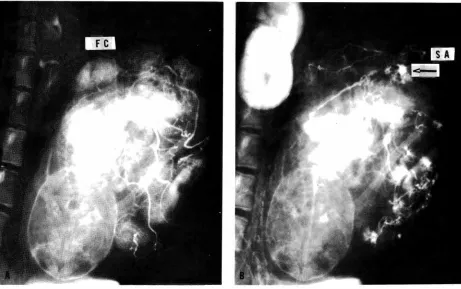
radiopaque medium (Ramsey, Martin, and
Don-ner, 1967), that is, into fetal and maternal circula-tions in rapid succession, permits visualization of the 1 : 1 relationship between maternal spiral arteries and fetal cotyledons (Fig. 9) .
Two points of clinical interest emerge from the foregoing. The first is that placental circulation
ceases during strong contractions. That this may present the fetus with periods of anoxia is clear and
should contractions be unduly prolonged, as the result of pathology or medication, it could indeed be critical. Second, and somewhat mitigating the implied threat of the cessation of flow, is the fact that the pool of placental blood is preserved throughout. Thus, under normal conditions, con-tinued maternal-fetal exchange is made possible.
And that exchange, of course, is the whole purpose of the long and elaborate procession of
Fig. 7-Photographs of X rays made at 2, 3, and 77\l seconds respectively after injection of contrast material into a femoral artery of a monkey. (R.a.) renal artery; (S.a.) endometrial spiral artery;->''spurts" into intervillous space. Carnegie Collection Monkey
60 /14, lOOth day of pregn~ncy. (Reprinted with permission from Ramsey, et al. Montanino Editore, Napoli. II: 1779-1784, 1962.)
RAMSEY: PLACENTAL CIRCULATION 67
Fig. 9- Spot films made during a combined fetal and maternal injection study. (A) taken 3 seconds after injection of contrast material into the fetal circulation; (B) taken 2 seconds after immediately subsequent maternal injection. (FC) fetal cotyledon;
(SA) endometrial spiral artery ;->''spurts" into the intervillous space. Carnegie Collection Monkey 65 /80, 152nd day of pregnancy.
(Reprinted with permission from Ramsey, et al. Am. J. Obstet. Gynec. 98: 419-423, 1967.)
REFERENCES
BARTELMEZ, G. W. The form and the functions of the uterine
blood vessels in the rhesus monkey. Carnegie Inst. Wash.,
Contrib. Embryo/. 36: 153-182, 1957.
DONNER, M. W., RAMSEY, E. M., AND CORNER, G. W., JR. Maternal circulation in the placenta of the rhesus monkey;
A radioangiographic study. Amer. J. Radio!. and Roentgen.
Therapy. 90:638-649, 1963.
HARRIS, J. W. S. AND RAMSEY, E. M. The morphology of
human uteroplacental vasculature. Carnegie Inst. Wash.,
Contrib. Embryo/. 38:43-58, 1966.
HERTIG, A. T. AND ROCK, J. Two human ova of the
pre-villous stage, having a developmental age of about seven and nine days respectively. Carnegie Inst. Wash., Contrib.
Embryo/. 31:65-84, 1945.
MARTIN, C. B., JR., MCGAUGHEY, H. S., JR., KAISER, I. H.,
DONNER, M. W., AND RAMSEY, E. M. Intermittent function-ing of the uteroplacental arteries. Am. J. Obstet, Gynec.
90:819-823, 1964.
MARTIN, c. B., JR. AND RAMSEY, E. M. Gross anatomy of the placenta of rhesus monkeys. Obstet. Gynecol 36: 167-177, 1970.
MARTIN, C. B., JR., RAMSEY, E. M., AND DONNER, M. W.
The fetal placental circulation in rhesus monkeys
demon-strated by radioangiography. Am. J. Obstet. Gynec. 95:943-947, 1966.
RAMSEY, E. M. The vascular pattern of the endometrium of
the pregnant rhesus monkey (Macaca mulatta). Carnegie
Inst. Wash., Contrib. Embryo/. 33: 113-147, 1949'.
RAMSEY, E. M. Placental circulation in rhesus and man.
Prenatal Life. Proceedings of the Third Annual Symposium
on the Physiology and Pathology of Human Reproduction. Harold C. Mack (ed.). Detroit: Wayne State University Press, 1970, pp. 37-53.
RAMSEY, E. M. AND HARRIS, J. w.
s
.
Comparison of utero-placental vasculature and circulation in the rhesus monkeyand man. Carnegie Inst. Wash., Contrib. Embryo/. 38:59-70, 1966.
RAMSEY, E. M., MARTIN, C. B., JR., AND DONNER, M. W. Fetal and maternal placental circulations. Am. J. Obstet.
Gynec. 98:419-423, 1967.
RAMSEY, E. M., MARTIN, C. B., JR., MCGAUGHEY, H. S., JR., KAISER, I. H., AND DONNER, M. w. Venous drainage of the
placenta in rhesus monkeys: radiographic studies. Am. J.
Obstet. Gynec. 95:948-955, 1966.
REYNOLDS, S. R. M. Uterine accommodation of the products of conception: physiologic considerations. Am. J. Obstet.
Gynec. 53:901-913, 1947.
WISLOCKI, G. B. AND STREETER, G. L. On the placentation of the macaque (Macaca mulatta), from the time of im-plantation until the formation of the definitive placenta.