Pancreatitis
FRANZ J. INGELFINGER*
The New England Journal of Medicine, Boston, Massachusetts 02115
I am, of course, most grateful to everyone responsible for arranging this-the 39th McGuire Lecture Series. It is a series distinguished by tradition: few medical lecture-ships can point to a continuity of nearly 40 years, and few can boast of the three M's of American medicine-Mayo, Mann and Minot. Incidentally, in going over the list of previous lectures, I could not help noticing that I am the eighth Bostonian in this series, which puts us well ahead of our nearest com-petitor, a clinic in a state with a name that also happens to begin with M.
My pleasure at being invited to give this Stuart McGuire Lecture has naturally been doubled-and I am speaking in logarithmic terms-by having on the same program 11 men who had, let us say, some ex-perience in our gastrointestinal sec-tion in Boston. Dr. Farrar, Dr. Caravati and others, in arranging this, were doubtlessly motivated by generosity but also probably felt a bit sorry for the old man who has entered what some regard as the retirement of an editorship.
The subject of my lecture re-quires better definition: I shall talk about pancreatitis and, chiefly, about the diagnosis of the recurrent variety of pancreatitis. How can we recognize the pain of this disease? This pain does tend to be in the upper abdomen, more often on the left than the right, and radi-ates to the back in about a quarter
* Thirty-Ninth Annual
McGuire Lecturer for the McGuire Lecture Series, November 9-10, 1967, Medical College of Virginia, Richmond.4
of the cases. It has, however, no characteristic quality, and the de-gree of the pain may range from the relatively painless to the ex-cruciating. Most often, its severity is between these two extremes.
In an essay on pain published in 1930, Dr. Stuart McGuire wrote as follows:
In considering the significance of pain which occurs in patients who are suddenly taken with belly-ache, the vital question to decide is whether it is caused by an acute intra-abdom-inal disease such as appendicitis, cholecystitis, perforating ulcer or in-testinal obstruction-conditions which demand prompt operative interven-tion-or whether it is reflex from extra-abdominal diseases such as tabes, angina, pneumonia, lead poi-soning, rheumatic conditions or dis-eases of the upper urinary tract-conditions which call for medical rather than surgical treatment. Note that, among the common causes of abdominal pain listed, pancreatitis does not appear. He certainly knew about pancreatitis, but my guess is that Dr. McGuire considered it an uncommon ail-ment. The Index Medicus in those years usually had less than 50 en-tries annually relating to pancrea-titis, and chronic or recurring pan-creatitis was hardly mentioned. In contrast, the 1966 Index Medicus contained over 350 entries, and as one scans the list, the preponder-ance of reports originating either from the United States or France is striking. Articles from Israel, on the other hand, are conspicuously absent. Indeed, when an experienced surgeon from Israel was recently shown one of Boston's many pa-tients with chronic alcoholic
pan-creatltls, he doubted the diagnosis. Why? Because, he said, it was such a very, very rare disease. He had never seen a case.
Alcohol the Villain
What do the United States and France have that Israel does not? Enthusiastic consumption of alco-hol. The evidence is very suggestive that the prevalence of pancreatitis and the consumption of alcohol show parallel trends in various areas of the world, even as cirrhosis and alcohol consumption show parallel trends. The beer-drinking Bantu of South Africa, for example, not only has much cirrhosis; he also has much chronic pancreatitis.
Dr. Gerald Klatskin of Yale likes to show a graph plotting deaths from cirrhosis against years. After the enactment of prohibition, the rate fell sharply from about 12 to 7 per 100,000, but after drinking became legal again, the death rate from cirrhosis steadily rose to a level of about 11(Klatskin,1961). Adequate figures for plotting the incidence of pancreatitis over the years are not available, but my guess is that this incidence has steadily gone up since the repeal of prohibition.
The relation of pancreatitis to alcohol consumption is not only of epidemiologic interest but also has a bearing on our subject: ab-dominal pains in a patient who drinks alcohol should make one think of pancreatitis. You may re-gard this as too obvious. But the following happens again and again in Boston, and I suspect elsewhere as well. A patient complains that he has vomited and has abdominal
pain coming on a few hours after some reasonably heavy drinking. The vomitus may or may not con-tain blood, but the chances are nearly 100% that a diagnosis of alcoholic gastritis will be made. The same story may repeat itself a few times until someone happens to take a serum amylase which, if ele-vated, helps establish the correct diagnosis. Serum amylase, however, is not always elevated under such conditions, and the correct diagno-sis may never be suspected until an abdominal x-ray one day re-veals calcification in the pancreas.
Not all abdominal pains in pa-tients who have been drinking are caused by pancreatitis, but the pos-sibility is a real one. That alcoholic gastritis is responsible for abdomi-nal pains appearing in the drinking patient, however, is not a good pos-sibility for the simple reason that the existence of such an entity is controversial and, in any case, its symptomatology quite obscure. No one has yet shown that the his-tologic appearance of the stomach in the patient who is drinking is different from that found in a con-trol population, or that the gas-troscopic appearance of the stomach in a patient vomiting because of alcoholism looks different from that of a patient vomiting for some other reason.
It is,. of course, silly to talk about drinking without defining its degree.
TABLE 1
Associates of Pancreatitis
Causal(%)
Alcohol Gallstones Peptic Ulcer Trauma Malnutrition Parasites Diabetes
In cirrhosis of t has to go on fc before the liver clinically manifes ing has to be ht ous. Pancreatifo hand, does not : a background his drinking, and alt ing may be he: be intermittent. P get their first at pancreatitis, usua been eating well, It is striking that alcoholic pancrea cirrhosis of the 1 monly seen in 1 Possibly the diffe individual suscei: also possible that it first hits, hits t nutritionally in g· Table 1 shows alcohol so insister delicious beverag< at least half the titis that you wil case of recurrent percentage is pre other words, awa sociation is cruci: tial diagnosis of pain.
Other Backgrou1
When Dr. McC abdominal pain,
Re
50 25 7 4
?
1 0 2 1
Other Metabolic Diseases Drugs
10 None of these
If "none": chances of pancreatitis
<
10%considered the primary cause of pancreatitis, responsible for some 80% of attacks. Their contribution to the etiology of pancreatitis, how-ever, is rapidly decreasing, partly on a relative basis because of the increasing frequency of alcoholic pancreatitis, and partly because symptomatic gallstones are so promptly removed these days that pancreat1tls caused by neglected biliary tract disease is becoming a rarity.
Whether peptic ulcer really ini-tiates pancreatitis by eroding into the pancreas, with subsequent spread to involve more of the organ, is controversial. In my opinion it is an uncommon but not rare an-tecedent of pancreatitis.
Trauma clearly appears to be an antecedent of pancreatitis, some-times on the table . and sometimes off. The "on the table" variety is usually precipitated by vigorous manipulation of the upper abdomi-nal cavity during the course of gas-troduodenal or biliary tract surgery. The "off the table" kind is caused by blunt and non-penetrating trauma to the belly such as occurs when the body is thrown against a steering wheel, or when someone dives into a swimming pool, not realizing that the water has been emptied out of it. Since this phe-nomenon admittedly is only apt to happen under certain conditions, the dilemma is posed whether the dive or the pdive alcohol is re-sponsible. As a matter of fact, with the exception of gallstones, the same question applies to most of the possible background conditions listed. Many a patient with pan-creatitis, for example, has both peptic ulcer and a tendency to drink alcohol in excess.
Malnutrition is often invoked as a background condition responsible for pancreatitis, because the pan-creas is histologically altered in kwashiorkor and because the me-thionine antagonist, eme-thionine, pro-duces a chronic pancreatic reaction in experimental animals. Direct evi-dence, however, is lacking. Many
natives of Uganda, to be sure, have chronic pancreatitis with calcifica-tion of the organ, but Ugandans appear to be as intemperate as Americans and French, and, hence, the problem is again one of select-ing the real culprit from at least two candidates.
Another disorder apt to affect malnourished populations is parasit-ism. The parasite most apt to cause pancreatitis is Ascaris lumbricoides, which crawls into the common duct to obstruct it. In our adult US population, especially in people of higher socio-economic status, ductal pancreatic obstruction by this mechanism is almost unheard of. On the other hand, if a population of children in an impoverished trop-ical country is under consideration, parasitism turns out to be the prin-cipal cause of pancreatitis. These facts underscore the difficulty of arriving at universally applicable percentage figures for the frequency of various etiologies of pancreatitis. The figures shown in Table 1 are approximations of the crudest type. Metabolic diseases other than diabetes have been well publicized as causes of pancreatitis, and the recent literature incriminates a num-ber of drugs that are also suspect (Table 2). Although fascinating because of their diagnostic and eti-ologic implications, metabolic dis-orders and drug reactions account,
TABLE2
Conditions and Drugs Suspected of Causing Pancreatitis
Metabolic Disorders:
Hyperlipemia Hyperparathyroidism
Amino-aciduria (cysteine
+
lysine)Drugs:
ACTH and Adreno- lsoniazid cortical Agents
Sulfamethizole Thiazides
Indomethacin
Opiates Carbon tetrachloride Methyl
alcohol
PANCREATITIS
at least in my opinion, for only a small proportion of all cases of pancreatitis. When hyperlipemia is associated with pancreatitis, either condition may be cart or horse. Pa-tients with essential hyperlipemia are, for unknown reasons, suscepti-ble to attacks of pancreatitis. Con-versely, hyperlipemia in other in-stances is the consequence of repeated attacks of pancreatitis, often alcoholically induced. Hyper-lipemia, thus, may be found incon-stantly in about 5 % of patients with chronic recurrent pancreatitis, and, when present, may in some mysterious way suppress the in-creases in serum amylase and lipase usually expected when pancreatic inflammation is active ( Greenber-ger et al., 1966).
The association of pancreatitis with hyperparathyroidism also tends to be emphasized, but the absolute number of such cases is small, ex-cept at the Massachusetts General Hospital, a magnet for patients with parathyroid dysfunction. At this famous institution, indeed, the significance of blood calcium levels is uncertain, for obviously the hy-percalcemia of hyperparathyroid-ism and the hypocalcemia of severe pancreatitis may cancel each other. Finally, pancreatitis appears to be associated in certain families with amino-aciduria, particularly of ly -sine and cysteine. In a number of these patients, however, alcoholic intake is heavy, and, again, the real criminal is hard to identify. The situation resembles that of hemochromatosis. Many think that hemochromatosis is a genetically transmitted disorder, but since so many hemochromatotics drink, one almost has to assume that a pen-chant for drinking alcohol is also genetically transmitted.
In about 10% of the cases, pan-creatitis affects a patient who has no recognizable background condi-tion.
Complications of Pancreatitis
Many of the disorders listed in Table 1 may be re~arded as the
consequences (right-hand column of percentage figures) rather than the causes of pancreatitis. Is it really true that 1 % of patients are alcoholic because they have pancreatitis? I doubt it, but, at one time, when alcoholism was not a socially acceptable diagnosis, it was suggested that some people took up drinking because their abdomi-nal pain was so bad. It reminds me of one of our patients who always made the two-glass test when he got one of his attacks of pancreatitis. With the onset of pain, he would promptly repair to the local bar and take two shots of whiskey. If this made the pain disappear, well and good-he could drink more. If it did not make the pain disappear, he had to keep on drinking so that he might forget the pain.
Can chronic pancreatitis cause gallstones? There is considerable evidence that one of the back-ground conditions necessary for precipitation of biliary sediment is stasis, and, hence, chronic pancrea-titis, to the extent that it obstructs the biliary passages, may lead to the production of gallstones. The 5 % is obviously a very rough guess. The precipitation of pancreatitis by peptic ulcer is, as I have indi-cated, debatable. On the other hand, there is very good evidence to sug-gest that peptic ulcer may result from chronic pancreatic insuffi-ciency. The patient who puts out pancreatic juice grossly deficient in bicarbonate because of pancreatic insufficiency obviously cannot neu-tralize gastric contents in a normal fashion, and, hence, his duodenal contents may be abnormally acid. In addition, it has been shown that the absence of normal pancreatic juice in the duodenum itself stimu-lates gastric secretion. Thus, both enhanced gastric acid secretion and decreased neutralization of the duo-denum join forces to make the pa-tient with chronic pancreatic di s-ease susceptible to peptic ulcer.
to argue that the driver seized by the belly pain of pancreatitis will smash into somebody else. If pan-creatitis is so severe that pain and vomiting interfere with eating, or if digestion is impaired by pancre-atic insufficiency, malnutrition ob-viously ensues. Similarly, diabetes is a frequent concomitant and result of chronic pancreatitis. Many pa-tients with chronic pancreatitis use narcotics and, after a while, it is hard to tell whether they are ad-dicted or really need the medica-tion. In any case, abuse of narcotic drugs is certainly the result in some patients with pancreatitis. To the extent opiates and meperidine cause further spastic ohstruction at the sphincter of Oddi, the use of these drugs may in turn aggravate the original disease.
The percentage figures in both columns of Table 1 are not only imprecise; they are also grossly affected by a great deal of overlap. Many patients with pancreatitis have both diabetes and malnutri-tion. If the percentages indicating prevalence on both sides are added, their total is 180%, as might be expected, since one patient with pancreatitis often has several asso-ciated conditions.
The diagnostic importance of Table 1 is that there are relatively few patients with pancreatitis who have no associated conditions what-soever. When the prevalence of con-ditions associated, for one reason or another, with pancreatitis is taken into account, one may logically conclude that less than 10% of patients with pancreatitis have pan-creatitis all by itself, without any of the conditions listed in Table 1. Put another way, this means that, if one sees a patient with upper ab-dominal pains of unexplained na-ture and he has none of these conditions associated with pancrea-titis, the chances are less than 10% that he is suffering from pancrea-titis. The converse is, of course, not true. Many patients with abdominal pain have alcoholism, gallstones, or diabetes and are not necessarily
suf-fering from pancreatitis. The list in Table 1 presents criteria that are negatively important: their absence militates against the diagnosis of pancreatitis.
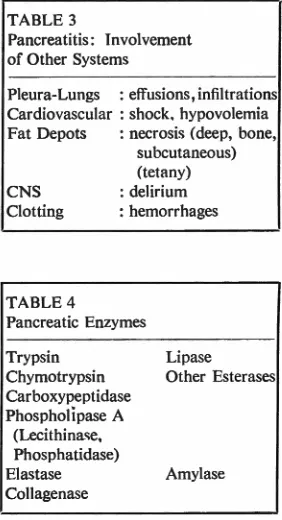
Extra-Pancreatic Manifestations
Recognition of pancreatic pain is not only aided by an awareness of those conditions that are asso-ciated historically with pancreatitis. Equally helpful are extra-pancre-atic phenomena often appearing during the acute episode (Table 3). The presence of such phenomena is related to the severity of the attack. The more drastic the attack, the more likely that other systems will be involved. Particularly fre-quent are radiologic signs of pleural or pulmonary changes; even in the moderate cases, one may expect them about a third of the time. The cardiovascular reactions to severe pancreatitis are well-known. Less emphasized are some of the unusual manifestations of fat necrosis such as aching in the bones, possibly related to necrosis of marrow fat, and, very rarely, subcutaneous nod-ules reminiscent of Weber-Christian syndrome. One of the explanations for the tetany that occurs a few days after an attack of severe pan-creatitis is liberation of free fatty acids which then, in turn, bind cal-cium to form soaps.
Central nervous system disorders associated with pancreatitis are sometimes striking. In my experi-ence they have been limited to those patients who have alcoholic pancreatitis and, consequently, are particularly susceptible, because of chronic cerebral damage, to the vascular and blood flow derange-ments that attend pancreatitis. In a few cases, in addition, large doses of atropine or of similar agents given for therapy aggravate the de-lirium.
Bleeding phenomena, although unusual, do take place, and one of the most striking is the appearance of a bluish-black discoloration on the flanks, the so-called
Grey-Tur-ner sign. How do you spell it? Is it with an e or with an a? Which-ever it is, is it hyphenated or not? Is it one person or two persons? Man or woman? I ask these ques-tions because journals and textbooks seem to be quite inconsistent. One textbook spells it with an a; an-other one seems to hyphenate it in some places and not in others. I emphasize this type of niggling be -cause it epitomizes the editor's life, and only editorial experience re-veals the intricacies of truth. It was just one person. There was no hyphen ever between the Grey and the Turner. However, it is appar-ently correct usage that if a man's two names are used as an adjective, then the hyphen is properly placed between them. Grey Turner, the man, is not hyphenated, but Grey-Turner sign is. ·
The Enzymatic Attack
What is responsible for all these phenomena, as well as for the ma-jor presenting complaint- namely, that of pain? Presumably it is the digestive enzymes of the pancreas
TABLE 3
Pancreatitis: Involvement of Other Systems
Pleura-Lungs : effusions, infiltrations Cardiovascular : shock. hypovolemia Fat Depots : necrosis (deep, bone,
subcutaneous) (tetany)
CNS : delirium
Clotting : hemorrhages
TABLE 4
Pancreatic Enzymes
Lipase Trypsin
Chymotrypsin Carboxypeptidase Phospholipase A
Other Esterases
(Lecithinase, Phosphatidase) Elastase Collagenase
attacking the patient's own tissues.
Proteolytic and lipolytic ferments,
listed in Table 4, are disrupting
the basic constituents of tissue.
In-terestingly enough, amylase, the
enzyme which we most depend
upon for diagnosis, presumably
does little damage when it accumu-lates in the bloodstream.
Since trypsin and its congeners
are so implicated in initiating and perpetuating both the local and
dis-tant deleterious effects of
pancrea-titis, it would seem reasonable to
measure the activity of these
pro-teolytic enzymes in the blood and other body fluids, not only for the
purpose of diagnosis, but also to
assess the severity of the
process-much as we use transaminases in
the diagnosis of acute liver disease.
A priori tests of proteolytic
ac-tivity should be easy to devise.
Trypsin was one of the first
mam-malian digestive enzymes to be
iso-lated and crystallized, practically 40
years ago. The brilliant research of
Neurath and others, furthermore,
has defined in a nearly complete
fashion both the chemical and steric
structure of trypsin and
chymotryp-sin, and of their inactive precursors,
trypsinogen and chymotrypsinogen
(Neurath, 1964).
Chymotrypsino-gen, the longer chain of the two, consists of 246 amino acids in a
curlicue chain interlinked at various
points by sulfide bridges. When the
terminal 15 amino acids are split
off, the enzyme becomes active, i.e.,
it is chymotrypsin, presumably
be-cause removal of the end chain
permits steric reorganization with
the creation of active enzymatic
sites. The chain of trypsinogen in
general looks about the same
ex-cept that it contains 229 rather
than 246 amino acids.
Though trypsin and
chymotryp-sin look alike, they are highly
fas-tidious in selecting where they
at-tack the proteins they digest. Amino
acids making up proteins are joined by bridges that form between amide
and carboxyl terminals of amino
acids. When trypsin splits the pep-tide linkages, it attacks only those
PANCREATITIS
that are next to an amino acid with
a positively charged side group,
such as arginine. Chymotrypsin
splits only bridges that are next to
amino acids with a six-carbon ring,
such as tyrosine.
So what more is needed? The
perpetrators of pancreatitis, trypsin
and chymotrypsin, are chemically
recognized, and specific substrates
are available to catch them. Indeed,
this substrate specificity has been
exploited by the synthesis of
rela-tively simple materials and by the
use of these, rather than of complex
proteins, for substrates. For
ex-ample, an amide of a derivative of
arginine can be made, such as
benzoyl argininamide. If this is
ex-posed to trypsin, ammonia is split
off, and this ammonia can be mea
-sured to obtain an expression of
tryptic activity. This principle was
first used in the 1930's but was
revived about ten years ago as
a means of measuring tryptic
ac-tivity in the blood. More recent
modifications have improved the
procedure technically. Trypsin and
chymotrypsin do not care about
the ends of the substrate molecule.
If an ester is formed by combining
an arginine derivative with methyl
or ethyl alcohol, tryptic activity can
be measured by titrating the acidity liberated by digestion of the ester.
If a tyrosine derivative is used,
chymotrypsinogen can be similarly
measured.
Problems of Testing Proteolysis
The method does not appear to
yield reliable and consistent results
when proteolytic activity in the
blood is assayed. There are two
major reasons for this. In the first
place, though trypsin is fastidious,
other substances are similarly
fastid-ious and attack the same amino
acid linkage. Thus, thrombin, plas-min or fibrinolysin, the first
com-ponent of complement, and a serum
esterase split ammonia from a
sub-strate such aJ; benzoyl argininamide.
Trypsin is specific in that it attacks
only a certain type of peptide
link-age, but the substrate is not specific
for trypsin.
The second difficulty is that the
body appears to react to proteolytic
activity as nature reacts to a
vac-uum. Whenever proteolytic
activ-ity starts to appear in excess,
in-hibitory substances appear with
equal rapidity in an effort to control
autodigestion. Some of the very
products of tryptic digestion may in
themselves be inhibitory. But even
in the absence of excess proteolytic
activity, body tissues and fluids are
well supplied with anti-tryptic
sub-stances. Indeed, pancreatic juice in
its native stage contains an inhibitor
of proteolytic activity, and it may
be that enhancement of tryptic
di-gestion is at times attributable less
to increased trypsin than to de-creased concentration of inhibitor. The difficulty of recognizing
pan-creatitis in its moderate and early
stages has encouraged a number of
ingenious diagnostive approaches.
Water, bicarbonate and enzyme
out-put by the pancreas, stimulated by
secretin and pancreozymin, can be
measured with reasonable
satisfac-tion in the intact human subject.
Though direct x-ray of the
pan-creas is still a goal to be achieved,
the organ can be outlined by
scan-ning after giving selenium-75
-me-thionine. Subselective angiography
may at times detail the small
branches of the arterial system
supplying the pancreas. All these
methods are highly successful in
the advanced cases of pancreatitis,
but in such cases, simpler means
suffice to make the diagnosis. The
usefulness of angiographic and
iso-topic methods in diagnosing early
or diffuse pancreatitis is still
cir-cumscribed, and I doubt that they
will improve markedly in this
re-spect within the next ten years.
The Amylase Test
Our diagnostic skill must thus
rest, as it has rested, on measuring
the pancreatic enzyme which
ap-parently does little harm to the
has been shown that under certain conditions the measurement of uri-nary amylase output-and I em-phasize output per unit time, not merely amylase concentration in the urine in a spot sample-may be increased when serum amylases are normal. One cannot say categori-cally whether measurement of serum or urinary amylase output is better, but if a patient is seen with abdominal pains, and the over-all clinical picture includes one or more of the conditions so fre-quently associated with pancreatitis, serial tests of both serum amylase concentration and of urinary amy-lase output before, immediately after, and days after the attack are the most successful laboratory means available to us, at present, for recognizing pancreatitis (Gambill and Mason, 1963).
Methods for measuring amylase in body fluids are, in addition, be-coming more discriminatory. By ap-propriate electrophoretic and chro-matographic techniques, amylases of salivary and pancreatic origin may be distinguishable both in the urine and in the serum. Thus, a recent English article has indicated that decreased output of urinary amylase may be found in patients with chronic pancreatitis, provided salivary amylase is separated from pancreatic amylase (Aw, Hobbs, and Wootan, 1967). The total uri-nary amylase in such patients, the authors claim, may be normal, but a decreased proportion contributed by the pancreas may be masked by a corresponding increase in salivary amylase.
Other studies, such as those which have appeared recently in The New Eng!and Journal
of
Med-icine (Berk et al., 1967), indicate that serum amylase, as well, may be separated into several compo-nents. Just how useful this will be in the recognition of most pancre-atic disease is not yet certain, but it appears likely that the source of elevated serum amylase-whether from pancreas, salivary glands or,perhaps, even liver-will be iden-tifiable.
Pending the development of s o-phisticated techniques that are less cumbersome and more reliable, the diagnosis of pancreatitis will con-tinue to depend on amylase deter-minations, especially those carried out serially.
References
Aw, S. E., J. R. HOBBS, AND I. D. P. WOOTAN. Urinary isoamylases in diagnosis of chronic pancreatitis. Gut 8:402-407, 1967.
BERK, J. E., H. Krzu, P. WILDING,
AND R. L. SEARCY.
Macroamylase-mia: a newly recognized cause for elevated serum amylase activity. New Eng. ]. Med. 277:941-946, 1967.
GAMBILL, E. E. AND H. L. MASON.
One-hour value for urinary amylase in 96 patients with pancreatitis: comparative diagnostic value of tests or urinary serum amylase and serum lipase. JAMA 186:24-28, 1963.
GREENBERGER, N. J., F. J. HATCH,
G. D. DRUMMEY, AND K. J.
lssEL-BACHER. Pancreatitis and hyperlipe-mia: study of serum lipid altera-tions in 25 patients with acute pancreatitis. Medicine 45: 161-174, 1966.
KLATSKIN, G. Alcohol and its relation to liver damage. Gastroenterology. 41 :443-51, 1961.
McGUIRE, S. Cause, character and
significance of pain. Southern Med. Surg. 92:788-790, 1930.
NEURATH, H. Protein-digesting