Bone marrow-derived mesenchymal stem cells attenuate the
subchronic adverse effect of lead acetate on the kidney of adult
albino rat
Heshmat S. W Haroun
1, Olfat G. Shaker
2, MahaKh. Abd El-wahed
3, Tarek I.Abd El-Galil
1, Eman
A.
Abdein
3*1Department of Anatomy and Embryology, Faculty of Medicine, Cairo University, Cairo, Egypt, 2Department of Biochemistry and Molecular Biology, Faculty of Medicine,
Cairo University, Cairo, Egypt, 3Department of Anatomy and Embryology, Faculty of Medicine, Fayoum University, Egypt.
Correspondence: Eman A. Abdein, Anatomy and Embryology, Faculty of Medicine, Fayoum University, Egypt, E-mail: [email protected]
ABSTRACT
There is deficient data in literature about the role of bone marrow-derived mesenchymal stem cells (BMSCs) against lead induced-nephrotoxicity. Also, the literature is deficient in correlation between the structural and functional alterations and improvements. The present study is designed to explore the therapeutic role of bone marrow-derived mesenchymal stem cells (BMSCs) against lead induced-nephrotoxicity as regarding the structural and functional changes. Twenty-one adult albino rats, 2-3 month old and weighing 180-200 g, were divided into 3equalgroups: control group, lead intoxicated group (30 mg/ kg b. wt/ three times a week orally for eight weeks) and lead intoxicated followed by single injection of BMSCs group. At the end of the experiment, the kidney functions were assessed and kidney specimens were processed for paraffin sections and stained with haematoxylin and eosin (H&E), Masson’s trichome (MT) and periodic Acid-Schiff (PAS) stains. Other sections were processed for immunohistochemical demonstration of CD24. Image analyzer was used to analyze the results morphometrically and statistically. BMSCs administration to lead intoxicated animals elicited significant reduction in serum urea and creatinine levels and kidney/body weight ratio also; there was significant increase of total antioxidant levels in comparison to lead intoxicated group. BMSCs improved shrinkage of glomeruli, widening of the urinary spaces, degeneration of convoluted tubules and interstitial fibrosis in lead intoxicatedanimals. BMSCs attenuated effectively some biochemical and histological changes in lead nephrotoxicity.
Keywords:Lead acetate, stem cells, kidney.
Introduction
Exposure to lead (Pb) is one of the environmental problems all over the world that can lead to an imbalance between the generation and the removal of reactive oxygen species (ROS) leading to many hazardous effects and celldamage [1]. The
exposure to Pb in the developing countries arises from
interaction with Pb based batteries, paints, automobiles and
fertilizers [2]. Impaired kidney function is one of the most silent
features of lead toxicity. The cortex is more affected than the medulla, the tubular changes occur earlier than glomerular and
interstitial changes and proximal convoluted tubules (PCT) are more damaged than the distal convoluted tubules (DCT) [3]. The
affection of kidney is mostly through oxidative stress [4].
Mesenchymal stem cells (MSCs) are multi-potent adult stem cells having immunomodulative and regenerative properties also; they are characterized by minimal side effects and lack of rejection. In addition, MSCs have the capability of proliferation and give rise to generations with a variable degree of differentiation that can substitute the injured areas in the body [5].
There is a controversy about the role of stem cells in protecting the renal damage. Morigi et al. [6] noticed that injection of
mesenchymal stem cells had protected mice from severe tubular injury and renal dysfunction. There is deficient data in the literature about the role of the BMSCs in protecting the kidney against the lead induced-nephrotoxicity. The mechanism of action the MSC remains controversial because some authors noticed that the injected BMSCs infiltrate the kidney and directly populate the injured renal tubule [6]and whereas others have
found no evidence for direct BMSC incorporation into tubules during the repair process [7] and they explained such repair by the
Access this article online
Website: www.japer.in E-ISSN: 2249-3379
How to cite this article:Heshmat S. W Haroun, Olfat G. Shaker, MahaKh. Abd El-wahed, Tarek I. Abd El-Galil, EmanA. Abdein, Bone marrow-derived mesenchymal stem cells attenuate the subchronic adverse effect of lead acetate on the kidney of adult albino rat. J Adv Pharm Edu Res 2018;8(1):45-52.
paracrine function of the stem cells [8]. CD24 (Cluster of
Differentiation) surface marker is a glycoprotein antigen expressed by the MSCs [9]. It is used to identify migration of the
stem cells toward injured tissues [10]. So, the present study is designed to detect the possible effects of the BMSCs in improving the subchronic adverse effect of lead on kidney and the mechanism of their action. This was done through histological, morphometric, immunohistochemical and biochemical studies of the adult albino rat.
Material and Methods
Ethical approval
All the ethical protocols for animal treatment were followed and supervised by the animal house, Faculty of Medicine, Cairo University. We followed the guidelines of the ethical standards of the National Institutes of Health guide for the care and use of Laboratory Animals (NIH Publications No. 8023, revised 1978).
Materials
Lead acetate was obtained in the form of white crystalline
powder (100 gm in a glass bottle - 99.5% purity) from Sigma-Aldrich Chemicals Company, Egypt. It was prepared for administration by dissolving 1.2 gm lead acetate in 100 ml distilled water to obtain concentration of 30 mg/ kg b. wt; thus each 0.5 ml of this solution contained 6 mg lead acetate and given orally. The calculated dose of lead acetate was based on preliminary data [11] on its nephrotoxic effect.
Bone marrow-derived mesenchymal stem cells
(BMSCs) were retrieved from the Biochemistry and Molecular
Biology Department, Faculty of Medicine, Cairo Universit.
Bone marrow-derived mesenchymal stem cells
(BMSCs) were retrieved from the Biochemistry and Molecular
Biology Department, Faculty of Medicine, Cairo University.
Preparation of BMSCs from rats:
Bone marrow was harvested by flushing the tibiae and femora of 6 weeks-old male albino rats with Dulbecco’s modified Eagle’s medium (DMEM, GIBCO/BRL) supplemented by 10% fetal bovine serum (GIBCO/BRL). Nucleated cells were isolated with a density gradient and resuspended in complete culture medium supplemented with 1% penicillin-streptomycin (GIBCO/BRL). The culture media were incubated at 37° C in 5% humidified CO2 for 12-14 days until formation of large colonies (80-90% confluence). The cultures were washed twice with phosphate buffered saline (PBS) and cells were released with 0.25% trypsin in 1 mm EDTA (GIBCO/BRL) for 5 minutes at 37◦ C. After centrifugation (at 2400 rpm for 20 minutes), the cells were resuspended with serum- supplemented medium and incubated in 50 cm2 culture flask (Falcon). The resulting cultures were referred to as first passage cultures [12]. BMSCs in culture were
identified by their adhesiveness and fusiform shape and by detection of CD24 [13]. The cells were then centrifuged, washed
twice in serum-free medium, pelleted and suspended in dye
solution, then injected into the rat tail veinat a dose of 1×10⁶cells per animal [14].
Animals
Twenty- one adult Wistaralbino rats, 2-3 month old and weighing 180-200 g and of both sexes, were obtained from the animal house, Faculty of Medicine, Cairo University. They were maintained under normal laboratory conditions, and were received free access of normal laboratory chow and water ad libitum.
Experimental protocol
Rats were randomly divided into three groups (7 rats each): -Group I (control group): received no medications.
-Group II (lead-administrated group): received lead acetate at a dose of 30 mg/ kg b. wt, three times a week for eight weeks, by gastric gavage [11]. The rats were sacrificed 24 hours after the last
dose.
-Group III (lead-administrated+BMSCs-treated group): receivedlead acetate at the same dose, route and period as group II followed 24 hours later by a single intravenous injection of BMSCs, through the tail vein, at a dose of 1×10⁶ cells per animal
[14]. The rats were left for four weeks then sacrificed.
At the end of the allocated duration for each group, blood samples were withdrawn from retro-orbital veins of experimental groups, using capillary tube and the serum was separated from each sample for assessing urea, creatinine and total antioxidant capacity levels.
Absolute and relative kidney weight
The mean weight of the rats was recorded; then the animals were sacrificed by decapitation. On performing a midline-ventral abdominal incision, both kidneys of each rat were excised, examined for any changes, washed with saline, and left to dry on a plot paper. Following fine removal of the suprarenal gland and perinephric fat, each kidney was weighed using a digital balance. Absolute and relative organ weights were determined.
Histological and immunohistochemical
techniques
Kidney specimens were separated and immediately fixed in 10% buffered formalin and processed for preparing histological sections 5 µm thick. They were stained with haematoxylin and eosin (H&E) and Masson’s trichrome (MT) stains and periodic Acid-Schiff’s reaction (PAS) [15].
Other sections were immunohistochemically stained by streptavidin-biotin peroxidase complex method for detection of CD24 expression [16]. Briefly, sections were deparaffinized,
(Santa Cruz, US) at the optimal working dilution of 1:100, we used substrate chromogen mixture (A biotinylated secondary anti-immunoglobulin (IgG) LSAB® System (k0679), a preformed Streptavidin biotinylated horseradish peroxidase complex and the chromogen used were 3-3` diaminobenzidinetetrahydro-chloride (D.A.B.) (DAKO, Denmark) and sections were counterstained with Mayer’s hematoxylin before mounting. Positive control was done using colon cancer. Negative control slides included a blank control and omission of primary antibody. Positive and negative controls were stained in the same settings (battery) of stain to standardize our techniqu.
Morphometric studies
(Leica Qwin 500 software). The following parameters were assessed: diameter of renal glomeruli and proximal convoluted tubules, width of renal space, height of tubular epithelial lining, area percentage of collagen fibers and immunohistochemical brown coloration, and optical density of proximal tubular membranes and brush borders. In hematoxylin and eosin-stained sections, at a magnification of 400, ten non-overlapping microscopic fields were randomly chosen for measuring the glomerular and proximal tubular diameters, width of the renal space and proximal tubular epithelial height. In PAS-stained sections, at a magnification of 400, optical density of membranes and brush borders of the proximal convoluted tubules were determined in ten non-overlapping microscopic fields. In Masson's trichrome-stained sections, at a magnification of 400, ten non-overlapping microscopic fields were randomly chosen for assessment of area percentage of collagen fibers. In immunohistochemical sections of the kidney (x400), the area percentage of brown coloration was also assessed in ten non-overlapping microscopic fields. The mean values were calculated.
Statistical analysis:
Comparison between different groups was statistically done using one-way analysis of variance (ANOVA) and then by multiple comparison test to evaluate the main difference between various groups. Differences were considered statistically significant when p < 0.05.
Results
Gross inspection
Kidneys of control rats (GI) revealed their bean-like shape, reddish brown coloration and smooth surfaces. Others of lead intoxication groups (GII and GIII), kidneys exhibited paleness, swelling, surface granulation and heterogeneous petechial hemorrhages.
Effect on BW and relative KW to BW
Lead intoxicated rats (group II) had significantly lower body weight (BW) than controls. However, lead intoxicated rats that additionally received BMSCs (group III) were significantly heavier than the rats receiving lead acetate alone. Moreover, a
significant increase in kidney weight (KW) was shown in lead intoxicated group. Animals injected with BMSCs showed a significant reduction in KW in comparison with group III. There was a significant increase in KW to BW (KW/BW) ratio in the lead intoxicated group (p < 0.05). BMSCs administration in group III decreased KW/BW ratio compared to lead intoxicated group as shown in Table 1.
Biochemical results
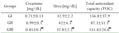
Table 2 illustrates effects on serum urea, creatinine and total antioxidants in rats receiving lead acetate with or without BMSCs. Lead intoxicated rats exhibited a considerable increase in serum urea and creatinine levels compared with apparent decrease of serum total antioxidant contents. However, lead intoxicated group followed by BMSCs injection showed marked amelioration but was still below the normal value.
Table1: Body weight, absolute and relative kidney weight (KW/BW) in different rat groups
Groups BW[g] KW[g] KW/BW GI 293±3.2 1.16±0.10 0.38±0.02
0.75±0.05* GII 219.5±5.2* 1.65±0.07*
0.43±0.03** GIII 280.7±5.4** 1.22±0.09**
Data are represented as mean ± standard deviation; p < 0.05 was considered significant
*A significant change in comparison with control (group I)
**A significant change in comparison with lead intoxicated group (group II)
Table 2: Renal function markers and total antioxidant capacity in different rat groups
Total antioxidant capacity (TOC) Urea [mg/dL]
Creatinine [mg/dL] Groups
156.8±37.9 32.9±2.2
0.71±0.13 GI
87.3±51.1⃰
42±4.5⃰
0.99±0.1⃰
GII
151.8±24.6⃰⃰
37.8±2.2⃰⃰
0.85±0.1⃰⃰
GIII
Data are represented as mean ± standard deviation; p < 0.05 was considered significant
*A significant change in comparison with control (group I)
**A significant change in comparison with lead intoxicated group (group II)
Histological and immunohistochemical results
Group I (control group). The control kidney had normal histological architecture, Examination of H&E stained sections of kidneys of this group showed renal cortex with renal corpuscle formed of a glomerulus surrounded by a parietal layer of Bowman’s capsule with the urinary space in between. The proximal convoluted tubules have narrow lumina and cuboidal or low columnar lining cells that have strong acidophilic cytoplasm and spherical basal nuclei. The distal convoluted tubules are identified by their wider lumina, lower cuboidal lining cells, faint acidophilic cytoplasm and rounded central nuclei (Fig. 1a).
convoluted tubules. Intraglomerular PAS positive material was also detected (Fig. 1b).
Collagen fibres were found to be of minimal amounts and were confined to the Bowman’s capsule, around the tubules and basal laminae of glomerular capillaries (Fig. 1c).
All structures in the renal cortex (glomeruli and tubules) had displayed a negative reaction for CD24 monoclonal antibody (Fig.1d).
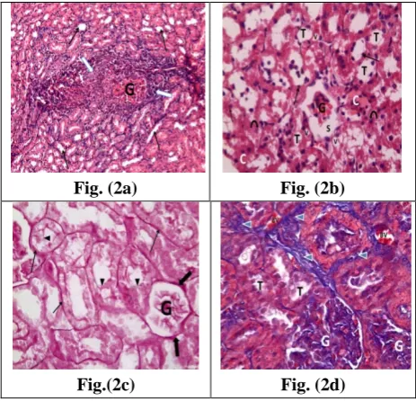
Group II (lead intoxicated). In lead intoxicated rats, areas of glomerular and tubular degeneration were seen among apparently normal ones. There was progressive shrinkage of the glomeruli, widening of the urinary spaces, massive inflammatory cell-infiltration as well as dilated renal tubules. The renal tubules, in this group, also revealed marked diminution of the cellular height, widened lumina, cytoplasmic vacuolations, pyknotic and ghost nuclei, intratubular exfoliation, and cast formation (Figs. 2a&2b).
The parietal layers of Bowman's capsules were mostly expanded by increase of PAS positive material. Some tubules showed strong PAS reaction in their basal laminae while others revealed weak PAS reaction in their brush border (Fig. 2c). Masson trichrome stained sections revealed increased intraglomerular and peritubular collagen fibres (Fig. 2d).
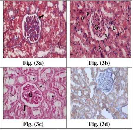
Group III (lead intoxicated + BMSCs injected). BMSCs supplementation to lead intoxicated group, showed reduction of the degenerative changes that were induced in group II. Manyrenal glomeruli and tubules were restored toward the control appearance. Some of the urinary spaces and tubular lumina were persistently widened. A moderate inflammatory cell- infiltration was also noticed (Fig.3a).
The parietal layers of Bowman's capsules and tubular basement membranes showed decreased intensity of PAS reaction together with partially regained apical brush borders of the proximal tubules (Fig. 3b). The intertubular collagen tissue was reduced in amount. There is also, reduced thickness of the parietal layer of Bowman’s capsule and the tubular basement membranes (Fig. 3c). The lining cells of Bowman’s capsules and renal tubules, in this group, demonstrated a diffuse positive reaction for CD24 monoclonal antibody (Fig. 3d).
Fig. (1a) Fig. (1b)
Fig. (1c) Fig. (1d)
Figure 1: Photomicrographs of sections in the kidney of a control rat (group I) showing: (a) Normal renal architecture: glomerulus (G), proximal (P) and distal (D) convoluted tubules haematoxylin and eosin x400; (b) Strong periodic acid-Schiff (PAS) reaction in the glomeruli (thick arrow), brush border of the proximal convoluted tubules (arrow heads), basal lamina (thin arrows) of tubules PAS x400; (c) Normal distribution of collagen fibres in the glomerulus (G), Bowman’s capsule(thick arrow), surrounding the tubules (thin arrows) and intertubular (arrow heads) Masson’s trichrome x400; (d) Glomerulus (G) and tubules (arrows) negative for CD24 monoclonal antibody Immunohistochemical expression of CD24 x 400.
Fig. (2a) Fig. (2b)
Fig.(2c) Fig. (2d)
Fig. (3a) Fig. (3b)
Fig. (3c) Fig. (3d)
Figure 3:Photomicrographs of sections in the kidney of lead intoxicated ratfollowed by a single injection of BMSCs (group III) showing: (a) Persistent widening of the urinary space (S) surrounding the glomerulus (G). The renal tubules (P&D) are mostly of a control appearance. A moderate cell-infiltration (arrows) haematoxylin and eosin x400; (b)Decreased periodic acid-Schiff (PAS) reaction in the glomeruli (G) and Bowman's capsule (thick arrow) and tubular basement membranes (thin arrows), apical brush borders (arrow heads) of the proximal convoluted tubules are partially regained PAS x400; (c)Reduced thickness of Bowman’s capsule (thick arrow), tubular basement membranes (thin arrows) with diminished amount of intertubular collagen fibers (arrow heads) Masson’s trichrome x400; (d) Diffuse positive reaction for CD24 monoclonal antibody in lining cells of Bowman’s capsule (black arrows) and in tubular cells (red arrows) Immunohistochemical expression of CD24 x 400.
Quantitative morphometric results
The quantitative morphometric histological results are summarized in Table 3. The mean values of the glomerular diameter of GII (lead intoxicated group) and GIII (BMSCs treated group) were significantly decreased when compared to GI (control group). BMSCs treatment (group III) increased significantly the glomerular diameter to 122.4±5.42 compared to group II (87.16±13.77). The mean values of the width of the urinary spaces in GII and GIII were significantly increased to 58.1±10.79 and 54.35±13.93, respectively when compared to the control group (36.33±3.03). However, non-significant difference was found between groups II and III.
The mean values of the tubular diameter were significantly decreased to 219.8±11.49 in group III when compared to group II (311.3±16.45). Moreover,the height of the lining epithelium of the proximal convoluted tubules showed significant decrease to 6.88±1.03 and 8.95±1.45, respectively in (GII and GIII) in comparison to the control group (10.79±2.04). Meanwhile, there was a significant increase in group III in comparison with group II.
Intensity of PAS-reaction in GII and GIII indicated significant increase to 66.67±1.79 and 60.99±3.08, respectively in comparison to GI (48.52±4.46). In BMSCs treated group, mean values of the intensity of PAS-reaction were significantly decreased than lead intoxicated group. The mean area percentage of the collagen fibers showed highly significant increase in lead intoxicated and BMSCs treated groups in comparison to the control group. But, BMSCs treatment decreased significantly percentage of fibrosis to 26.198±4.666 than lead intoxication (37.01±4.166).
Table 3: Morphometric histological results in different rat groups
G
roups
G
lo
m
er
ul
ar
di
am
et
er
Uri
na
ry
sp
ac
e
w
idth
Tub
ula
r di
am
ete
r
Ep
ith
el
ial
h
ei
gh
t
O
pti
ca
l de
ns
ity
of
PAS
-r
ea
cti
on
Per
cen
ta
ge
of
fibr
osi
s
GI ±10.06 132.70 ±3.03 36.33 ±8.83 224.3 ±2.04 10.79 ±4.46 48.52 ±1.025 3.035
GII ±13.7787.16 ⃰ ±10.7958.1 ⃰ ±16.45311.3 ⃰ ±1.036.88 ⃰ ±1.7966.67 ⃰ ±4.16637.01 ⃰
GIII ±5.42122.4 ⃰⃰ ±13.93 54.35 ±11.49219.8 ⃰⃰ ±1.458.95 ⃰⃰ ±3.0860.99 ⃰⃰ ±4.66626.198 ⃰⃰
Data are represented as mean ± standard deviation; p < 0.05 was considered significant
*A significant change in comparison with control (group I)
**A significant change in comparison with lead intoxicated group (group II)
Table 4 demonstrated the mean percentage of epithelial cells revealing positive reaction to CD24 monoclonal antibody, in rats injected with BMSCs, was significantly higher in glomerular mesangium and proximal tubules of GIII than the control rats (GI) that lacked cells with positive reaction to CD24 monoclonal antibody.
Table4: Morphometric immunohistochemical results in different rat groups
Tubular expression of CD24 Glomerular expression of
CD24 Groups
0.0±0.0 0.0±0.0
GI
95.00±5.0⃰
78.40±3.78⃰
GIII
Data are represented as mean ± standard deviation; p < 0.05 was considered significant
*A significant change in comparison with control (group I)
Discussion
Chronic nephropathy is a slowly progressive disease of both glomerulo-tubular and interstitial regions that are caused by a number of toxins and drugs. Chronic lead exposure and intoxication is thought to be a common ailment that particularly affects the kidneys which form the major route of excretion of this metal. The hazardous effects of lead on the renal tissue are mediated through the generation of reactive oxygen radicals [17].
Yuan et al. [18] mentioned that most of the acute and chronic
proximal convoluted tubules. Therefore, the current work studied the possible protective effect of BMSCs treatment against lead acetate induced nephrotoxicity.
In this study, the kidneys extracted from lead intoxicated rats whether followed or not by BMSCs, have revealed pale, granular and hemorrhagic surfaces. The kidneys were also swollen. Suradkar et al. [19] mentioned that rat kidneys were grossly
congested and enlarged on exposure to lead. In the present work, the rat kidney weight / body weight ratio in lead intoxication groups has revealed significant increase when compared to control group. These increased values could be attributed to the swelling and congestion of the kidneys, reduction of rat body weight or both of them. Amjad et al. [20] related the increase in
relative kidney weight to both accumulation of lipids in the kidneys and nutritional disturbances induced by lead acetate intoxication. A significant reduction in rat body weight has been claimed to be due to anorexia and decreased muscle mass caused by leadintoxication [21]. However, Yuan et al.[22] noticed
non-significant changes in rat body weight following lead and cadmium intoxication for 90 days.
The biochemical findings in this work were in agreement with those obtained by Alya et al. [17] and Sujatha et al. [11] who
reported that lead administration led to impairment in kidney function as shown by increase in creatinine and urea. Moreover, there was a significant reduction in total antioxidant capacity which was also observed by Abdel-Moniem et al. [23] who
recorded impaired antioxidant status in lead intoxicated rats. Nephrotoxicity induced by lead was confirmed by histological changes including progressive shrinkage of renal glomeruli, widening of urinary spaces and dilatation of renal tubules. In addition, massive inflammatory cell-infiltration and focal loss of cortical architecture have been also detected. These results coincide with the observations of glomerular collapse and sclerosis mentioned by Sabolic[24]. The injurious effects of lead
intoxication on the renal tubules, observed herein, have included marked diminution of the cellular height of the tubular epithelium, widening of the tubular lumina, and intratubular exfoliations, and cast formation. Such findings run in accordance with similar observations mentioned by Begum et al. [25]. The
derangement effect of lead intoxication on the proximal convoluted tubules has been statistically confirmed in this study. There were significant increases in the diameter of the proximal renal tubules and significant decrease in their cellular height. The current observation of cytoplasmic vacuolations of most of the proximal tubular lining cells in kidneys of lead intoxicated group supported by Alwin and Arthur[26] who reported that an
increased osmotic gradient across the cellular plasma membranes had led to water withdrawal, swelling and vacuolation of the tubular cells. Mohamed and Saleh[27] postulated that vacuolations
of the proximal and distal tubular cells were most probably a cellular defensive mechanism against the injurious effect of lead. The nuclear changes observed in the tubular cells of lead intoxication rat group included pyknotic and ghost nuclei also reported by Navarro-Moreno et al. [28] who explained these
nuclear changes to be due to the ability of lead to inhibit protein synthesis and to interfere with nucleic acid formation.
The current work revealed dilatation and congestion of the renal blood vessels together with intertubular hemorrhages as a result of lead intoxication. Prozialeck et al. [29] assumed that these
vascular phenomena might be due to direct or indirect vasoactive effect of lead on the vascular endothelium via increased production of reactive oxygen radicals. Histological sections from lead intoxicated rats in this study showed intensive PAS reaction of the parietal layers of Bowman's capsules and tubular basement membranes with a progressive loss of the apical brush border of their epithelial lining, these observations supported by Begum et al.[25] who attributed these findings to accumulation of
glycoproteins in the glomerulo-tubular basement membranes that drastically influenced the renal functions. Furthermore, Mohamed and Saleh[27] concluded that heavy metals intoxication
led to shortening and loss of the microvilli of the proximal convoluted tubules.
This study demonstrated statistically significant increase in the area percentage of fibrosis in lead intoxicated group. Macrophages are mentioned to play an important role in interstitial renal fibrosis through the production of a fibrogenic growth factor that induces my fibroblastic cells to produce extracellular matrix [30].
Bone marrow–derived stem cells (BMSCs) are promising therapy for the repair of various tissues including the kidneys. Presently, injection of BMSCs after eight weeks of lead acetate intoxication apparently reduced its deleterious effects and attenuated the renal damage. This improvement was clearly reflected by a significant decrease in KW relative to BW and serum creatinine. Also, serum urea and total antioxidant capacity returned nearly to its normal levels. These findings were also reported by Chen et al.[31]. The authors proposed that mesenchymal stem cells had
inhibited inflammatory reactions and suppressed oxidative stress through increasing the level of antioxidant enzymes. Yadav et al.
[32] detected improvement of kidney weight in BMSCs-treated
mice following mercuric chloride-induced nephrotoxicity. In the current study, histological and immunohistochemical recovery findings in the form of glomerular and tubular regeneration together with restoration of the brush borders and positive expression of CD24 monoclonal antibody in glomeruli and tubular basement membranes were observed in BMSCs injected animals. Sadek et al.[33] supported the previous findings.
The mechanism of action of mesenchymal stem cells (MSCs) in the process of regeneration has been explained by several investigators. MSCs have paracrine activity that leads to secretion of a number of growth factors and cytokines which are essential for angiogenesis and cytoprotection, including vascular endothelial growth factor (VEGF), transforming growth
factor-β1 (TGF-β1), and hepatocyte growth factor (HGF). Fox et al. [34]
found that mesenchymal cells could migrate to glomeruli, tubules, peritubular capillaries, and interstitium in both acute and chronic kidney injury models. Broekema et al. [35] stated that
renal ischemia. Milwid et al. [36] concluded that BMSCs had
prevented tubular apoptosis and necrosis in injured kidneys through maintenance of the glomerular filtration rate and preservation of renal architecture; thus improving renal functions. On the contrary, Moghadasali et al. [37] found
non-significant changes in the renal functions on intravenous injection of stem cells in animal models with chronic kidney disease. Our morphometric results supported the role of BMSCs injection in attenuating lead induced nephrotoxicity, in the form of, significant increase in the mean values of glomerular diameter with decrease in width of urinary space when compared to lead intoxicated group. Also, there was marked improvement in mean values of proximal tubular diameter and the height of epithelial lining. Moreover, there is a significant regression of the interstitial fibrous tissue formation and collagen deposition in the kidneys of BMSCs treated group. Semedo et al. [38] observed
similar reduction of fibrosis in rats treated with BMSCs in case of chronic renal failure.
Conclusions
In conclusion, administration BMSCs was found to attenuate the renal damage seen in lead acetate intoxicated rats. BMSCs have a tendency to preserve most of histological, immunohistochemical and biochemical parameters towards normal values. Human trials are essential for limitation of environmental lead pollution. Further studies are required to prove the therapeutic role of BMSCs.
References
1. Zhang Z, Gao X, Guo M, Jiang H, Cao Y, Zhang N (2017) The protective effect of baicalin against lead-induced renal oxidative damage in mice. Biol Trace Elem Res; 175:129– 135
2. Dewanjee S, Sahu R, Karmakar S, Gangopadhyay M (2013) Toxic effects of lead exposure in Wistar rats: Involvement of oxidative stress and the beneficial role of edible jute (Corchorusolitorius) leaves. Food ChemToxicol 55:78–91. 3. Sharma S and Singh B (2014) lead acetate induced
histopathological alterations in renal tissue of balb-c mice. Int J applBiol pharm; 5 (3):23-28.
4. Ahamed M and Siddiqui MKJ (2007) Low level lead exposure and oxidative stress: Current opinions. ClinChimActa; 383(1-2):57-64.
5. Togel F and Westenfelder C (2011) The role of multipotent marrow stromal cells (MSCs) in tissue regeneration. Organogenesis; 7:97-100.
6. Morigi M, Imberti B, Zoja C, Corna D, Tomasoni S, et al. (2004) Mesenchymal stem cells are renotropic, helping to repair the kidney and improve function in acute renal failure. J Am Soc Nephrol; 15:1794-1804.
7. Duffield J S, Park K M, Hsiao L L, Kelley V R, Scadden D T (2005) Restoration of tubular epithelial cells during repair
of the postischemic kidney occurs independently of bone marrow-derived stem cells. J Clin Invest; 115:1743-1755. 8. Togel F, Weiss K, Yang Y, Hu Z, Zhang P, Westenfelder C
(2007) Vasculotropic, paracrine actions of infused mesenchymal stem cells are important to the recovery from acute kidney injury. Am J Physiol Renal Physiol; 295: 1626 – 1635.
9. Kobayashi T, Tanaka H, Kuwana H, Inoshita S, Teraoka H, Sasaki S, et al. (2005) Wnt4-transformed mouse embryonic stem cells differentiate into renal tubular cells. BiochemBiophys Res Commun; 336:585-595.
10. Vlkova M, Fronkova E, Kanderova V (2010)
Characterization of lymphocyte subsets in patients with common variable immunodeficiency reveals subsets of naive human B cells marked by CD24 expression. J Immunol; 185: 6431-6438.
11. Sujatha K, Srilatha CH, Anjaneyulu Y, Amaravathi P (2011) Lead acetate induced nephrotoxicity in wistar albino rats. A pathological, immunohistochemical and ultrasructural studies. Int J Pharma Bio sci; 2(2): 459-469.
12. Alhadlaq A and Mao J J (2004) Mesenchymal stem cells: isolation and therapeutics. Stem Cells Dev; 13:436-448. 13. Rochefort G, Vaudin P, Bonnet N, Pages J, et al. (2005)
Influence of hypoxia on the domiciliation of mesenchymal stem cells after infusion into rats: possibilities of targeting pulmonary artery remodelling via cells therapies? Respir Res., 6:125.
14. Asanuma H, Vanderbrink B A, Campbell M T, Hile K L, Zhang H, Meldrum D R, Meldrum K K (2011) Arterially delivered mesenchymal stem cells prevent obstruction-induced renal fibrosis. J Surg Res; 168: 51–59.
15. Bancroft J and Gamble M (2008) Theory and practice of histological techniques. 5th ed. Churchill-Livingstone,
London, Edinburgh, New york, Philadelphia, St Louis, Sydney and Toronto; 126-127,150,171&601-612.
16. Hsu S M, Raine L, Fauger H (1981) Use of avidin- biotin peroxidase complex (ABC) in immunoperoxidase technique: a comparison between ABC and unlabeled antibody (PAP) procedure. J Histochem; 29: 577-580. 17. Alya A, Ines B D, Montassar L, Najoua G h, Saloua E
F (2015) Oxidative stress, biochemical alterations and hyperlipidemia in female rats induced by lead chronic toxicity during puberty and post-puberty periods. Iran J Basic Med Sci; 18:1034‐1043.
18. Yuan H E X, Yun Y L, Tao L Y, Meng L I, et al. (2016) Cytotoxic responses and apoptosis in rat kidney epithelial cells exposed to lead. Biomed Environ Sci; 29(7): 529-533. 19. Suradkar S G, Vihol P D, Patel J H, Ghodasara D J, Joshi B
P, Prajapati K S (2010) Patho-morphological changes in tissues of Wistar rats by exposure of lead acetate. Vet World; 3(2): 82-84.
21. Djebli N, Slimani M, Aoues A (2004) Effect of lead exposure on dopaminergic transmission in the rat brain. Toxicol; 207: 363-368.
22. Yuan G, Dai S, Yin Z, Lu H, Jia R, Xu J (2014) Sub-chronic lead and cadmium co-induce apoptosis protein expression in liver and kidney of rats. Int J Clin Exp Pathol; 7(6):2905-2914.
23. Abdel-MoneimA E, Dkhil M A, Al-Quraishy S (2011) The protective effect of flaxseed oil on lead acetate-induced renal toxicity in rats. J Hazard Mater; 30 (194): 250-255. 24. Sabolic I (2006) Common mechanisms in nephropathy
induced by toxic metals. Nephron physiol; 104(3):107-114. 25. Begum B, Rana I N, Majeed M (2014) Lead induced morphological changes in the kidneys of albino mice. J Rawal Med Coll; 18(1):75-79.
26. Alwin H and Arthur H (2009) Drug-induced kidney disease. Pathology and current concepts. Ann Acad Med; 38: 240 -250.
27. Mohamed N A and Saleh S M (2010) Effect of pre and postnatal exposure to lead acetate on the kidney of male albino rat: A light and electron microscopic study. Egypt J Histol; 33(2): 365-379.
28. Navarro-Moreno L G, Quintanar-Escorza M A, González S (2009) Effects of lead intoxication on intercellular junctions and biochemical alterations of the renal proximal tubule cells. Toxicology in Vitro; 23(7): 1298–1304.
29. Prozialeck W C, Edwards J R, Nebert D W, Woods J M, et al. (2008) The vascular system as a target of metal toxicity. Toxicol Sci; 102(2):207-218.
30. Vallon V and Thomson S C (2012) Renal function in diabetic disease models: the tubular system in the pathophysiology of the diabetic kidney. Annu Rev phsiol; 74:351-375.
31. Chen Y T, Sun C K, Lin Y C, Chang L T, Tsai T H, et al (2011) Adipose-derived mesenchymal stem cell protects kidneys against ischemia-reperfusion injury through suppressing oxidative stress and inflammatory reaction. J Transl Med; 9(51): 1479-1496.
32. Yadav N, Rao S, Bhowmik D M, Mukhopadhyay A (2012) Bone marrow cells contribute to tubular epithelium regeneration following acute kidney injury induced by mercuric chloride. Indian J Med Res; 136: 211-220. 33. Sadek E M, Afifi N M, Abd-El Fattah L I, Abd-El Mohsen
(2013) Histological study on effect of mesenchymal stem cell therapy on experimental renal injury induced by ischemia/reperfusion in male albino rat. Int J Stem Cells; 6(1): 55-66.
34. Fox J M, Chamberlain G, Ashton B A (2007) Recent advances into the understanding of mesenchymal stem cell trafficking. Br J Haematol; 137:491-502.
35. Broekema M, Harmsen M C, Koerts J A, et al (2005) Determinants of tubular bone marrow-derived cell engraftment after renal ischemia/reperfusion in rats. Kidney Int; 68(6): 2572-2581.
36. Milwid J, Ichimura T, Parekkadan B, Tilles A, Bonventre J, Yarmush M (2012) Secreted factors from bone marrow stromal cells upregulate IL-10 and reverse acute kidney injury. Stem Cells Int; 2012:12.
37. Moghadasali R, Hajinasrollah M, Argani H (2015) Autologous transplantation of mesenchymal stromal cells tends to prevent progress of interstitial fibrosis in a rhesus Macacamulatta monkey model of chronic kidney disease. Int Society Cellular Therapy; 17: 1495-1505.