M
AŁGORZATAP
ORĘBA1, L
IDIAU
SNARSKA−Z
UBKIEWICZ2, B
OŻENAJ
AŹWIEC2,
R
AFAŁP
ORĘBA3, K
AZIMIERZK
ULICZKOWSKI2Expression of CD105 Antigen
in Patients with Acute Leukemia,
Malignant Lymphoma, and Multiple Myeloma
in Active Phase of the Disease
Wysoka ekspresja antygenu CD105 w aktywnej fazie choroby
u chorych na ostre białaczki, chłoniaki złośliwe i szpiczaki
1Department of Pathophysiology, Silesian Piasts University of Medicine in Wrocław, Poland
2Department of Hematology, Blood Neoplasms, and Bone Marrow Transplantation, Silesian Piasts University
of Medicine in Wrocław, Poland
3Department of Internal Medicine, Occupational Disease, and Hypertension, Silesian Piasts University
of Medicine in Wrocław, Poland
Adv Clin Exp Med 2006, 15, 6, 1023–1028 ISSN 1230−025X
ORIGINAL PAPERS
© Copyright by Silesian Piasts University of Medicine in Wrocław
Abstract
Background.CD105 (endoglin) is a proliferation−associated protein abundantly expressed in angiogenic endothe− lial cells. CD105 is a receptor for TGF (transforming growth factor)−β1 and −β3. It is important for the develop− ment of normal vascular architecture and may be associated with tumor angiogenesis. CD105 was strongly expressed in the endothelium of various tumor tissues compared with normal tissues and has been shown to be a useful marker to identify tumor angiogenesis.
Objectives.Determining the expression of CD105 in blood of patients with acute leukemia, malignant lymphoma and multiple myeloma using the cytofluorimetric method. This should help to evaluate the role of CD105 in the development of hematopoietic malignancies. There are few available data in medical literature on this topic.
Material and Methods.The study group consisted of three subgroups of patients with hematopoietic malignan− cies (20 with acute leukemia, 21 with malignant lymphoma, and 20 with multiple myeloma) and a control group of 30 healthy people. Blood samples were obtained from consecutive patients admitted to the clinic with the newly diagnosed disease before cytostatic treatment was introduced. Cells were prepared, stained with monoclonal anti− bodies, and analyzed by flow cytometry.
Results.High expression of CD105 was present in patients with ALL (acute lymphoblastic leukemia) and AML (acute myelogenous leukemia) and was significantly higher than in the control group. In the patients with LM (lym− phoma malignum) and MM (multiple myeloma) the mean expression was higher than in healthy individuals, but not statistically significantly. No statistically significant correlations were observed between CD105 expression and age, gender, LDH concentration, chromosomal aberrations typical for patients with acute leukemia, or the number of blasts. In blood of patients with acute leukemia, CD105 expression was present on leukemic blast cells. In patients with LM an MM, CD105−positive cells were identified mainly within the population of neoplastic lym− phocytes.
Conclusions.These results may suggest a connection between CD105 expression and the pathogenesis of blood neoplasms. The finding of a very high endoglin expression especially in patients with ALL was limited to a small group of patients and needs to be confirmed in a larger group. The mechanisms underlying the role of CD105 action in hematological malignancies still need to be fully elucidated (Adv Clin Exp Med 2006, 15, 6, 1023–1028).
CD105 (endoglin) is a proliferation−associated and hypoxia−inducible transmembrane protein abundantly expressed in angiogenic endothelial cells. It is a receptor for transforming growth fac− tors (TGF)−β1 and −β3. The human CD105 gene is located on chromosome 9q34 [1]. The exact mechanisms for CD105 regulation of vascular development have not been fully elucidated. A variety of important functions of CD105 have been uncovered. most of which are likely to be associated with TGFβsignalling. CD105 suppress− es TGFβsignaling in many cells in vitro [2]. CD105 expression increases during angiogenesis, wound healing, and inflammation, all of which are associated with TGFβsignalling and alterations in vascular structure. It is also important for normal vascular architecture. CD105 null mice exhibit multiple vascular and cardiac defects, leading to death at early embryonic stages [3]. From embry− onic day 9, the primitive vascular plexus of the yolk sac failed to remodel into mature vessels, causing vascular channel dilation, rupture, and hemorrhage; vessel fragility resulted in the inter− nal bleeding. These severe vascular impairments observed in CD105 null mice suggest that CD105 is required for the formation of mature blood ves− sels in the extra−embryonic vasculature. Failed endocardial cushion formation, essential for valve development and heart septation, and pericardial edema were also noticed in the CD105 null mice,
indicating another crucial role of CD105 in cardiac development. CD105 knock−out mice die from malvascularisation by 11.5 day p.c. [4].
The role of CD105 may be associated with tumor angiogenesis. CD105 was strongly ex− pressed in the endothelium of various tumor tis− sues compared with normal tissues [5, 6]. It has been shown to be a more useful marker in identi− fing tumor angiogenesis than panendothelial mar− kers such as CD31 [7]. Studies performed in dif− ferent laboratories using various antibodies to CD105 have revealed CD105 upregulation in a wide range of tumor endothelia, including those within the colon, breast, brain, lung, prostate, and cervical cancer, suggestive of the possible involve− ment of CD105 in tumor angiogenesis [5, 8]. In one study, concentrations of CD105 measured in plasma samples were significantly increased in patients who developed distant metastasis com− pared with disease−free patients and with healthy individuals [9].
The aim of this study was to determine the expression of CD105 in the blood of patients with acute leukemia, malignant lymphoma, and myelo− ma multiplex with the use of the cytofluorymetric method. The studies may help to evaluate the role of CD105 in the development of hematopoietic malignancies, for there are few data available on this topic in medical literature.
Streszczenie
Wprowadzenie.CD105 jest białkiem związanym z proliferacją, które ulega obficie ekspresji na powierzchni ko− mórek śródbłonka. CD105 jest elementem receptora wiążącego się z TGF β−1 i 3 (transforming growth factor be− ta). Endoglina bierze udział w rozwoju prawidłowego unaczynienia i może być związana z angiogenezą nowotwo− rową. Wykazywano wysoką ekspresję CD105 w porównaniu ze zdrowymi tkankami w śródbłonku pochodzącym z unaczynienia różnych typów nowotworów i antygen ten okazał się użyteczny jako marker angiogenezy nowo− tworowej.
Cel pracy.Oznaczenie ekspresji CD 105 we krwi chorych na ostre białaczki, chłoniaki złośliwe i szpiczaka mno− giego z zastosowaniem metody cytofluorymetrii przepływowej i podkreślenie roli CD105 w powstawaniu nowo− tworów krwi.
Materiał i metody.Grupa badana składała się z trzech podgrup chorych na nowotwory krwi: 20 chorych na ostrą białaczkę, 21 chorych na chłoniaka złośliwego, 20 chorych na szpiczaka mnogiego i z grupy kontrolnej obejmują− cej 30 zdrowych ludzi. Próbki krwi pobierano od kolejnych pacjentów przyjmowanych do Kliniki z nowo rozpo− znaną chorobą, przed włączeniem leczenia cytostatycznego. Po przygotowaniu do zawiesiny komórek dodano przeciwciała monoklonalne, a następnie analizowano za pomocą cytofluorymetrii przepływowej.
Wyniki.U chorych na ALL (acute lymphoblastic leukemia) i AML (acute myelogenous leukemia) obserwowano wysoką ekspresję CD105 i była ona istotnie statystycznie wyższa niż u osób zdrowych. We krwi chorych na LM (lymphoma malignum) i MM (myeloma multiplex) stwierdzono wyższą ekspresję niż u osób zdrowych, ale nie była to różnica istotna statystycznie. Nie wykazano istotnych zależności między ekspresją CD105 a wiekiem, płcią, stę− żeniem LDH, obecnością typowych zmian chromosomalnych chromosomalnych chorych na ostre białaczki lub licz− bą blastów. We krwi chorych na ostre białaczki ekspresja CD105 była obecna na komórkach białaczkowych. U cho− rych na LM i MM komórki CD105−dodatnie znajdowały się głównie w populacji limfocytów nowotworowych.
Wnioski.Wyniki badań mogą sugerować istnienie związku między ekspresją CD105 a patogenezą chorób nowo− tworowych krwi. Wykazanie dużej ekspresji endogliny u chorych na ALL jest ciekawym faktem, wymagającym jednak potwierdzenia na większej grupie chorych. Mechanizmy, z którymi jest związana rola CD105 w powstawa− niu nowotworów krwi wciąż nie są dobrze poznane (Adv Clin Exp Med 2006, 15, 6, 1023–1028).
Material and Methods
The study group consisted of three subgroups with hematopoietic malignancies and a control group. We investigated blood samples of patients with acute leukemia (n = 20), 14 men and 6 wo− men with an average age of 52.55 ± 17.17 years. In this subgroup, 15 patients were identified as having AML and 5 as having ALL. Twenty−one persons were in the subgroup of patients with LM: 14 men and 7 women (mean age 52.0 ± 14.71 years old). The characteristics of this subgroup according to the Ann Arbor classification are shown in Table 1. The subgroup of patients with MM (n = 20) was composed of 11 women and 9 men with an average age of 61.8 ± 11.81 years. Staged by Salmon and Durie criteria, 4 patients had stage II A, 9 patients had III A, and 7 patients had III B. The control group consisted of 30 healthy people without any chronic disease (19 men and 11 women, mean age: 54 ± 16.4 years).
In the study subgroups the mean concentration of lactate dehydrogenase LDH (a well−known marker of progression in blood neoplasms) was 1345 ± 218 U/l in the patients with acute leukemia, 856.05 ± 153 U/l in the patients with LM, and
406 ± 63 U/l in the patients with MM. The per− centage of blast cells in the bone marrow in patients with acute leukemia was from 20 to 98%, mean 65 ± 6%, and the mean percentage of blast cells in peripheral blood was 58 ± 5%. In patients with grade IV of LM, bone marrow was infiltrated in all cases by neoplastic cells as were liver, spleen, all palpable lymph nodes, and retroperi− toneal lymph nodes. In three of the patients, infil− trations in the mediastinum were present and in one in the facial region (septum of the nose and pharyngeal and palatine tonsils).
Fasting blood samples were obtained from consecutive patients admitted to the clinic with newly diagnosed disease before cytostatic treat− ment had been introduced. In each patient, punc− ture of a peripheral vein was carried out and
10 cm3 of blood was collected. Preparations of samples for flow cytometric analysis were per− formed by centrifugation, then the cell suspen− sions were stained for the surface expressions of various markers using FITC−(green), Cyanine 5 (RPE−Cy5, red) fluorochrome, and phycoerythrin (PE, orange) conjugated with mAbs (monoclon− al antibodies). R−phycoerythrin−Cyanine5 (RPE− −Cy5)−conjugated anti−CD45 mAb (antibody against CD45 leukocyte common antigen) was purchased from Dako (Glostrup, Denmark). Monoclonal antibody against CD105 FITC was obtained from Ancell (Bayport, MN, USA). Mouse IgG1 FITC/PE/RPE−Cy5 negative control mAb (DAKO, Glostrup, Denmark) was used as a negative control. In blood samples from patients with acute leukemia, cells were also stained with CD34 RPE (DAKO, Glostrup, Denmark) anti− body, routinely used in diagnosing acute leukemias.
The cell suspensions were incubated with the monoclonal antibodies for 45 minutes on ice with 80 µl of anti−CD105/FITC at a 1:50 dilution (10 mg/ml). The cells were then washed three times and analyzed using a FACS (Partec, Germany). For analysis, the cells were gated on the basis of forward and side scatter. Cells stained positive were compared with the Mouse IgG1 negative control at a similar concentration. Cells were shown as dots in separate quadrants and charts as a percentage of the gated cells.
Statistical analysis was performed with the commercially available package STATISTICA, version 6.0. The results were presented as a mean (X) ± one standard deviation (SD). The distribu− tion of data was examined using the Shapiro−Wilk test. Differences between the analyzed parameters were examined using the non−parametric ANOVA Kruskal−Wallis test. Correlations between the vari− ables were tested using the Spearman correlation coefficient r. For the determination of survival analysis, the Kaplan−Meier estimator was used. Apvalue less than 0.05 was considered statistical− ly significant.
Results
High expression of CD105 was present in patients with ALL and AML (mean ± SD: 36.2 ± ± 27.6% and 17.8 ± 11.3%, respectively). The expression of CD105 in the group with ALL and AML was significantly higher than in the control group (1.15 ± 0.45%; p= 0.002 for ALL and p= = 0.4 for AML). In LM and MM patients the mean expressions were 6.35 ± 4.31% and 4.03 ± 2.03%. These results were not statistically significant in Stage of lymphoma Number of patients
(Stadium chłoniaka) (Liczba chorych) n = 21
IIB 1
IIIA 1
IIIB 2
IVB 17
comparison with healthy individuals (Table 2). In patients with ALL and high CD105 expression, a high percentage of blasts in bone marrow (93–97%) was observed. These patients were found to be in poor clinical condition, with high levels of lactate dehydrogenase and the extremely increased levels of D−dimer. However, no statisti− cally significant correlations were observed between CD105 expression and age, gender, LDH concentration, typical chromosome aberrations, or blast cells in bone marrow or peripheral blood for patients with acute leukemia. In patients with AML, CD105 expression was diverse and ranged from several to 30% (Fig. 1).
There were no statistically significant differ− ences in CD105 expression between patients with higher (above median) and lower (below median) concentrations of LDH in any of the studied sub− groups (Fig. 2). No correlations were found be− tween advanced stage of disease in patients with LM (IV) and increased CD105 expression. How− ever, the relatively small number of patients and
the heterogeneous character of the subgroup could make statistical analysis in this matter inaccurate.
In the group of patients with LM, a significant correlation between the expression of CD105 and fibrinogen concentration was noted (r= 0.81, p< 0.05). In this group, a negative correlation between CD105 expression and hemoglobin concentration was also present (r= –0.61, p< 0.05).
Analysis with two−color flow cytometry enabled determining in which part of CD45−posi− tive cells (a marker characteristic for leukocytes) CD105 was co−expressed. In the blood of patients with acute leukemia the expression of CD105 was present on leukemic blast cells. There was no sta− tistically significant correlation between a high (above median) percentage of blast cells and high expression of CD105 antigen in these patients.
In the subgroups of patients with LM an MM, CD105−positive cells were identified mainly with− in the population of neoplastic lymphocytes.
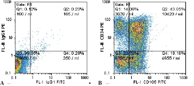
In a group with acute leukemia, staining with marker CD34, typically used for the routine diag− nosis of AML and ALL, revealed a high co− expression with CD105 in patients with ALL, ranging from 25 to 43% (Fig. 3).
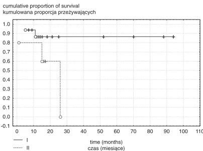
The highest statistically significant rate of mo− rtality was present in patients with LM and a high level of CD105 (above median) in comparison with patients with lower expression (p< 0.02).
Discussion
In these studies it was revealed that the expres− sion of CD105, a well−known marker of neopastic angiogenesis, is relatively high in patients with
Disease CD105 expression
(Choroba) (Ekspresja CD105)
%
AML 36.2 ± 27.6
ALL 17.8 ± 11.3
LM 6.35 ± 4.31
MM 4.03 ± 2.03
Table 2. CD105 expression in the study subgroups
Tabela 2. Ekspresja CD105 w badanych podgrupach chorych
Fig. 1. Comparison of CD105 expressions in patients with acute leukemia, malignant lymphoma, myeloma multiplex, and the control group
Ryc. 1.Porównanie ekspresji CD105 u chorych na ostrą białaczkę, chłoniaka złośliwego, szpiczaka mno− giego i w grupie kontrolnej
±standard deviation odchylenie standardowe ±standard error błąd standardowy mean średnia –10
0 10 20 30 40 50 60 70
AML ALL LM MM controls
grupa kontrolna CD105
Fig. 2. Kaplan−Meier survival analysis according to the median expression of CD105 (I – below median, II – above median) in patients with lymphoma malignum (p= 0.025)
Ryc. 2. Krzywe przeżycia Kaplana−Meiera w zależno− ści od mediany ekspresji CD105 (I – poniżej mediany, II – powyżej mediany) u chorych na chłoniaka złośli− wego (p = 0,025)
I
II czas (miesiące)time (months) −0.1
0.0 0.1 0.2 0.3 0.4 0.5 0.6 0.7 0.8 0.9 1.0
0 10 20 30 40 50 60 70 80 90 100 110
acute leukemia, especially those with ALL. Moderately high expressions were also noted in patients with malignant lymphomas and multiple myeloma, although these were not statistically sig− nificant. The results correspond with data present− ed by Calabro and colleagues [10], who found increased serum levels of CD105 in patients with acute leukemia and also those with CMD (chronic myeloproliferative disorders). However, the pre− sent experiments enabled the measurement of CD105 not as a soluble molecule in serum or plas− ma, but as a molecule directly expressed on the surface of CD45−positive cells (leukocytes). The fact that the highest expression of CD105 was found in the population of blast cells suggests a connection with the pathogenesis of the disease. Unfortunately, the interesting finding of a very high CD105 expression in patients with ALL was limited to a small group of five individuals. In the medical literature no reports about the expression of CD105 in ALL were found. These interesting data need to be confirmed in larger groups of patients. The blood samples were collected from consecutive patients with acute leukemia, so the number of patients with ALL was random.
Endoglin is a prognostic marker in patients with prostate cancer [11]. High levels of soluble sCD105 were found in women with breast cancer compared with healthy people, especially in patients with metastases [9] and those with cancer of the large intestine [12]. In the studies of Pruneri et al., anti−CD105 monoclonal antibody was very sensitive in the determination of increased angio− genesis in bone marrow samples from patients with myeloma multiplex [13]. The same authors reported increased CD105 expression in bone mar− row samples with hairy cell leukemia, one of the types of lymphoma [14].
There are only a few reports supporting some connections between CD105 and the development of hematolological malignancies and the mecha− nisms of it. It is known that CD105 antagonizes the inhibitory effect of TGFβ1 on human and murine endothelial cells [15]. TGFβis a cytokine with an
inhibitory effect on lymphoma cells in animal stud− ies [16]. It is possible that the complex system of cytokines and their interactions with CD105 have an important influence on lymphoma development. Miller et al. revealed connections between the expression of endoglin and proliferation markers such as cyclin A and Ki−67 protein in lung cancer, which suggests an influence on the cell cycle [17]. Preliminary studies done by Guo et al. showed that the role of CD105 is not limited to being a receptor for TGFβ, but also includes participation in adhe− sion, migration, and survival of cells [18]. TGFβ inhibits EC proliferation, migration, and the forma− tion of microvessels, whereas CD105 counteracts these actions, thereby promoting angiogenesis.
CD105 is a promising target that can be used for tumor imaging and prognosis and it possesses a therapeutic potential in patients with solid tumors and other neoplastic diseases with increa− sed angiogenesis. Increased CD105 levels may be useful as an indicator of disease progression and to identify patients at risk of recurrence and/or metas− tasis, especially in women with breast cancer. Additionally, endoglin could serve to monitor the effects of treatment, because the level of CD105 decreased after effective treatment in patients with neoplastic diseases [3, 9].
CD105 represents an ideal target for antiangio− genic therapy and a good marker for tumor prog− nosis. However, the mechanisms underlying the pro−angiogenic action of CD105 have not been fully elucidated. Studies have demonstrated long− lasting complete abrogation of human breast tumors in SCID mice using CD105 mab (anti− CD105 antibodies) that has been conjugated with immunotoxins [19] and growth suppression of human solid tumors using a radio−labeled mab to CD105 [12]. Endoglin is not a fully specific mark− er of neoplastic vascularity [20]. Slight amounts of CD105 were identified within normal vessels and in stroma. In future, the use of monoclonal anti− bodies should be thoroughly considered. The results of this study support the important role of CD105 in angiogenesis and in tumor progression.
Fig. 3.CD105 expression by flow cytometric analysis in a patient with ALL (A – negative control, B – co−expression of CD105 and CD34)
Ryc. 3. Ekspresja CD105 w bada− niu na cytofluorymetrze
przepływowym u chorych na ALL (A – kontrola ujemna, B – ko− ekspresja CD105 i CD34)
References
[1] Fernandez−Ruiz E, St.−Jacques S, Bellon T, Letarte M, Bernabeu C:Assignment of the human endoglin gene (END) to 9q34−qter. Cytogenet Cell Genet 1993, 64, 204–207.
[2] Li C, Hampson I, Hampson L, Kumar P, Bernabéu C, Kumar S:CD105 antagonizes the inhibitory signaling of transforming growth factor β1 on human vascular endothelial cells. FASEB J 2000, 15, 55–64.
[3] Arthur H M, Ure J, Smith A, Renforth G, Wilson D, Torsney E, Charlton R, Parums D, Jowett T, Marchuk D et al.:Endoglin, an ancillary TGFβreceptor, is required for extraembryonic angiogenesis and plays a key role in heart development. Dev Biol 2000, 5, 42–53.
[4] Li D, Sorensen L, Brooke B, Urness L, Davis E, Taylor D, Boak B, Wendel D: Defective angiogenesis in mice lacking endoglin. Science 1999, 284,1534–1537.
[5] Burrows F, Derbyshire E, Tazzari, P, Amlot P, Gazdar A, King S, Letarte M, Vitetta E, Thorpe P: Up−reg− ulation of endoglin on vascular endothelial cells in human solid tumors: implications for diagnosis and therapy. Clin Cancer Res 1995, 1, 1623–1634.
[6] Wang J, Kumar S, Pye D, van Agthoven A, Krupinski J, Hunter R:A monoclonal antibody detects hetero− geneity in vascular endothelium of tumours and normal tissues. Int J Cancer 1993, 54, 363–370.
[7] Saad R, El−Gohary Y, Memari E, Liu Y, Silverman J:Endoglin (CD105) and vascular endothelial growth fac− tor as prognostic markers in esophageal adenocarcinoma. Hum Pathol 2005, 36, 955–961.
[8] Bodey B, Bodey B, Siegel S, Kaiser H: Overexpression of endoglin (CD105): a marker of breast carcinoma− induced neo−vascularization. Anticancer Res 1998, 18, 3621–3628.
[9] Li C, Guo B, Wilson P, Stewart A, Byrne G, Bundred N, Kumar S:Plasma levels of soluble CD105 correlate with metastasis in patients with breast cancer. Int J Cancer 2000, 89, 122–126.
[10] Calabro L, Fonsatti E, Bellomo G, Alonci A, Colizzi F, Sigalotti L, Altomonte M, Musolino C, Maio M:
Differential levels of soluble endoglin (CD105) in myeloid malignancies. J Cell Physiol 2003, 194, 171–175.
[11] Wilkstrom P, Lissbrant I, Stattin P, Egevad L, Bergh A:Endoglin (CD105) is expressed on immature blood vessels and is a marker for survival in prostate cancer. Prostate 2002, 268–275.
[12] Takahashi N, Kawanishi−Tabata R, Haba A, Tabata M, Haruta Y, Tsai H Seon, B: Association of serum endoglin with metastasis in patients with colorectal, breast, and other solid tumors, and suppressive effect of chemotherapy on the serum endoglin. Clin Cancer Res 2001, 7, 524–532.
[13] Pruneri G, Ponzoni M, Ferreri AJ, Decarli N, Tresoldi M, Raggi F, Baldessari C, Freschi M, Baldini L, Goldaniga M, Neri A, Carboni N, Bertolini F, Viale G: Microvessel density, a surrogate marker of angiogene− sis, is significantly related to survival in multiple myeloma patients. Br J Haematol 2002, 118, 817–820.
[14] Pruneri G, Bertolini F, Baldini L, Valentini S, Goldaniga M, Soligo D, Carboni N, Viale G, Lambertenghi− Deliliers G:Angiogenesis occurs in hairy cell leukaemia (HCL) and in NOD/SCID mice transplanted with the HCL line Bonna−12. Br J Haematol 2003, 120, 695–698.
[15] Warrington K, Hillarby MC, Li C, Letarte M, Kumar S:Functional role of CD105 in TGF−beta1 signalling in murine and human endothelial cells. Anticancer Res 2005, 25, 1851–1864.
[16] Buske C, Hannig H, Schneider EM, Blaschke S, Hunsmann G, Bodemer W, Hiddemann W: Transforming growth factor beta is a growth−inhibitory cytokine of B cell lymphoma in SIV−infected macaques. AIDS Res Hum Retroviruses 1999, 15, 1477–1485.
[17] Miller DW, Graulich W, Karges B, Stahl S, Ernst M, Ramaswamy A, Sedlacek H, Muller R, Adamkiewicz J:
Elevated expression of endoglin, a component of the TGF−beta−receptor complex, correlates with proliferation of tumor endothelial cells. Int J Cancer 1999, 81, 568–572.
[18] Guo B, Rooney P, Slevin M, Li C, Parameshwar S, Liu D, Kumar P, Bernabeu C, Kumar S:Overexpression of CD105 in rat myoblasts: role of CD105 in cell attachment, spreading and survival. Int J Oncol 2004, 2, 285–291.
[19] Matsuno F, Haruta Y, Kondo M, Tsai H, Barcos M, Seon B: Induction of lasting complete regression of pre− formed distinct solid tumors by targeting the tumor vasculature using two new anti−endoglin monoclonal antibod− ies. Clin Cancer Res 1999, 5, 371–382.
[20] Balza E, Castellani P, Zulstra A, Neri D, Zardi L, Siri A: Lack of specificity of endoglin expression for tumor blood vessels. Int J Cancer 2001, 94,579–585.
Address for correspondence:
Lidia Usnarska−Zubkiewicz
Department of Hematology, Blood Neoplasms, and Bone Marrow Transplantation
Silesian Piasts University of Medicine Pasteura 4
50−367 Wroclaw Poland
Tel.: +48 071 784 25 76 E−mail: [email protected]
Conflict of interest: None declared
Received: 17.02.2006 Revised: 20.09.2006 Accepted: 9.11.2006
Praca wpłynęła do Redakcji: 17.02.2006 r. Po recenzji: 20.09.2006 r.