ISSN 0015–5659 www.fm.viamedica.pl
Endodontium — together or separately?
A. Kleinert
1, L. Kleinert
1, M. Ozimirska
2, R. Chałas
31Private Dental Practice, Jestetten, Germany 2Private Dental Practice, Warsaw, Poland
3Chair and Department of Conservative Dentistry with Endodontics, Medical University of Lublin, Poland
[Received: 8 December 2017; Accepted: 2 January 2018]
Endodontium, otherwise referred to as pulp-dentin complex or endodont. This term includes two tooth tissues: dentin and pulp, which constitute a structural and fun-ctional unity. These tissues have a huge, inseparable influence on each other — the pulp (inter alia) nourishes the dentine, while the dentin forms a protective barrier for the pulp. They develop from the papillary tissue (Latin: papilladentis) from mesen-chymal tissue. Nevertheless, in clinical practice this structural-functional complex is often treated as two separate tissues, and not as a whole. Adequate knowledge of the structure, function and protective mechanisms of the endodontium produces successful results in the treatment. The appropriate choice and application of the therapeutic methods and materials to the dentin secures vitality of both tissues of this complex. (Folia Morphol 2018; 77, 3: 409–415)
Key words: dentin, pulp, endodontium, dentin-pulp complex, types of dentin, endodontium functions, pulp capping materials
DENTIN
Structure and morphology
Dentin (Latin: dentinium) constitutes the largest mass in tooth both in the crown and root of the tooth. In the crown part it is covered by enamel, while in the root part by the root cement. In the permanent teeth it is of a yellowish colour and a bluish-white in primary teeth. It is harder than the dense bone — its hardness corresponds to 4–5 degrees in a ten-degree Mohs scale. At the same time dentin has the elasticity which increases the mechanical resistance of the teeth to injuries. In chemical terms, dentin is composed of: inorganic material — 70%, organic material — 18% and water — 12% [9].
Organic compounds
The dentin organic compounds are 90% collagen
fibres type I, called Ebner fibres. The remaining 10%
are mainly, a phosphoprotein, and a small amount of glycoproteins and proteoglycans, and enzymatic proteins of hydrolase type.
The Ebner fibres are cemented with a calcified ground substance, are in collagen fibre bundles ap -proximately 4 μm thick, almost parallel to the surface of the tooth, and perpendicular to the dentinal tubules. On the dentin surface the bundles cross with each other at an acute angle. Near the tooth chamber the angle is close to the right angle. At the dentinal
tu-bules, collagen fibres are archwise arranged, forming
a collagen-free strip.
The main dentine-specific non-collagen proteins are
phosphoproteins: phosphite (DPP, dentin phosphopro-tein) and dentin sialoprotein (DSP). They are formed by enzymatic cleavage of the precursor protein of the dentin sialophosphoprotein (DSPP).
Phosphites play a major role in initiating and con-trolling dentin mineralisation — the shape and size of the hydroxyapatite crystal depend on them. These
proteins have a specific affinity for calcium ions and combine with chemical bonds with collagen. Defi -ciency or lack of phosphite causes a dentinogenesis imperfecta.
Dentin sialoproteins are needed in the processes of formation of dentin and control mineralisation process [3, 9].
Inorganic compounds
The inorganic part of dentin is composed primar-ily of calcium phosphate (95%) in the form of dihy-droxyapatite crystals, with a small amount of carbon-ate and trace amounts of Mg2+, K+, Na+, Fe2+, Cl– [3]. Dihydroxyapatite crystals are deposited during
the dentin calcification in the form of calcospherites
— spherical bodies that are connected to one another [9]. In the place where calcospheritic structures have not fully merged, there are areas of non-mineralised
or-ganic substance called interglobular dentin. Calcifi -cation/mineralisation of dentine is a rhythmic and layered process. Rhythmic process of dentin growth and calcium salts depositing in it is observed in the
form of incremental lines of von Ebner. Sometimes the
incremental lines are very broad and they are referred to as the Owen contour lines, the interglobular dentin is arranged according to their arrangement. In the milk teeth there is a neonatal line that separates the den-tin layer produced in the foetal life from the denden-tine produced after birth and is the result of mineralization disorders during this period [3].
Ground substance
In ground substance of dentin there are proteins found in bone and cartilage tissue (osteopontin, os-teonectin, ostecalcin), bone morphogenetic proteins (BMP), as well as peptide growth and differentiation
factors — fibroblast growth factor (FGF), transform
-ing growth factor (TGF), insulin-like growth factors (IGF) [1].
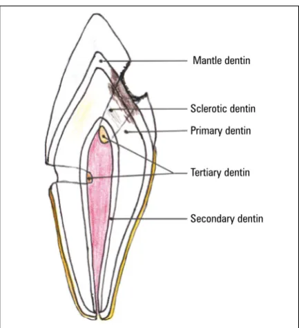
Types of dentin (Fig. 1)
Mantle dentin — is the first layer of dentin formed
during odontogenesis. It is located at the dentin-enam-el junction — constituting the outermost part of the dentin [6].
Primary dentin
During the development of the tooth or in the process of odontogenesis, primary dentin is formed. It is formed by odontoblasts until complete formation of the tooth root. It has a typical tubular structure with odontoblast processes inside the tubules. This is a physiological dentin [6].
Predentin
It is the layer of dentin closest to the pulp above the odontoblast layer — it is the innermost layer. It is a thin layer of freshly made, yet not mineralised dentin, consisting of a basic ground substance and collagen
fibres. It is deposited throughout the tooth’s life until
the pulp is vital [6, 9].
Secondary physiological dentin (secondary dentin) After root formation is completed and tooth is
erupted, the pulp continues to deposit dentin. Un
-der the influence of physiological processes, mainly
related to chewing, secondary dentin develops. As a result of its deposition in the pulp periphery, the tooth chamber lumen and the tubules lumen gradually decrease. The dentin has less regular structure and is more mineralised. Clinically, the colour is from honey to dark brown [6, 9].
Secondary pathological dentin (tertiary dentin) Tertiary dentin is produced in response to external irritants — pathology (caries, abrasion, attrition,
ero-sion, managing and filling of cavities). When there is
stimulus, the tertiary dentin is created as soon as the enamel-dentin border is affected. It has an irregular structure. The degree of irregularities depends on the dynamics of the pathological process (in acute pro-cesses it is more irregular than in chronic propro-cesses),
Mantle dentin
Sclerotic dentin
Primary dentin
Tertiary dentin
Secondary dentin
and on the severity of pathological irritants. The dentin resulting from the pathological irritants, formed by the primary odontoblasts, is called reactionary dentin. If odontoblasts are destroyed by a pathological process, they release into the intercellular space biologically active substances that are strongly stimulating
mes-enchymal cells and possibly fibroblasts. This results in
the transformation of these cells into odontoblast-like cells capable of producing dentin. The dentin that was formed in this way is a reparative dentin. It is formed by the odontoblast-like cells under external stimulation (most often pulp capping naterials). If damage to the pulp was severe, and it was exposed to direct
patho-logical factors — the dentin must first be formed by fibroblasts and it has a non-tubular structure, forming
so called dentin bridge [6, 9, 17].
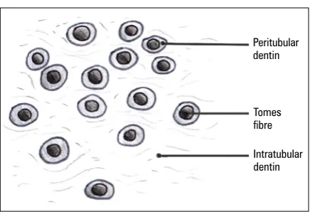
Dentin tubules
Dentin tubules are characteristic structures in the dentine structure (Fig. 2). Microscopic tubules extend to the dentin-enamel junction, while within the root they rarely reach the dentin-cementum junction. They
are perpendicular to the tooth surface. Within the
crown, these tubules follow an S-shaped path, and in the root they run parallel to each other, slightly wavy. The diameter of the tubules is 3–5 μm, but in the outermost parts is about 1 μm. Dentin tubules have many branches by which they can connect. A few of the dentinal tubules enter between the enamel prisms at various depths creating the so-called enamel cobs. In the dentinal tubules there are odontoblastic dentin
processes termed Tomes fibres, the non-myelinated nerve fibres, and tissue fluids filling the space. Od -ontoblasts processes have a lot of lateral branches passing into tubular side branches. In the cytoplasm
of Tomes fibres there are microtubules, microfilaments,
mitochondria and matrix vesicles.
Peritubular dentin forms the wall of each den-tinal tubule. The main mass of the dentin between the tubules is the intertubular dentin (Fig. 3). Peri-tubular dentin is more mineralised and has fewer
collagen fibres than intertubular dentin, and also
has a slightly different composition of matrix and smaller hydroxyapatite crystals. As a result of greater mineralisation, peritubular dentin is more likely to be damaged during the early phase of caries, and toxic substances can easier penetrate directly into the pulp. The walls of the tubules near the odonto-blasts are lined with a thin layer of non-mineralised substance called the Neumann lining. The formation
of a peritubular dentin is a continuous process that can be accelerated by external stimuli. Then, so-called sclerotic dentin is formed in the primary dentin. The stimuli cause progressive obliteration of the tubule and even its complete closure [9]. The odontoblast cytoplasmic processes withdraw to the pulp or
de-generate leading to calcification. As a result of these processes, a layer of dead tubules filled with gases, fluid or cell debris is formed creating death tracts. Its
presence facilitates the penetration of bacteria into the dentin after enamel destruction [9].
Types of dentinal tubules
The vast majority of dentinal tubules are tubules of classical structure, but there are also tubules of different structure. These include bifurcate, clavate, Figure 2. Dentin tubules and odontoblasts.
Peritubular dentin Intratubular dentin
Odontoblast process
Mantle dentin
Primary dentin
Predentin
Peritubular dentin
Intratubular dentin Tomes fibre
feather-like and giant tubules (Fig. 4). Straight tubules form bundles near the tooth chamber, disappear in the outer dentin. These bundles are separated by spaces
to form niches and fissures within the calcified den -tin ground substance. In these niches or slits there are bifurcate tubules. They are numerous, thick and have twisted branches. These tubules rarely reach the
dentin-enamel junction, usually ending in half or 1/3 of the outer dentin. They are most often found in root dentin. A variation of bifurcate tubules are feather-like tubules of more delicate structure. These tubules are less frequent and not only in niches, but also between classical dentin tubules. They are characterised by a large number of lateral branches. The rarest among dentinal tubules are clavate tubules. They are charac-terised by considerable thickness and unevenness of the surface. They have lateral, short branches, con-necting and merging together, this way making the tubule thicker. These clavate extensions of the tubules are only at a distance from the tooth chamber. There are also sometimes giant tubules, which are considered to be places where odontoblasts are trapped. They are found in root dentin and vary in size and shape [6, 9].
Dentin innervation
Dentin as compared to enamel is an innervated tissue sensitive to various stimuli. Non-myelinated
nerve fibres extend into dentin from the subodonto -blastic neural plexus. They initially form delicate plexuses between the odontoblasts and near the predentin. Then they proceed to the predentin and dentinal tubules, where they run along the Tomes
fibres. These fibres do not reach the surface of the
dentin, but to the depth of 0.5–0.7 mm, and the odontoblast processes are the supporting elements [6, 9].
PulP
Structure and morphology
Pulp (Latin: Pulpa dentis) is a tissue filling the tooth
(chamber, canals) (Fig. 5). Histologically, it resembles the gelatine-like matured connective tissue, containing a large number of ground substance, rich in
proteo-glycans, a network of loosely arranged collagen fibres,
and numerous stellate cells. On the peripheral strip of
the pulp there are thick silver-absorbent fibres called Korff spiral fibres.
Within the pulp, three zones can be distinguished: the odontoblastic zone, a cell-free zone of Weil be -neath the odontoblasts, and the core pulp or cell-rich zone [6].
Odontoblasts
These cells are located most peripherally in the pulp, arranged in a palisade. In the coronal part they
form 2–3 rows of cylindrical cells. Within the root pulp, there is one row of cubic or flat cells. Odontoblast
Straight
Bifurcate
Feather-like
Clavate
Figure 4. Types of dental tubules.
Figure 5. Pulpal dentin junction.
Dentin
Predentin
Odontoblasts
Cell-free zone of Weil
Cell-rich zone
has three types of processes: dentinal, pulpal and lat-eral. The dentinal process is long and penetrates into
the dentinal cavity to form a Tomes fibre. The pulpal
process is shorter and passes through the basal zone
of Weil into the pulp. Odontoblasts also have short
lateral processes that are connected with other lateral processes of adjacent odontoblasts.
Odontoblasts are involved in the formation of the dentin ground substance and its mineralisation. The pre-dentin layer is deposited in the apical part between dentin processes and then is mineralised. These cells also produce procollagen, which at the secretory part of cells is released into the matrix of dentin to form
col-lagen fibres. In addition to procolcol-lagen, glycoproteins
and proteoglycans are also synthesised.
Odontoblasts have the capability to produce dentin throughout their lifespan. As a result of damage, the
new odontoblasts can be formed from pulp fibroblasts
or differentiated mesenchymal cells to form a so-called pseudoodontoblasts [9].
Cell-free zone of Weil
This layer contains pulpal processes of odontoblast,
fibroblast processes, Korff spiral fibres, and non-myeli
-nated nerve fibres from the nerve plexus that penetrate
into the dentinal tubules. In this layer there is also a subodontoblastic capillary plexus [6, 7].
Core pulp — cell-rich zone
This layer is formed of mesenchymal cells and
star-shaped, spheroidal and spindle-shaped fibroblasts.
These cells join together to form a network [7]. Both
types of cells, under the influence of stimuli (physi -ological or path-ological), can divide and differentiate
into fibroblasts or (via the transitional form — pre -odontoblasts) into odontoblasts. In addition, there
are infiltrating cells in the pulp involved in immune
responses. These include lymphocytes, granulocytes, plasma macrophages, mast cells, and antigen
present-ing cells. Their number grows in the inflammatory state
of the tooth [6, 7].
Nerves and vascularity of the pulp
Innervation
Pulp is nourished by sensory fibres (myelinated) and by vascular fibres (non-myelinated vegetative fi
-bres). Along with blood vessels, nerve fibres (in 2–3
bundles containing several hundred axons) penetrate into the pulp through the apical opening. In the
cen-tral part of the pulp, the fibres are myelinated and
non-myelinated. They then branch into several smaller bundles. Directly under the odontoblast layer, both
types of fibres form subodontoblastic plexus (nerve
plexus of Raschkow).
Sensory nerves are the receptors that receive differ-ent stimuli, transmitting the sensation of pain. Sensory nerves run from the trigeminal ganglion. The sensory
fibres penetrate as myelin fibres and then branch off
and lose the myelin sheath. From the subodontoblastic plexus, they proceed to the odontoblast layer into very
thin non-myelinated fibres and further to the dentinal tubules. Vasomotor fibres are fibres of the sympathetic
nervous system and are responsible for vascular
circula-tion, regulating the flow of blood in blood vessels by
their narrowing or dilating [6, 7, 9].
Vascularity
Arteries of the pulp are branches of periodon-tal vessels; they penetrate as small arterioles (3 or more) and branch out within the coronal pulp. These branches are of the capillary type in the number of about 150–180 capillaries. At the base of the odon-tocytes, they form a network of broad capillaries, often creating loops, that merge into venules. There
are two types of capillaries in the pulp. The first one
— fenestrated capillaries, the thinnest, having pores in basal membrane of 50 nm in diameter, facilitating the exchange of various substances between blood and the cells. The second type are continuous capil-laries that have continuous endothelial lining and are typical capillaries. Both of these types of vessels are linked together, forming a subodontoblastic capillary network. Pulp capillaries change into wide thin-walled venules that extend through the central portion of the pulp. These venules, one or two, taper within the root canal and leave the pulp through the apical opening.
All pulp vessels have very thin walls due to muscu-laris mucosa reduction. This makes the pulp very
sensi-tive to pressure changes. During rapid flow of blood,
vessels surrounded by hard tissue of the tooth cannot
increase their volume. Also the blood outflow through the narrow apex is difficult. Long-term elevated vas -cular pressure, resulting in pulp congestion, can lead to pulp necrotizing changes [9–11].
ENDoDoNTIum fuNcTIoNs
Nutrition of tooth
Through periodontium and cement, the exchange
-tional path of the tooth. This is done by a very well de-veloped blood vessel system. Subodontoblastic plexus is particularly important in the delivery of nutrients to odontoblasts. Nutrition of dentin is achieved by
a network of dentinal tubules, filled with tissue fluids and odontoblast processes. Tomes fibres penetrate
the organic dentin and enamel to provide nutrients to the tissue [9].
Dentin formation
During the tooth formation, primary dentin is formed constituting the main bulk of the tooth. Once the development of the tooth is complete, dentin formation continues, but at a slower rate, and such
dentin is called a secondary dentin. Under the influence
of various pathological irritants (e.g. caries, attrition, abrasion), the pulp deposits the secondary pathologi-cal dentin, also pathologi-called the tertiary dentin [9].
Defensive role
Primary and secondary physiological dentin sur-rounds the pulp from all sides, protecting it from
the external environment. Under the influence of
pathological factors, secondary pathological dentin is formed, which has a more irregular structure than physiological dentin. Sclerotic dentin is also a protec-tive reaction to the harmful effect of pathological stimulus and develops in the primary dentin between the carious lesion and the pulp. Pulp and dentin protect themselves against harmful factors by building a new dentin or sealing primary dentin. Another protective reaction is the dentin bridge created by the pulp cap-ping materials at the site of exposure or pulp injury [9].
Sensory function
The tooth pulp through innervation has the ability to record sensation of pain regardless of the type or location
of the stimulus. The first path of transmitting physical and chemical stimuli to the pulp is odontoblast fibres in dentinal tubules. The other path is the nerve fibres
that surround the odontoblast processes in the dentinal tubules that come from the subodontoblastic plexus [9].
AgE-rElATED chANgEs
IN ENDoDoNTIum
With age, the tooth tissue degenerates like other
tissues in the human body. As a result of deposition of the secondary dentin into the tooth pulp (chamber, canals, lateral and pulp-dentin canals), the lumen is reduced. Obliteration of lateral canals, however, is
preferable for endodontic treatment due to the in-ability to spread the infection through these gates. Dentinal tubules become obliterated by mineralised tissue, causing even their complete occlusion. As a result of apoptosis, odontoblasts disappear, there is a reduced number of the remaining cells, and the
number of fibrous elements increases. Nerve fibres and
blood vessels, especially capillaries and precapillaries commonly calcify. All these changes cause weakening of the protective and regenerative properties of
endo-dontium, and consequently, difficulties in endodontic
treatment [6, 9, 12].
DENTINE-PulP comPlEx rEAcTIoN
To PulP cAPPINg mATErIAls
A specific type of dentin is a repair dentine. It is
formed by odontoblast-like cells as a result of external stimulation with preparations having odontotropic effect after the destruction of the odontoblasts. If the
pulp damage was severe, dentin is formed by fibro -blasts as a dentin bridge of non-tubular structure j [7]. Odontotropic preparations used in the daily dental practice as a direct pulp capping include calcium hy-droxide and mineral trioxide aggregate (MTA) [9, 11].
Under the odontotropic influence of calcium hydrox -ide, the pulp under the produced dentin bridge shows
no signs of inflammation [1, 3, 10, 17]. According to
some authors, calcium hydroxide is not a long-term barrier to bacteria, and after several years, symptoms of infection and necrosis can be observed [16]. A newer formulation used for direct covering of pulp is MTA. It has a tighter adhesion than calcium hydroxide and
lower incidence of the symptoms of inflammation,
congestion or necrosis of the pulp. Thicker dentin bridge was formed and more often pseudoblastic layers were formed [2]. Most authors show a greater success rate of direct pulp therapy with MTA than Ca(OH)2. Although MTA has better treatment results, it is not devoid of defects. The application of the prepa-ration is simpler, while the bonding time is 4–6 h and requires two visits. The cost of using this material is also much higher. Biodentine is another formulation used in practice in biological therapy, perforation, or as a substitute for dentin without the need for
etching [19]. This preparation, like MTA, is classified
shortens the binding to a few minutes and improves the mechanical properties of the formulation. The studies of other authors have shown similar effects of MTA when used to cover pulp directly [13]. In both groups, total dentin bridge formation was observed
and no pulp inflammation was present, suggesting
Biodentine as an alternative to MTA [4, 5]. Attempts are also made to cover wounded and open pulp with bonding systems [14, 18], glass ionomer cement [8] or enamel matrix proteins [7]. The effects are
debat-able, because inflammation, including necrosis, and
no dentin bridge has been found [12, 15, 16].
coNclusIoNs
Endodontium is a structural-functional unity, creat -ed by the pulp and dentin. It has an indispensable role in the processes occurring in vital tissues of the tooth and in the periapical tissues. This dentin-pulp complex has four functions: nutritive, formative, protective and sensory. As a structural-functional unit it deserves to be treated as a whole. Complete knowledge of its structure, function, and age-related changes gives an image of complexity and shows how important it is in daily practice to treat these two tissues not separately, but as a closely interrelated complex. The use of the
pulp capping materials is beneficial for prolonging the life of the tooth, as well as reducing inflammation.
Accordingly, the medical procedures should lead to the preservation of protective and repair processes of the tooth.
Acknowledgements
All figures are drawn by Adriana Kleinert.
rEfErENcEs
1. About I, Mitsiadis TA. Molecular aspects of tooth patho-genesis and repair: in vivo and in vitro models. Adv Dent Res. 2001; 15: 59–62, doi: 10.1177/08959374010150011 501, indexed in Pubmed: 12640742.
2. Aeinehchi M, Eslami B, Ghanbariha M, et al. Mineral trioxide aggregate (MTA) and calcium hydroxide as pulp-capping agents in human teeth: a preliminary report. Int Endod J. 2003; 36(3): 225–231, indexed in Pubmed: 12657149. 3. Bjørndal L, Mjör IA. Pulp-dentin biology in restorative
den-tistry. Part 4: Dental caries--characteristics of lesions and pulpal reactions. Quintessence Int. 2001; 32(9): 717–736, indexed in Pubmed: 11695140.
4. Chalas R, Mielko E, Zubrzycka-Wrobel J, et al. A chemical activity evaluation of two dental calcium silicate-based
ma-terials. Curr Iss Pharm Med Sci. 2015; 28(2), doi: 10.1515/ cipms-2015-0051.
5. Chałas R, Mielko E, Bachanek T, et al. Assessment of den -tin reaction after Bioden-tine application. Curr Iss Pharm Med Sci. 2013; 26(4): 435–439, doi: 10.12923/j.2084-980x/26.4/a.19.
6. Hellwig E, Klimek J, Gattin T. Einführung in die Zahner-haltung. Deutscher Zahnärzte Verlag Köln. 2009; 8-11: 311–315.
7. Kiatwateeratana T, Kintarak S, Piwat S, et al. Partial pul-potomy on caries-free teeth using enamel matrix deriva-tive or calcium hydroxide: a randomized controlled trial. Int Endod J. 2009; 42(7): 584–592, doi: 10.1111/j.1365-2591.2009.01552.x, indexed in Pubmed: 19467054. 8. Lan WH, Lan WC, Wang TM, et al. Cytotoxicity of conven
-tional and modified glass ionomer cements. Oper Dent. 2003; 28(3): 251–259, indexed in Pubmed: 12760696. 9. Lapińska J, Dąbrowska E, Stokowska W. Możliwości napraw
-cze i obronne miazgi w procesie próchnicowym – przegląd piśmiennictwa. Nowa Stomatol. 2004; 2: 83–86.
10. Magloire H, Romeas A, Melin M, et al. Molecular regulation of odontoblast activity under dentin injury. Adv Dent Res. 2001; 15: 46–50, doi: 10.1177/08959374010150011201, indexed in Pubmed: 12640739.
11. Makowiecki P. Trusewicz M, Tyszler Ł, Buczkowska-Radlińska J. Leczenie biologiczne miazgi zębów stałych. Rocznik PAM w Szczecinie. 2014; 60(2): 80–88.
12. Mjör IA. Pulp-dentin biology in restorative dentistry. Part 7: The exposed pulp. Quintessence Int. 2002; 33(2): 113–135, indexed in Pubmed: 11890026.
13. Nowicka A, Lipski M, Parafiniuk M, et al. Response of human dental pulp capped with biodentine and mineral trioxide aggregate. J Endod. 2013; 39(6): 743–747, doi: 10.1016/j. joen.2013.01.005, indexed in Pubmed: 23683272. 14. Onur MA, Taşman F, Cehreli ZC, et al. Effect of a
fifth-generation bonding agent on vascular responses in rats. J Endod. 2000; 26(7): 407–409, doi: 10.1097/00004770-200007000-00007, indexed in Pubmed: 11199766. 15. Postek-Stefańska L, et al. Leki i materiały stosowane do
bezpośredniego pokrycia miazgi zębowej - przegląd piś -miennictwa. Prz Stom Wieku Rozw. 2001; 1: 12–16. 16. Schuurs AH, Gruythuysen RJ, Wesselink PR. Pulp capping
with adhesive resin-based composite vs. calcium hydroxide: a review. Endod Dent Traumatol. 2000; 16(6): 240–250, indexed in Pubmed: 11202889.
17. Smith AJ, Murray PE, Sloan AJ, et al. Trans-dentinal stimu -lation of tertiary dentinogenesis. Adv Dent Res. 2001; 15: 51–54, doi: 10.1177/08959374010150011301, indexed in Pubmed: 12640740.
18. Taşman F, Cehreli ZC, Onur MA, et al. Effect of different single-bottle dentin adhesives on vascular responses in rat carotid artery. Am J Dent. 2000; 13(6): 337–339, indexed in Pubmed: 11764129.