O R I G I N A L A R T I C L E
The morphological conditions
of the permanent pacemaker lead extraction
Dariusz Kozlowski
1, Ada Dubaniewicz
2, Edward Kozluk
3, Marek Grzybiak
2,
Wojciech Krupa
1, Piotr Kolodziej
2, Anna Pazdyga
1, Monika Adamowicz-Kornacka
4,
Ewa Walczak
4, Franciszek Walczak
312nd Department of Cardiac Diseases, Institute of Cardiology, Medical University of Gdansk, Poland 2Department of Clinical Anatomy, Medical University of Gdansk, Poland
3Department of Electrophysiology, National Institute of Cardiology, Warsaw, Poland 4Department of Pathological Anatomy, Medical University of Warsaw, Poland [Received 15 November 1999; Accepted 3 December 1999]
Pacemaker lead extraction is the treatment of choice in infectious complica-tions regarding implantation procedure. The purpose of this study was to esti-mate the safety of the extraction in relation to the morphological changes of the pacing electrode. Research was carried out on materials consisting of 60 human hearts from 45 to 95 years of age (average 63 ± 15 yrs), with VVI or DDD pacing (pacing duration 84 ± 26 months) fixed in a formalin solution. Classical macro-scopic anatomical methods were applied.
In 44 hearts (73.3%) from the investigated group the posterior tricuspid leaflet was thickened only, and in 24 of these hearts the process regarded not only posterior leaflet but also the septal one and especially commissure between them. In 52 hearts (86.6%) inflammatory reaction spread also to the neighbour-ing part of the electrode. The length of the neointima-inflammatory tissue ranged from 4 to 8 mm (average 5 ± 2 mm). On the tip of the electrode in the right ventricle cavity in 56 hearts (93.3%) we observed that endocardial leads were surrounded by fibrous thickening , and partially covered by endocardial tissue.
We concluded that from the anatomical point of view the extraction of the pacing electrode seems to be questionable, especially in long-term permanent pacing. The experimental traction shows that only recently implanted electrodes were removed without any complications and in others with fraction of the tip, myocardial tissue avulsion or such removal was not successful at all.
key words: anatomical conditions of extraction, morphology of the per-manent pacing electrodes, functionless chronic leads
INTRODUCTION
Percutaneous extraction of chronic pacemaker leads has traditionally been difficult, with potential for myocardial perforation and other serious com-plications [13]. Many investigators have developed
several transvenous techniques of lead extraction trying to avoid thoracotomy and cardiopulmonary bypass surgery. Current extraction techniques range from simple, weighted tractions to a variety of snare devices, retrieval forceps and finally countertraction
[3,5,10,11]. A special technique mode is used, based on clinical history, time after implantation and the type of the electrode. There are numerous articles in medical literature describing lead removal in the context of the lead type. We found only one article reporting the biological reaction of the cardiovascu-lar system to the electrode with an implication for the extraction possibility [12] and describing micro-scopic changes caused by pacing [5,9]. However there are no further data regarding the morpholog-ical changes of the heart and implanted electrodes in extraction aspect, which seems to be of great im-portance in clinical practice. In relation to this, it was decided to examine the anatomical conditions of the implanted pacing leads in order to eventually define the possibility of their extraction.
MATERIALS AND METHODS
Research was carried out on autopsy material con-sisting of 60 adult human hearts of both sexes (24 female, 36 male) from 45–95 years of age (average 63 ± 15 years) fixed in formalin-ethanol solution in which no macroscopic pathological (e.g. not related to the age) changes were found. All investigated hearts were from patients with VVI or DDD pace-makers implanted as a treatment for II and III de-gree atrioventricular block. There were 20 electrodes with silicone rubber insulation (Sorin, Biotronik) and40 polyurethane-insulated leads (Biotronik, Siemens) implanted. The time from the implant to death ranged from 2 to 120 months (average 84 ± 26 months).
Classical macroscopical anatomical methods were applied with special attention paid to the relation-ship between the electrode and the anatomical struc-tures of the right ventricle (e.g. tricuspid valve, wall relief of the ventricle). We looked at the course of the electrode at the level of the tricuspid ring, the inferior part of the ventricle and the reaction of the cardiovascular system to the electrode (fibrosis, ad-herence, and traction possibility). Multivariate anal-ysis was performed by Cox proportional hazards re-gression model, F-Snedecor test and t-Student test of unpaired data. Differences were considered sig-nificant at p value < 0.05.
RESULTS
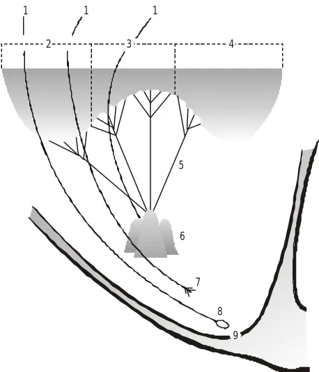
On the basis of our study we stated that the po-sition of the electrode at the level of the right atrio-ventricular orifice varies. In order to specify the ex-act location of the lead in relation to the tricuspid valve, we divided the valve into the following parts: the posterior leaflet, the posteroseptal junction (e.g. commissure — without any form of cusp or leaflet-additional cusp within commissure), septal leaflet (Fig. 1) and anterior leaflet. In 41.6% of examined
5
6
7
8
2 3 4
1 1 1
9
hearts (25 hearts) the pacing leads were positioned at the level of the posterior leaflet of the tricuspid valve, in 31.6% (19 hearts) just over the posterosep-tal junction (commissure — 13 hearts, leaflet — 6 hearts), in 23.3% (14 hearts) at the level of the sep-tal leaflet and finally in 3.5% (2 hearts) at the anteri-or level. The proximity of the leads to the valve ap-paratus caused the thickening of its structures (pos-terior leaflet in 33.3%, septal leaflet in 16.6% and commissure or additional leaflet in 23.3%). As re-gards the course of the electrode downwards to the ventricle, we confirmed that in 86.6% (52 hearts), leads were placed between the attachments of the chordae tendinae running from the posterior papil-lary muscle to the valve (Fig. 2). In 44 of these hearts the lead caused thickening of the chordae tendinae. These changes depended on the time that passed from the implantation to our examination (p < 0.05). In hearts with leads implanted 4–8 months before the autopsy we did not observe any fibrosis around the electrode, although extensive fibrin depositions were present. The extensive coat of fibrin ensheathed the distance of 4–8 mm (average 5 ± 2 mm) adher-ing to part of the electrode (Fig. 3). We also observed that if the commissural leaflet was present the reac-tion of the endocardium to the electrode was much
more pronounced (3–6 mm fibrin sheath about the lead), compared to the samples without such a leaf-let (only 2–4 mm fibrin sheath). Those differences were statistically not significant. The experimental traction on the electrode resulted in tearing its fi-brous attachments to the endocardium while the position of the electrode remained unchanged. In 5% of the cases we refragmented the valve appara-tus tearing the chordae tendinae.
The position of the electrode within the ventri-cle also varied. The tip of the electrode was posi-tioned exactly in the apex in 51.6% (31 hearts) only, in 36.7% (22 hearts) between the apex and the base of the posterior papillary muscle and finally in 11.7% (7 hearts) at the base of this muscle. In 86.6% (52 hearts) we observed fibrous thickening and partial coverage of the electrode tip by endocardial tissue. The degree of this fibrous reaction of the heart to the electrode tip varied with the time the lead was in place. The most extensive fibrosis occurred in the heart with the lead inserted 10 years ago, to a lesser degree fibrosis was present in hearts with a 1–9 year-old lead (Fig. 4). Fibrin deposition but no fibrosis was present in hearts with recent implantation (3–5 months) and only slight fibrosis was noted 6–14 months after implantation. Fibrous tissue formed
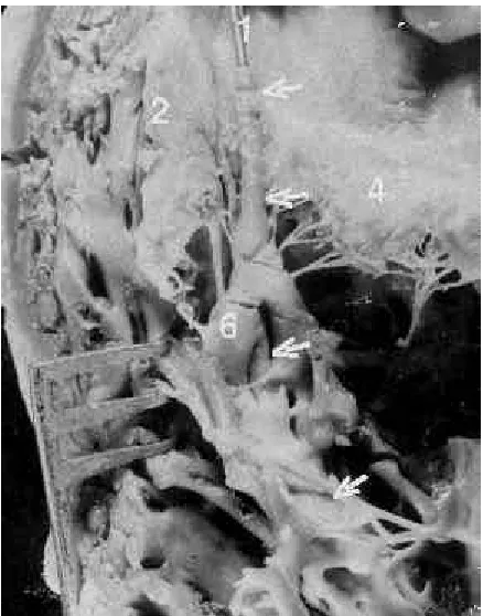
Figure 2. Polyurethane-insulated electrode without any fibrous tissue positioned between chordae tendinae. Denotations see Fig. 1 ( 42 year-old, 2 months after implantation)
around the lead was related also to the type of the tip. We were able to confirm that fibrocollagenous reaction that surrounds the lead tip was more ex-tensive in electrodes with a bulbous tip, than with those with a tined tip. We also observed that by our experimental traction we were able to extract with-out complications electrodes implanted within 3–14 months (average 5 ± 7). In older leads such a trac-tion caused many complicatrac-tions, such as fractrac-tion of the lead, myocardial tissue avulsion or we were un-able to remove it at all.
DISCUSSION
The issue of chronic non-functional pacemaker lead removal has been a controversial one. On the one hand extraction of such leads is recommended to prevent venous thrombosis, migration and possi-ble perforation of the heart wall [4], on the other hand they cannot be safely removed and could be abandoned in the cardiovascular system [1]. The fac-tors that lead to the failure of extraction of the pac-ing leads and eventually complications associated with it, are complex. Tined electrodes, used routine-ly for permanent pacing, give a marked decrease in the early lead dislodgement rate, although this has been rapidly accomplished by extraction [10].
In the light of our study, the failure of the re-moval can be connected with the degree of the fi-brotic process involving the electrode. Madigan et al. [10] confirmed that in their experience the long-tined ventricular leads are significantly more diffi-cult to extract (implanted > 3 months) than the old-er non-tined leads. In contrast to that, Myold-ers et al. [11] concluded that leads with bulbous or finned tips are particularly resistant to extraction and could be dangerous. On the basis of our results we were able to confirm that the fibrocollagenous reaction that sur-rounds the lead tip was more extensive in electrodes with a bulbous tip than in those with a tined tip. There-fore during our experimental extraction and the clin-ical extraction performed by Byrd et al. [2,3]. in ca 8– –10% this technique included the inability to remove the lead due to fragmentation near the electrode tip. The success of the chronic lead removal also depends on the insulation material of the lead. Ebe et al. [5] looked for the presence of any changes in the lead during a 1–4 month pacing in mongrel dogs. They concluded that fibrous tissue was formed around the polyurethane lead within the same period of pacing as had also been previously reported with silicone rubber leads. Unfortunately, however, they did not specify the degree of that fibrosis. Based on our study we were able to state that polyurethane-insulated pacemaker leads have a minor degree of fibrous thickening in contrast to silicone rubber in-sulation. We can confirm the tendency only, because differences were statistically not significant. This is in contrast to the paper of Robboy et al. [12], who performed autopsy studies on seven patients with permanent transvenous pacemakers. They found no differences between fibrous tissue reactions to the Medtronic and Cordis leads. We suppose that in the past when the studies were done we could use sili-cone rubber electrodes only, therefore the differences were not confirmed. Also the time between implan-tation and removal is an important factor. Our mor-phological research confirms that the fibrous sheath that forms along the course of a transvenous lead and at the tip of the electrode varied roughly with the time the electrode was placed. The most exten-sive fibrotic changes occurred in the patient whose pacemaker system was implanted 10 years ago in contrast to the leads implanted 5 months before our study was done, where fibrotic changes were mini-mal. Byrd et al. [3] concluded that leads implanted at least 8 years earlier are the most difficult to re-move, and leads implanted less than 1 year are most easily removable. This is concordant with the other Figure 4. Polyurethane-insulated electrode with marked fibrous
research regarding the extraction of the lead in paced patients [10–12].
The placement of the electrode in various parts of the right ventricle and the possibility of adher-ence (contact) to its structures may also play an im-portant role in extraction possibility. In our autopsy material we observed that in more than half of the cases the lead was entrapped within the valve appa-ratus. All leads in this situation become firmly ad-herent to the endocardium of the valve and the pro-cess of fibrosis was present. The same observations were made in our previous study [8] and by Robboy et al. [12] in contrast to Lagergren et al. [9]. The latter did not find any adherence of the lead to the valve although they observed the passing of the elec-trode through the valve leaflet itself. We did not confirm such a situation and additionally we did not observe any perforation of the valve. We observed, however, fibrotic tissue between the electrode and the valve’s leaflets that could be misinterpreted as the perforation of the valve. Ebe et al. [6] and Fear-not et al. [7] stated that the catching of the tip’s tines in the tricuspid valve during extraction signifi-cantly complicated this procedure. The traction of such an electrode, in our experimental traction, caused fragmentation of the tricuspid valve and es-pecially their chordae tendinae.
Summing up, on the basis of our morphological study we stated that fibrotic reaction occurs at the contact sides of the electrodes with the endocardi-um of the valve apparatus (e.g. valve and chordae tendinae) or the myocardium of the right ventricle (apex, free wall, papillary muscle). The fibro-collag-enous sheath, which is a result of the tissue response to the lead, forms along the course of the electrode. The amount of that tissue greatly depends on the time of contact. Therefore, percutaneous removal of old pacing lead could be difficult, dangerous or impossible. We must consider all the risks and bene-fits of such a lead removal and maybe leaving it in situ is the best option.
We concluded that from the anatomical point of view:
1) In 74% of examined hearts fibrosis occurs at sites of lead contact with the endocardium of the tri-cuspid valve apparatus or myocardium of the right ventricle.
2) The experimental traction shows that only recent-ly implanted electrodes were removed without any
complications. In contrast to that, older lead re-moval caused fraction of the tip, myocardial tissue avulsion or such a removal was not successful. 3) From the anatomical point of view the extraction
of the pacing electrode seems to be questionable, especially in long-term permanent pacing.
REFERENCES
1. Barbetseas J, Lalos S, Kyriakidis M, Aggeli C, Toutou-zas P (1998) Role of transesophageal echocardiography in the diagnosis of infected retained pacing lead. Pac-ing Clin Electrophysiol, 21: 1159–1161.
2. Byrd CL, Schwartz SJ, Hedin NB, Goode LB, Fearnot NE, Smith HJ (1990) Intravascular lead extraction us-ing lockus-ing stylets and sheats. Pacus-ing Clin Electrophys-iol, 13: 1871–1875.
3. Byrd CL, Schwartz SJ, Hedin N (1991) Intravascular tech-niques for extraction of permanent pacemaker leads. J Thorac Cardiovasc Surg, 101: 989–997.
4. Colavita PG, Zimmern SH, Gallagher JJ, Fedor JM, Aus-tin WK, Smith HJ (1993) Intravascular extraction of chronic pacemaker leads: efficacy and follow-up. Pac-ing Clin Electrophysiol, 16: 2333–2336.
5. Ebe K, Funazaki T, Aizawa Y, Shibata A, Fukuda T (1991) Experimental study about removal of the implanted tined polyurethane ventricular lead by radiofrequency waves through the lead. Pacing Clin Electrophysiol, 14: 1222–1227.
6. Espinosa ER, Hayes DL, Vlietstra RE, Osborn MJ, Mc-Goon MD (1993) The dotter retriever and pigtail cath-eter: efficacy in extraction of chronic transvenous pace-maker leads. Pacing Clin Electrophysiol, 16: 2337–2342. 7. Fearnot NE, Smith HJ, Goode LB, Byrd CL, Wilkoff BL, Sellers TD (1990) Intravascular lead extraction using locking stylets, sheats, and other techniques. Pacing Clin Electrophysiol, 13: 1864–1870.
8. Kozlowski D, Dubaniewicz A, Kozluk E, Adamowicz M, Grzybiak M, Walczak E (1997) Possible mechanism of the tricuspid valve insufficiency in the permanent right ventricular pacing. A morphological study. Cardiac Pacing, Monduzzi Editore, Bologna, 31–35.
9. Legergren H, Dahlgren S, Nordenstam H (1966) Car-diovascular tissue response to intracardiac pacemak-ing. Acta Chir Scand, 132: 696–673.
10. Madigan NP, Curtis JJ, Sanfelippo JF, Murphy TJ (1984) Difficulty of extraction of chronically implanted ventric-ular endocardial leads. J Am Coll Cardiol, 3: 724–731. 11. Myers MR, Parsonnet V, Bernstein AD (1991) Extraction
of implanted transvenous pacing leads: a review of per-sistent clinical problem. Am Heart J, 121: 881–888. 12. Robboy SJ, Harthorne JW, Leinbach RC, Sanders CA,
Austen WG (1969) Autopsy findings with permanent pervenous pacemakers. Circulation, 39: 495–501. 13. Sloman G, Strathmore N (1993) Permanent