Tanaffos (2003) 2(8), 23-30
2003 NRITLD, National Research Institute of Tuberculosis and Lung Disease, Iran
Pulmonary Function Tests and Their Reversibility in
Smokers
Mohammad Hossein Boskabady, Hamideh Dehghani, Mehdi Esmaeilzadeh
Department of Physiology, Ghaem Medical Centre, Mashhad University of Medical Sciences and Health Services, MASHHAD-IRAN
ABSTRACT
Background: Smoking is known as the major cause of chronic obstructive pulmonary disease (COPD). In COPD, most of
pulmonary function tests (PFTs) specially those indicating the diameter of airways are reduced. There are reports that
bronchodilator drugs have no or a very little effect on PFT of COPD patients. Therefore, in this study PFTs of smokers were
compared with those of nonsmokers, and the effect of bronchodilator inhaler (salbutamol) on PFTs of smokers were also examined.
Materials and Methods: Pulmonary function tests were measured in 97 male smokers (height 171.71±6.68 cm, age
36.49±13.06 years old) and compared with 95 male nonsmokers (height 171.79±8.81 cm, age 35.56±12.83 years old). The
subjects underwent measurement of spirometric flow and volume. The following variables were measured: forced vital capacity (FVC), forced expiratory volume in one second (FEV1), maximal mid-expiratory flow (MMEF), peak expiratory flow
(PEF), maximal expiratory flow at 75%, 50%, and 25% of the FVC (MEF75, MEF50, and MEF25 respectively). In addition,
pulmonary function tests of 33 male smokers (height 172.79±11.94 cm, age 38.30±6.65 years old) before and 10 minutes
after administration of 200 µg salbutamol inhaler were measured.
Results: The results showed that most values of PFTs in smokers were significantly lower than those of non-smokers
(p<0.001 for FVC, FEV1, PEF, MEF75, p<0.01 for MMEF, and p<0.02 for MEF50). However, there were not significant
differences in MEF25 of smokers and non-smokers. There were significant correlations between the smoking duration and
FEV1, PEF, MEF75, and MEF50 (p<0.05 to p<0.01), but correlations between smoking quantity and values of PFTs were not
significant. The results also showed that all values of PFTs were significantly increased after salbutamol administration
(p<0.05 to p<0.01). The enhancement in PEF, MEF75, and MEF50 was around 12% and that of MEF25 was 17%.
Conclusion: The profound effect of smoking on PFT showed that smoking leads to constriction of large and medium sized
airways which is mostly due to duration not to quantity of smoking. The airway constriction in smokers was reversible which, was mostly seen for medium sized airways. (Tanaffos 2003; 2(8): 23-30)
Key words: Pulmonary function tests, Smoking, Reversibility, Smoking duration, Smoking quantity
Correspondence to: Boskabady MH
INTRODUCTION
Chronic obstructive pulmonary disease (COPD) is a major cause of chronic morbidity throughout the world. Many people suffer from this disease for years and die prematurely from it or its complications. COPD is currently the fourth leading cause of death in the world (1), and further increases in its prevalence and mortality can be predicted in the coming decades (2).
Cigarette smoking is by far the most important risk factor for COPD and the most important way that tobacco contributes to the risk of COPD (3). Cigarette smokers have a higher prevalence of respiratory symptoms, pulmonary function abnormalities, greater annual rate of decline in FEV1, and a greater COPD mortality rate than those of non-smokers (4). These differences between cigarette smokers and non-smokers increase in direct proportion to the quantity of smoking. Smoking leads to rapid decline in pulmonary function tests (PFTs) specially those indicating diameter of airways such as forced expiratory flow in one second (FEV1) (5). Even in teenagers who have smoked only a few years, maximum expiratory flow-volume curves demonstrate decreases in flow rates at small lung volumes (6) yet another expression of small airway obstruction. If smoking causes changes in small airway calibre at such an early age , one might expect that smoking also causes acute changes in these small airways. Until now, the only well-documented acute effect of smoking on the airways was the decrease of airway conductance demonstrated by Nadel and Comroe (7).
The obstruction to airflow that develops in 15 to 20% of heavy smokers is thought to be due to abnormalities in airways with less than 2 mm internal diameter (8). Previous studies from several laboratories have shown that this airway obstruction is associated with chronic inflammatory process in the membranous and respiratory bronchioles (9-10).
It is believed that the airway constriction in COPD and decline in PFT are not reversible. Therefore, in the present study the pulmonary function tests of smokers was compared with those of non-smokers. The effect of quantity and duration of smoking on PFT and the reversibility of PFT were also evaluated in the present study.
MATERIAL AND METHODS Subjects
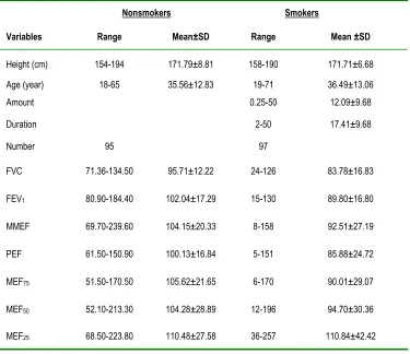
Ninety seven smokers (age 36.49±13.06, range 19-71 years old and height 171.71±6.68 range 158-190) and ninety five non-smoker men (age 35.56±12.83, range 18-65 years old and height 171.79±8.81 range 154-194) were randomly selected from the visitors of Ghaem Medical Centre (Table 1). All subjects had no history or symptoms of cardiovascular or respiratory diseases that required treatment (excluding the common cold). The protocol was approved by the “Ethics Committee” of our institution, and each subject gave informed consent.
Measurement
clips (11). All tests were carried out between 1000 and 1700 hours.
In 33 smokers, PFTs were repeated 10 min after 200 µg inhaled salbutamol. Pulmonary function tests were performed three times in each subject with an acceptable technique. The highest levels for forced vital capacity (FVC), forced expiratory volume in one second (FEV1), maximal mid-expiratory flow
(MMEF), peak expiratory flow (PEF), and maximal expiratory flow at 75%, 50%, and 25% of the FVC (MEF75, MEF50, and MEF25 respectively) were taken
independently from the three curves.
Data analysis
The data of height, age, and pulmonary function parameters were expressed as mean±SD. PFTs of
smokers were compared with those of nonsmokers using unpaired t-test. PFTs obtained after inhaled salbutamol were compared with the baseline values using paired t-test. The duration and quantity of smoking were related to decrease in their PFT values, using the least square regression. The criterion of significance was p<0.05.
RESULTS
Duration and quantity of smoking
Mean duration of smoking was 17.41±4.68 years (range 2-50 years) and mean quantity of smoking was 12.09±9.68 Cigarettes per day (range 0.25-50), (Table1).
Table 1: Characteristics of studied population.
Nonsmokers Smokers
Variables Range Mean±SD Range Mean ±SD
Height (cm) 154-194 171.79±8.81 158-190 171.71±6.68
Age (year) 18-65 35.56±12.83 19-71 36.49±13.06
Amount 0.25-50 12.09±9.68
Duration 2-50 17.41±9.68
Number 95 97
FVC 71.36-134.50 95.71±12.22 24-126 83.78±16.83
FEV1 80.90-184.40 102.04±17.29 15-130 89.80±16.80
MMEF 69.70-239.60 104.15±20.33 8-158 92.51±27.19
PEF 61.50-150.90 100.13±16.84 5-151 85.88±24.72
MEF75 51.50-170.50 105.62±21.65 6-170 90.01±29.07
MEF50 52.10-213.30 104.28±28.89 12-196 94.70±30.36
MEF25 68.50-223.80 110.48±27.58 36-257 110.84±42.42
Pulmonary function tests
All values of pulmonary function tests in smokers were significantly lower than those of nonsmoker subjects (p<0.02 to P<0.001) except MEF25 (Table-2).
Table 2: Pulmonary function tests (PFTs) among smoker and non-smoker subjects and statistical differences between two groups.
Nonsmokers Smokers Statistical
PFTs Mean±SD Mean±SD Differences
FVC 95.71±12.22 83.78±16.83 P<0.001
FEV1 102.04±17.29 89.80±16.80 P<0.001 MMEF 104.15±20.33 92.51±27.19 P<0.01 PEF 100.13±16.84 85.88±24.72 P<0.001 MEF75 105.62±21.65 90.01±29.07 P<0.001 MEF50 104.28±94.70 94.70±30.36 P<0.02 MEF25 110.48±27.58 110.84±42.42 NS
Correlation between duration and quantity of smoking with PFTs in smokers
There were significant negative correlation
between duration of smoking and decrease in FEV1, PEF, MEF75, and MEF50 (p<0.05 to p<0.01).
However, the correlations between the quantity of smoking and values of PFT were not significant.
Low PFTs among smoker and nonsmoker subjects
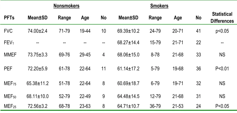
The percentage of low values of most PFTs (lower than 80% predictive values) among smoker was significantly more than those of normal subjects (Table 3). Only 0-10.6% of non-smokers had low PFT values while in 21.6-42.3% of smokers PFT values were lower than normal range.
Effect of sulbutamol on PFTs of smokers
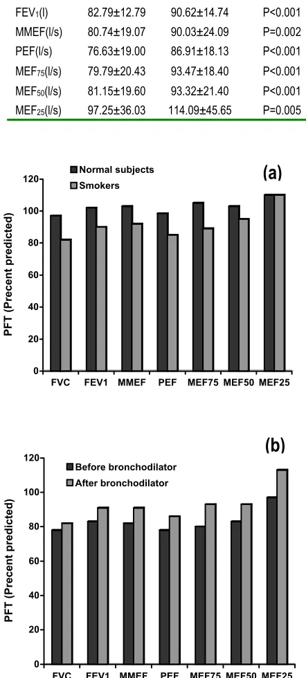
Pulmonary function tests of 33 male smokers (height 172.79±11.94 cm, age 38.30±6.65 years) before and 10 min. after administration of 200 µg salbutamol inhaler were measured. All values of PFT in smokers significantly increased 10 min. after 200 µg inhaled sulbotamol (p= 0.005 to p<0.001). The enhancement in PEF, MEF75, and MEF50, was around
12% and that of MEF25 was 17% (Table 4).
Table 3: The percentage and range of low PFTs (lower than 80% predicted values among smoker and nonsmoker subjects.
Nonsmokers Smokers
PFTs Mean±SD Range Age No Mean±SD Range Age No DifferencesStatistical
FVC 74.00±2.4 71-79 19-44 10 69.39±10.2 24-79 20-71 41 p<0.05
FEV1 -- -- -- -- 68.27±14.4 15-79 21-71 22
--MMEF 73.75±3.3 69-76 29-45 4 68.06±15.0 8-78 21-68 33 NS
PEF 72.20±5.9 61-78 22-64 11 61.14±17.2 5-79 19-68 36 P<0.01
MEF75 65.38±11.2 51-78 22-64 8 60.69±18.7 6-79 19-71 32 NS
MEF50 68.11±10.0 52-79 22-49 9 64.48±14.5 12-79 21-68 31 NS
Table 4: Pulmonary function tests (PFTs) of smoker subjects before and 10 min. after inhalation of 200 µg salbutamol.
PFTs Before After Statistical
Differences
FVC(l) 76.76±13.23 81.68±16.32 P<0.001 FEV1(l) 82.79±12.79 90.62±14.74 P<0.001 MMEF(l/s) 80.74±19.07 90.03±24.09 P=0.002 PEF(l/s) 76.63±19.00 86.91±18.13 P<0.001 MEF75(l/s) 79.79±20.43 93.47±18.40 P<0.001 MEF50(l/s) 81.15±19.60 93.32±21.40 P<0.001 MEF25(l/s) 97.25±36.03 114.09±45.65 P=0.005
Figure 1: (a): Comparison of pulmonary function tests between smokers and nonsmokers, and (b): comparison of PFTs before and after 200 µg inhaled salbutamol in smokers
DISCUSSION
This study has shown reduction of all values of pulmonary function tests in smokers compared to those of nonsmoker subjects. Although the mean values of PFTs in smokers was in normal range (83.78±16.83 to 110.84±42.42), they were significantly lower than PFT values in normal subjects. However, in 21.6-42.3 % of smokers, the values of PFT were lower than normal range, while only 0-10.6% of normal subjects had low values of PFT. In addition, relatively younger smoker subjects had low values of PFT comparing to normal subjects. Pervious studies (12-20) also showed reduction of different values of PFT among smokers comparing to normal subjects. The result of the present study showed the reduction in PEF and MEF75 among
smoker subjects was significantly more than other values of PFT.
These results may indicate that in smoker subjects medium and large airways are affected more than other airways. The results of our study were supported by previous studies indicating reduction of PFTs in smokers (17-18). However, there is some evidence that small airways are affected more by smoking (14).
The results of the present study also showed negative correlation between decrease in most values of PFT and duration of smoking. However, the relationships between decrease in PFTs and quantities of smoking were not significant. These results showed that duration of smoking has more profound effect on airways than quantity of smoking. The studies of Sherrill et al.(21) and Verschakelen et al. (22) also showed correlation between smoking and reduction in most values of PFT which support the results of the present study. In addition, Burrows et al. also showed quantitative relationship between cigarette smoking and reduction in values of PFT (5). Furthermore, the results of the present study, showed that the values of PFT of smokers were
0 20 40 60 80 100 120
PFT (Precent predicted)
FVC FEV1 MMEF PEF MEF75 MEF50 MEF25 Normal subjects
Smokers
0 20 40 60 80 100 120
PFT (Precent predicted)
FVC FEV1 MMEF PEF MEF75 MEF50 MEF25 Before bronchodilator
After bronchodilator
significantly increased due to 200 µg inhaled salbutamol indicating some degree of reversibility of the airway constriction in smokers. Although the mean value of MEF25 among smokers was normal,
increase in this value of PFT due to salbutamol administration was more than other values of PFT. This may indicate that in smokers small airways are more liable to reversible constriction. It is believed that airway constriction of COPD patients is not reversible, or there is very small reversibility of airways in these patients. However, the results of our study demonstrated a relatively large component of reversibility of airways in smokers, which is a novel finding of the present study.
In conclusion, the results of the present study demonstrated the profound effect of smoking on PFT and, therefore, indicated that smoking leads to constriction of large and medium airways, which is mostly due to duration, not to quantity of smoking. The airway constriction in smokers was reversible which was mostly seen for medium and small sized airways.
REFERENCES
1. World Health Organization. World health report. Geneva: WHO; 2000.
2. Murray CJ, Lopez AD. Evidence-based health policy--lessons from the Global Burden of Disease Study. Science 1996; 274(5288): 740-3.
3. Auerbach O, Hammond EC, Garfinkel L, Benante C. Relation of smoking and age to emphysema. Whole-lung section study. N Engl J Med 1972; 286(16): 853-7.
4. Lebowitz MD, Burrows B. Quantitative relationships between cigarette smoking and chronic productive cough. Int J Epidemiol 1977; 6(2): 107-13.
5. Burrows B, Knudson RJ, Cline MG, Lebowitz MD. Quantitative relationships between cigarette smoking and ventilatory function. Am Rev Respir Dis 1977; 115(2): 195-205.
6. Seely JE, Zuskin E, Bouhuys A. Cigarette smoking: objective evidence for lung damage in teen-agers. Science
1971; 172(984): 741-3.
7. Nadel JA, Comroe JH Jr. Acute effects of inhibition of cigarette smoke on airway conduction. J Appl Physiol 1961; 16: 713-6.
8. Hogg JC, Macklem PT, Thurlbeck MW. Site and nature of airway obstruction in chronic obstructive lung disease. N Engl J Med 1968; 278(25): 1355-60.
9. Berend N, Wright JL, Thurlbeck MW, Marlin GE, Woolcock AJ. Small airways disease: reproducibility of measurements and correlation with lung function. Chest 1981; 79(3): 263-8.
10. Wright JL, Lawson LM, Pare PD, Kennedy S, Wiggs B, Hogg JC. The detection of small airways disease. Am Rev Respir Dis 1984; 129(6): 989-94.
11. Standardization of spirometry, 1994 Update. American Thoracic Society. Am J Respir Crit Car Med 1995; 152(3): 1107-36.
12. Zuskin E, Mitchell CA, Bouhuys A. Interaction between effects of beta blockade and cigarette smoke on airways. J Appl physiol 1974; 36(4): 449-52.
13. Lange P, Groth S, Nyboe GJ, Mortensen J, Appleyard M, Jensen G, Schnohr P. Effects of smoking and changes in smoking habits on the decline of FEV1. Eur Respir J 1989; 2(9): 811-6.
14. Bosken CH, Wiggs BR, Pare PD, Hogg JC. Small airway dimensions in smokers with obstruction to airflow. Am Rev Respir Dis 1990; 142(3): 563-70.
15. Eidelman DH, Ghezzo H, Kim WD, Hyatt RE, Cosio MG. Pressure-volume curves in smokers. Comparison with alpha-1-antitrypsin deficiency. Am Rev Respir Dis 1989; 139(6): 1452-8.
16. Aparici M, Fernandez Gonzalez AL, Alegria E. Respiratory function tests. Differences between smokers and non-smokers. Effects of withdrawal. Rev clin Esp 1993; 192(4): 169-72.
17. Nemery B, Moavero NE, Brasseur L, Stanescu DC. Changes in lung function after smoking cessation: an assessment from a cross -sectional survey. Am Rev Respir Dis 1982; 125(1): 122-4.
in young men. Pneumonol Alergol pol 2000; 68(5-6): 226-31.
19. Goodman RM, Yergin BM, Landa JF, Golivanux MH, Sackner MA. Relationship of smoking history and pulmonary function tests to tracheal mucous velocity in nonsmokers, young smokers, ex-smokers, and patients with chronic bronchitis. Am Rev Respir Dis 1978; 117(2): 205-14.
20. Welty C, Weiss ST, Tager IB, Munoz A, Becker C, Speizer FE, Ingram RH Jr. The relationship of airways responsiveness to cold air, cigarette smoking, and atopy to respiratory symptoms and pulmonary function in adults. Am
Rev Respir Dis 1984; 130(2): 198-203.
21. Sherrill DL, Lebowitz MD, Knudson RJ, Burrows B. Longitudinal methods for describing the relationship between pulmonary function, respiratory symptoms and smoking in elderly subjects: the Tucson Study. Eur Respir J 1993; 6(3): 342-8.