Evaluation of Levels of Macro- and Micro-Nutrients in Workers
Exposed to Electromagnetic Fields and Comparison with Levels of
Patients with Leukemia
Issa Nourmohammadi
*1, Hassan Ahmadvand and Mohammad Taghikhani
21Dept. of Biochemistry, College of Medicine, Iran University of Medical Sciences, Center for Cellular and Molecular Biology Research;2School of Medical Sciences, TarbiatModarres University, Tehran, Iran
ABSTRACT
There have been much speculation and debate concerning the effect of electromagnetic fields (EMF) on body systems. Various reports have implicated excessive exposure to EMF to certain forms of leukemia. It has also been reported that EMF may cause alteration in the levels of certain macro- and micro-nutrients such as copper, zinc, selenium, calcium and iron. We have undertaken this study to determine the status of these elements in workers exposed to EMF for more than 10 years and compare these levels with those of patients with leukemia and both groups to a set of matched controls. Statistical analysis revealed meaningful differences in the serum levels of the nutrients under study both in terms of comparison of workers and patients to controls and in comparison of workers to patients. Mean Zn levels in both patient (P0.0002)and worker (P 0.006) groups were significantly higher than that of controls. However, statistical analysis of patients to workers revealed no meaningful variation. Selenium in both study groups also showed decreased levels when compared to controls. These results were statistically significant in comparison of patients to controls (P 0.0001) and workers to controls (P0.0001). Evaluation of patients to workers also resulted in a significant finding (P0.05). While we do not claim these results to be definitive, they do reflect the possibility that regular evaluation of the status of these elements in workers consistently exposed to EMF may be beneficial in terms of determining the heightened risk of these workers to development of leukemia. Iran. Biomed. J. 5 (2 & 3): 79-85, 2001
Keywords: Electromagnetic fields, Leukemia, Workers, Trace elements, Calcium
INTRODUCTION
n recent years, there has been a scientific debate regarding the effects of electromagnetic fields (EMF) on biological systems [1]. Although the basic mechanism of interaction between EMF and cellular function still remains uncertain, there are however practical and beneficial applications of EMF in medicine [2]. Numerous experimental and epidemiological observations show that human populations in industrialized communities experience a universal exposure to radiation from power lines and the low voltage of the home and working environments [3]. Exposure to this environmental phenomenon has prompted concern that it could increase biological disorders including cancer and leukemia in children [4, 5].
Although there are considerable controversies over the validity of any link between EMF and
disturbances on a biological system [6, 7], the number of reports on this newly developed concern continues to make it a reasonable undertaking to develop a definite understanding of the mechanism and its possible consequences at the molecular or even total body level.
The key events arising from exposure to EMF may include alterations in cell membrane activity, effects on various enzyme systems [8] and protein and DNA synthesis [9, 10], interference with the regulation of protein kinase-C [11], alterations in melatonin and hormone levels [12], Ca homeostasis [13] as well as variation on other important ions such as Cu and Zn which are necessary co-factors for many biological reactions [14].
This study was designed to evaluate the status of macronutrients such as Ca and Fe and micro-nutrients such as Cu, Zn and Se in subjects exposed to electromagnetic fields and compare the levels of
I
these same elements in leukemic patients who had not been exposed to EMF in the workplace. This study was undertaken firstly based on the observations stated above that indicate EMF may affect different aspects of the biochemistry of cells. Secondly, some studies have indicated that EMF may cause leukemia [3, 4]. Thus, it is important to determine the status of trace elements to ascertain if EMF workers are predisposed to leukemia. Thirdly, our preliminary study on several clinical parameters (unpublished data) showed interesting results in workers exposed to EMF (60 Hz, 40 T). Plasma vitamin C level (meanS.D.) of these workers was 0.82 0.2 mg/dl, significantly lower than that of control group which was 1.44 0.27 mg/dl. Total hemoglobin for EMF-exposed workers was 12.5 0.8 mg/dl and that of control group 14 0.7 mg/dl which was also statistically significant. Total protein in workers was recorded as 7.5 0.66 g/dl and in controls 9.10.68 g/dl.
This study thoroughly evaluated micro- and macro-nutrients in these subjects. To the best of our knowledge, there have been rare evaluations and comparisons of patients with leukemia and workers exposed to EMF.
MATERIALS AND METHODS
A group of power line station workers (n 40) continuously exposed to electromagnetic fields (60 Hz, 40 T) for at least 10 years and patients (n 80) with leukemia were studied. Leukemia patients were diagnosed by expert physicians and on the basis of clinical, morphological, hemato-logical and cytogenetics evaluation, categorized as acute lymphoblastic leukemia (ALL, n 30), acute myeloid leukemia (AML, n 40) and other
A solution of strontium was used in the Ca determination to prevent phosphate interference. Se concentration was determined with hydride-generation atomic absorption spectrophotometry (Perkin-Elmer Norwalk, CT) with the following conditions: Lamp current: 10 mA; type of flame: air C2H2; wavelength: 190 nm; and slit width: 2 nm.
All data were analyzed using the Student’s t-test and analysis of variance (ANOVA) with software SPSS.
RESULTS
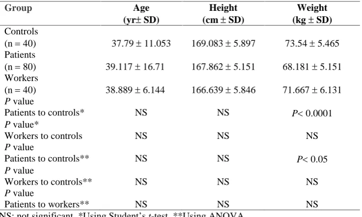
As can be seen in Table 2, the mean age for patients and controls was not significantly different nor was that of workers to controls, indicating a matched group in relation to age. Mean height of the patients to controls and workers to controls did not differ significantly. Mean weight of the patients to controls indicated a significant difference with P0.0001, however workers compared to controls indicated no significant difference.
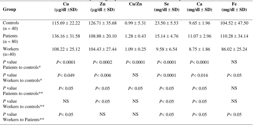
Results of the analysis of elements under study are summarized in Table 3. The mean concentration of Cu in our patients was higher than that of controls. This difference was statistically significant (P0.0001). A lower serum level was found in our EMF worker group compared to controls which was also significantly different with P0.049. Using analysis of variance (ANOVA) to evaluate the data showed the higher level of Cu in patients to be statistically significant (P0.05), however, the difference between workers and controls was not significant. In evaluation of worker to patient levels, the results indicated a significant difference with P0.05.
A significant difference in serum Zn levels was
Table 1. Conditions for atomic absorption analysis of elements under study.
Element Halode cathode lamp current
Type of flame Wavelength nm
Slit-width nm
Ca 6 Air-C2H2 422.7 0.5
Fe 8 Air-C2H2 248.3 0.2
Cu 3 Air-C2H2 324.8 0.5
Zn 4 Air-C2H2 213.9 0.5
Table 2. Comparison of age, height and weight of workers, patients and controls.
Group Age
(yrSD)
Height (cmSD)
Weight (kgSD)
Controls
(n40) 37.7911.053 169.0835.897 73.545.465
Patients
(n80) 39.11716.71 167.8625.151 68.1815.151
Workers
(n40) 38.8896.144 166.6395.846 71.6676.131
P value
Patients to controls* NS NS P0.0001
P value*
Workers to controls NS NS NS
P value
Patients to controls** NS NS P0.05
P value
Workers to controls** NS NS NS
P value
Patients to workers** NS NS NS
NS: not significant, *Using Student’st-test, **Using ANOVA
The Cu to Zn ratio in patients compared to controls was higher with P0.0001 indicating a significant difference. This same ratio in comparison of workers to controls was not statistically significant. ANOVA evaluation of results showed the higher Cu/Zn of patients to be significant (P0.05). In comparison of workers to controls and workers to patients, the difference was not significant in either group.
The serum level of Se in leukemia patient group was significantly lower than the controls. Analysis of these data indicated a significant difference with P0.0001. Mean Se level in workers was also
There was no significant difference found in serum Fe levels between patients and controls, even though the level of patients was higher. The mean serum Fe level in workers was lower than controls with P0.05, indicating a significant difference. Using ANOVA to evaluate Fe data demonstrated that there was no significant difference between controls and patient or workers, but the higher level of workers compared to patients was significant with a P0.05.
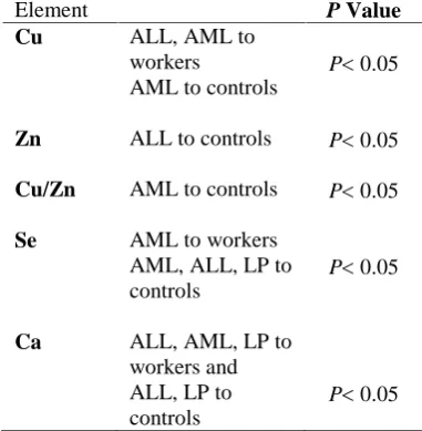
The significant results of ANOVA evaluation of our leukemia subgroups to both workers and controls are subgroups shown in Table 4.
Table 3. Mean levels of elements under study in all groups and results of statistical analyses.
Group
Cu (g/dlSD)
Zn (g/dlSD)
Cu/Zn Se
(mg/dlSD)
Ca (mg/dlSD)
Fe (mg/dlSD)
Controls (n40)
115.6922.22 126.7135.68 0.995.31 23.505.53 9.651.96 104.5247.50
Patients (n80)
136.1631.58 108.8820.10 1.280.43 15.144.76 11.072.96 110.2834.14
Workers (n=40)
108.2225.12 104.4327.44 1.090.25 9.586.54 8.751.86 86.0225.24
P value
Patients to controls*
P0.0001 P0.0002 P0.0001 P0.0001 P0.0001 NS
P value
Workers to controls*
P0.049 P0.006 NS P0.0001 P0.016 P0.05
P value
Patients to controls**
P0.05 P0.05 P0.05 P0.05 P0.05 NS
P value
Workers to controls**
NS P0.05 NS P0.05 P0.05 NS
P value
Workers to Patients**
P0.05 NS NS P0.05 P0.05 P0.05
NS: not significant, *Using Student’st-test, **Using ANOVA
Metals such as Fe, Cu, and Zn in mammalian lymphocytes are essential for cell growth and synthesis of DNA [16]. They also help to regulate the intracellular concentrations of transferrin, ferritin, ATP, proteins and cell surface interleukin-2 receptors [17]. Research has shown that leukemic cells have elevated concentrations of Fe and Cu and decreased levels of Zn [18]. We set out to detect variations of the trace and non-trace elements Cu, Zn, Se, Ca and Fe in serum in workers exposed to EMF with the objective to assess whether EMF can cause any variation in mineral concentrations in workers and compare these results to the same levels of these elements in patients with leukemia.
In answer to the two reasons outlined in the introduction, our higher mean level for serum Cu concentration is consistent with other reports for leukemic patients and confirms that Cu levels are elevated in persons with leukemia especially those with AML. Beguinet al. [19] also reported this increase with chronic lymphocytic leukemia and also for chronic myelogenous leukemia. In contrast, comparing to controls, workers exposed to EMF showed a slightly decreased level of Cu. Perhaps exposure time to EMF has not been sufficient to adversely effect Cu-containing proteins such as ceruloplasmin to release copper as seen in leukemic patients. We have found no human studies in this respect but Burchard’s study with diary cows in a controlled environment also showed lower Cu plasma levels during and after exposure to EMF [20].
Serum Zn levels in our patients with ALL and workers were significantly lower than controls. Beguinet al. [19] also saw this result with leukemic patients. This decrease in plasma Zn concentrations was also reported in dairy cows exposed to 60 Hz and 30 T, during and after exposure, but cerebrospinal fluid showed increased levels of Zn [20]. However, cerebrospinal fluid does not alter appreciably, as it maintains a relatively steady state. It is difficult to speculate on the physiological implication of such variations as reported by Burchardet al. [20], especially in human studies. This low concentration of Zn in leukemic patients has been attributed to anorexia, tissue catabolism and other forms of metabolic stress [21], since Zn has been proven to be an essential element for metabolic functions, especially under conditions of stress. In a similar fashion, Zn as an antioxidant, may be trying to combat the detrimental effects caused by EMF with the Zn pool going to tissue in order to increase immunocompetence in both cases. In addition, as Beguin and others [19, 21, 22] have postulated, low dietary intake, intercurrent infections and loss through sweat and urinary excretion could also partially account for lower Zn levels.
In relation to trace elements, Cu/Zn ratio, because of the antagonistic actions of Cu and Zn, is an important marker in showing the status of these two elements. According to our results, Cu/Zn ratio in leukemia patients, especially those with AML, and workers increased, supports the hypothesis that a high concentration of Cu and a high Cu/Zn ratio denote the extent of leukemic disease [19]. The high Cu/Zn ratio in our patients and workers may be due to an increase in Cu absorption and a decreased Zn absorption or competition of these two elements for binding to metallothionein or elimination of Zn in combating free radicals.
Se as an antioxidant exerts a protective effect against some human cancers and its levels have been reported to be lowered in some neoplasia [23]. Our leukemia patients, especially those with AML and our worker group had significantly decreased Se values. This low level of Se may be due to its scavenging properties, its function in eliminating free radicals as a reducing agent and acting as an important property of enzymes such as glutathione peroxidase. The important implication of these results is that Se could possible be used as an indicator for the extent of disease. This matter points toward the need for a more meticulous and comprehensive study of Se in relation to EMF exposure and leukemic disease.
Table 4. Results of ANOVA comparison of
leukemia subgroups to workers and controls.
Element P Value
Cu ALL, AML to
workers
AML to controls
P0.05
Zn ALL to controls P0.05
Cu/Zn AML to controls P0.05
Se AML to workers
AML, ALL, LP to controls
P0.05
Ca ALL, AML, LP to
workers and ALL, LP to controls
P0.05
Note: Only significant results are shown. For Fe, all comparisons were non-significant, ALL: acute lymphoblastic leukemia, AML: acute myeloid leukemia, LP: other leukemia
Role of cellular Ca is in control of cell signals and events such as cell movement and division [13]. If EMF influences the cellular activity then this Ca homeostasis can be disturbed. Several in vitro and in vivo studies on Ca disturbance and EMF showed that low-intensity EMF can alter Ca distribution in cells probably due to an effect on receptor operated Ca and or ion channels [24]. There have been reports on the efflux of Ca-ions from the brains of chickens and cats as well as neuroblastoma cells when exposed to EMF [25, 26]. On the other hand, several others reported that Ca uptake by cells exposed to EMF increased in rat thymocytes and in embryonic chicken tibia [27, 28]. Ca-ion uptake by normal and leukemia lymphocytes increased with weak EMF of 13.6 Hz, 20 T, a result indicating that EMF affects the Ca-dependent functions of normal and leukemia lymphocytes as reported by Lyle et al. [29] and such persistent alteration of Ca-ions by magnetic fields could influence the process of cancer promotion and effect the growth control in normal and malignant cells. The lower serum Ca concentrations in our workers may be due to increased uptake or may be influenced by the conductance of the cell membrane as well as the permeability of intercellular stores. Unlike our findings, Ca concentration of plasma and cerebrospinal fluid tends to increase in diary cows exposed to EMF [20]. Mean levels of Ca in our patient group were significantly higher. This increase in the level of Ca has been shown in other disorders such as breast cancer, lymphoma and leukemia [30, 31].
Results of our study on Fe concentration indicated a decrease in Fe levels in workers compared to controls. Even though little data are available in human studies, concentration of Fe decreased also in cerebrospinal fluid of dairy cows exposed to EMF [20]. Fe concentration of the patients did not
working environments. It is essential that these workers undergo routine checks for changes in biochemical indexes. Nutritional supplementation, including increased Zn and Se, could be beneficial in helping to alleviate some of the biochemical stress exposure to EMF causes and increase cellular immunity.
ACKNOWLEDGEMENTS
The authors wish to thank Judy Garland for editing and typing of the manuscript.
REFERENCES
1. Adey, W.R. (1993) Biological effects of EMF. J.
Cell Biochem. 51: 410-416.
2. Liboff, A.R., Williams, T., Strong, D.M. and Wistar, R. (1984) Time-varying magnetic fields: effect on DNA synthesis. Science 223: 817-819.
3. Tornqvist, S., Knave, B., Ahlbom, A. and Persson, T. (1991) Incidence of leukemia and brain tumors in
Some “Electrical Occupations”. Br. J. Indust. Med. 48: 597-603.
4. Bowman, J.D., Thomas, D.C., London, S.J. and Peters, J.M. (1995) Hypothesis: the risk of childhood leukemia is related to combinations of
power-frequency and static magnetic fields.
Bioelectromagnetics 16:48-59
5. Stevens, R.G., Davis, S., Thomas, D.B., Anderson, L.E. and Wilson, B.W. (1992) Electric power, pineal function, and the risk of breast cancer. FASEB J.
6:853-860.
6. Stevens, R.G. and Savitz, D.A. (1992) Is electromagnetic fields and cancer and issue worthy of study? Cancer 69: 603-606.
7. Jauchem, J.R. (1990) Electromagnetic fields: Is there a danger? Lancet 336: 538.
unit workers during work and leisure. J. Occup.
Environ. Med. 38: 1108-1110.
13. Wolke, S., Neibig, U., Elsner, R., Gollnick, F. and Meyer, R. (1996) Calcium homeostasis of isolated heart muscle cells exposed to pulsed high-frequency electromagnetic fields. Bioelectromagnetics 17:
144-153.
14. Brugere, H., Pupin, F. and Lambrozo, L. (1995) Effect of short term exposure to 50 Hz EMFs on plasma zinc and copper in rats. In: The annual
review of research on biological effects of electric and magnetic fields from the generation, delivery and use of electricity. (Frederick, M.D. ed.), W/L
Associates, Ltd.; Palm Springs, CA., p. 112.
15. Thomas, D.C., Bowman, J.D., Jiang, L., Jiang, F. and Peters, J.M. (1999) Residential magnetic fields
predicted from wiring configurations:
II. relationships to childhood leukemia
Bioelectromagnetics 20: 414-422.
16. Oblender, M. and Carpentieri, U. (1991) Control of the growth of leukemic cells (L1210) through manipulation of trace metals. Anticancer Res.
11:1561-1564.
17. Oblender, M. and Carpentieri, U. (1990) Effects of iron, copper and zinc on the activity of ribonucleotidereductase in normal and leukemic human lymphocytes. Anticancer Res. 10: 23-128. 18. Carpentieri, U., Myers, J., Thorpe, L., Daeschner,
C.W. III and Haggard, M.E. (1986) Copper, zinc and iron in normal and leukemic lymphocytes from children. Cancer Res. 46: 981-984.
19. Beguin, Y., Brasseur, F., Weber, G., Bury, J., Belbrouck, J.M., Roelandts, I., Robaye, G. and Fillet, G. (1987) Observations of serum trace elements in chronic lymphocytic leukemia. Cancer
60: 1842-1846.
20. Burchard, J.F., Nguyen, D.H. and Block, E. (1999) Macro- and trace element concentrations in blood plasma and cerebrospinal fluid of dairy cows exposed to electric and magnetic fields.
Bioelectromagnetics 20: 358-364.
21. Cuthbertson, D.P., Fell, G.S., Smith, C.M. and Tilstone, W.J. (1972) Metabolism after injury: I. effects of severity, nutrition, and environmental temperature on protein, potassium, zinc and creatine.
Br. J. Surg. 59: 925-931.
22. Allen, J.I., Bell, E., Boosalis, M.G., Martin, M.O., McClains, G.J., Levine, A.S. and Morley, J.E. (1985) Association between urinary zinc excretion and lymphocyte dysfunction in patients with lung cancer. Am. J. Med. 79: 209-215.
23. Clark, I.C. (1985) The epidemiology of selenium and cancer. Fed. Proc. 44: 2584-2589.
24. Kim, Y.V., Conover, D.L., Lotz, W.G. and Cleary, S.F. (1998) Electric field-induced changes in agonist-stimulated calcium fluxes of human HL-60 leukemia cells. Bioelectromagnetics 19: 366-376. 25. Blackman, C.F., Benane, S.G., Elliott, D.J., House,
D.E. and Pollock, M.M. (1988) Influence of electromagnetic fields on the efflux of calcium ions from brain tissue in vitro: A three-model analysis consistent with the frequency response up to 510 Hz.
Bioelectromagnetics 9: 215-227.
26. Adey, W.R., Bawin, S.M. and Lawrence, A.F. (1982) Effects of weak amplitude-modulated microwave fields on calcium efflux from awake cat cerebral cortex. Bioelectromagnetics 3: 295-307. 27. Walleczek, J. and Liburdy, R.P. (1990) Nonthermal
60 Hz sinusoidal magnetic-field exposure enhances
45Ca2+ uptake in rat thymocytes: dependence on
nitrogen activation. FEBS 271: 157-160.
28. Colacicco, G. and Pilla, A.A. (1983)
Electromagnetic modulation of biological processes: chemical, physical, and biological correlations in the ca-uptake by embryonal chick tibia in vitro.
Bioelectrochem. Bioenerg. 10: 119-131.
29. Lyle, D.B., Wang, X., Ayotte, R.D., Sheppard, A.R. and Adey, W.R. (1991) Calcium uptake by leukemic and normal t-lymphocytes exposed to low frequency magnetic fields. Bioelectromagnetics 12: 145-156. 30. Ishibashi, K. (1991) Tumor necrosis factor-beta in
the serum of adult t-cell leukemia hypercalcemia.
Blood 77: 2451-2455.
31. Sakakibara, M. and Morin, T. (1993) Hypercalcemia associated with all-trans-retinoic acid in the treatment of acute promyele leukemia. Leuk. Res.
17: 441-443.
32. Anntila, H.M., Salo, M.S. and Kirvela, O. (1992) Serum trace element concentration and iron metabolism in allogenic bone marrow transplant recipients. Ann. Med. 24: 55-59.