R E S E A R C H A R T I C L E
Open Access
Morphological and Transcriptomic Analysis
of a Beetle Chemosensory System Reveals a
Gnathal Olfactory Center
Stefan Dippel
1†, Martin Kollmann
2†, Georg Oberhofer
3, Alice Montino
1, Carolin Knoll
2, Milosz Krala
2,
Karl-Heinz Rexer
4, Sergius Frank
2, Robert Kumpf
5, Joachim Schachtner
2*and Ernst A. Wimmer
1*Abstract
Background:
The red flour beetle
Tribolium castaneum
is an emerging insect model organism representing the
largest insect order, Coleoptera, which encompasses several serious agricultural and forest pests. Despite the
ecological and economic importance of beetles, most insect olfaction studies have so far focused on dipteran,
lepidopteran, or hymenopteran systems.
Results:
Here, we present the first detailed morphological description of a coleopteran olfactory pathway in
combination with genome-wide expression analysis of the relevant gene families involved in chemoreception. Our
study revealed that besides the antennae, also the mouthparts are highly involved in olfaction and that their
respective contribution is processed separately. In this beetle, olfactory sensory neurons from the mouthparts
project to the lobus glomerulatus, a structure so far only characterized in hemimetabolous insects, as well as to a so
far non-described unpaired glomerularly organized olfactory neuropil in the gnathal ganglion, which we term the
gnathal olfactory center. The high number of functional odorant receptor genes expressed in the mouthparts also
supports the importance of the maxillary and labial palps in olfaction of this beetle. Moreover, gustatory perception
seems equally distributed between antenna and mouthparts, since the number of expressed gustatory receptors is
similar for both organs.
Conclusions:
Our analysis of the
T. castaneum
chemosensory system confirms that olfactory and gustatory
perception are not organotopically separated to the antennae and mouthparts, respectively. The identification of
additional olfactory processing centers, the lobus glomerulatus and the gnathal olfactory center, is in contrast to
the current picture that in holometabolous insects all olfactory inputs allegedly converge in the antennal lobe.
These findings indicate that Holometabola have evolved a wider variety of solutions to chemoreception than
previously assumed.
Keywords:
Tribolium castaneum
, olfaction, insect, chemoreception, gustation, neuroanatomy, lobus glomerulatus
* Correspondence:[email protected]; [email protected]
†Equal contributors
2Department of Biology–Neurobiology/Ethology, Philipps-University Marburg, Karl-von-Frisch-Str. 8, 35032 Marburg, Germany
1
Department of Developmental Biology, Göttingen Center for Molecular Biosciences (GZMB), Georg-August-University Goettingen,
Johann-Friedrich-Blumenbach-Institute of Zoology and Anthropology, Ernst-Caspari-Haus, Justus-von-Liebig-Weg 11, 37077 Göttingen, Germany Full list of author information is available at the end of the article
© 2016 The Author(s).Open AccessThis article is distributed under the terms of the Creative Commons Attribution 4.0 International License (http://creativecommons.org/licenses/by/4.0/), which permits unrestricted use, distribution, and reproduction in any medium, provided you give appropriate credit to the original author(s) and the source, provide a link to the Creative Commons license, and indicate if changes were made. The Creative Commons Public Domain Dedication waiver (http://creativecommons.org/publicdomain/zero/1.0/) applies to the data made available in this article, unless otherwise stated.
Dippelet al. BMC Biology (2016) 14:90
Background
Insects use chemical cues for most tasks they encounter
during their life history. Over long distances, airborne
chemical stimuli guide insects to food sources, mates,
and places for oviposition [1
–
6]. Within close range,
olfaction as well as gustation are used to discriminate
between different food qualities, to avoid toxins or
harmful microbes, to communicate intra- or
interspecifi-cally, to identify suitable mating partners, and to find
appropriate egg-laying sites [6
–
15]. Because of insects
’
devastating impact on agriculture and stored food
products, as well as their ability to serve as vectors for
detrimental diseases, insect olfaction has become an
im-portant research field in biology [4].
Chemical signals are typically perceived within
special-ized antennal and palpal cuticular structures, the
olfac-tory or gustaolfac-tory sensilla. These chemosensory sensilla
form a hollow structure filled with aqueous lymph and
harbor the dendritic branches of the chemosensory
neu-rons (CSNs), namely the olfactory (OSNs) or gustatory
sensory neurons (GSNs) [16, 17]. They are divided into
several sub-types according to their different
morph-ology [16]. The volatile molecules enter the cavity
through wall pores finally to reach and activate the
che-moreceptors on the dendrites of the OSNs. To enhance
olfactory sensitivity and specificity, odorant binding
proteins (OBPs) or potentially chemosensory proteins
(CSPs) facilitate the translocation of many, mostly
hydrophobic, chemicals through the aqueous lymph
[18]. In insects, typically three different receptor families
are involved in chemoreception [4]: the ionotropic
glutamate-like receptors (IRs) [19, 20], the gustatory
re-ceptors (GRs) [13, 21], and the odorant rere-ceptors (ORs)
[22
–
24]. The IRs are evolutionarily highly conserved
chemoreceptors involved in protostome olfaction [19].
They contain three transmembrane domains and form
functional heteromers between an odor-specific IR and a
co-receptor (IR8a and IR25a) The GRs are seven
trans-membrane receptors found across arthropods [25
–
28]
whose quaternary structure [13, 29
–
31], as well as the
signal transduction mechanism [32, 33], are still under
debate. The typical ORs are seven transmembrane
re-ceptors found in pterygote insects [24] that form
func-tional heteromers with the atypical (general) odorant
receptor co-receptor (Orco) [22, 23, 34
–
36]. Their signal
transduction mechanism is currently discussed and they
may either form an ionotropic receptor complex that is
regulated by second messengers or be functional
metab-otropic receptors [22, 23, 34, 37
–
40]. The described
in-fluence of G-proteins and affiliated second messengers
on insect olfaction supports both mechanisms [41
–
47].
Moreover, sensitive pheromone detection requires the
OR/Orco complex to interact with a sensory neuron
membrane protein (SNMP) related to the scavenger
receptor CD36 [48
–
50]. Besides the perireceptor events
involved in effective activation, the high temporal
resolution of olfactory reception probably also requires
signal termination, which is supposedly mediated by
se-creted or membrane-bound odorant-degrading enzymes
(ODEs) [51
–
55].
Activation of the described chemoreceptors elicits
ac-tion potentials in the CSNs that are further transmitted
via the antennal nerve to the antennal lobe (AL), the first
integration center of the olfactory pathway in the brain, or
for GSNs, to the primary gustatory center of the gnathal
ganglion (GNG) [56]. The AL of insects consists typically
of spherical sub-compartments, the olfactory glomeruli
[57]. Usually OSNs express only one typical (specific) OR
gene and all antennal OSNs expressing the same typical
OR converge into the same olfactory glomerulus, creating
a chemotropic map-like representation of chemical coding
in the AL [58
–
60], known as the central dogma of
olfac-tion [61, 62]. In
Drosophila melanogaster
, the OR/Orco
and IR derived sensory information from the antennae
and the maxillary palps is processed in the AL [63],
whereas in several hemimetabolous insects, CSNs from
the palps converge typically in the lobus glomerulatus
(LG), next to but outside the AL [57, 64
–
66]. In the AL,
olfactory information from the OSNs, is processed by a
complex network of local interneurons [57, 67
–
69]. The
processed odor information is further relayed by distinct
antennal lobe tracts (ALTs) formed by the projection
neu-rons (PNs) to the mushroom body (MB) and the lateral
horn (LH) [57, 70]. The MBs are higher-order integration
centers for multiple processed sensory information and are
responsible for odor discrimination, associative learning, as
well as memory storage and retrieval. The LHs receive odor
input directly from the ALs or indirectly from the MBs,
de-code the quality and intensity of the information, and finally
trigger immediate odor-driven behavior [71
–
77].
Despite the evolutionary success and ecological as well
as economic importance of beetles [78, 79], little is
known on the neuroanatomy, genetics, or biochemistry
of their olfactory pathway. Within the Coleoptera, the
red flour beetle
Tribolium castaneum
has become the
prime model organism for developmental biology and
pest management [80]. With its fully annotated genome
[81
–
83] and the multiple powerful genetic tools
–
such
as systemic RNA interference [84, 85], insertional
muta-genesis [86], and transgene-based misexpression systems
[87, 88]
–
T. castaneum
represents an eligible beetle
gnathal input into olfaction is provided by genome-wide
expression analysis of gene families involved in
chemo-reception (e.g., ORs, GRs, IRs, SMNPs, and ODEs) and
CSPs and OBPs, which have recently been published [89].
Results
The Antenna of
Tribolium castaneum
To determine the distribution and number of CSNs, we
used immunohistochemistry (IHC) with a cross-reactive
antibody against Orco, fluorescent
in situ
hybridization
with an
Orco
-specific probe, and a transgenic line,
EF1-B-DsRed
, that labels almost all and only CSNs in the adult
an-tenna (see
‘
Methods
’
for a detailed characterization).
More-over, we generated an
Orco-Gal4
line that partially covers
the Orco pattern, which we refer to as the partial
Orco-Gal4 line (see
‘
Methods
’
for a detailed characterization of
reagents). These different approaches unequivocally
con-firm that CSNs are restricted to the distal three segments
(9
–
11) that form the enlarged club of the antenna [90]
(Fig. 1a; Additional file 1: Figure S1a; and Additional file 2:
Figure S2a). To improve on previous data in respect to the
characterization, location, and exact number of antennal
sensilla [90], we used in addition to the confocal
laser-scanning microscopy (CLSM) approaches also laser-scanning
electron microscopy (SEM) (Figs. 1b
–
h and 2a
–
g). This
morphologically verified the presence of chemosensory
sen-silla exclusively on the three club segments [90], with the
highest number and diversity on the apical part of the
ter-minal segment 11 (Fig. 1b
–
b''; Additional file 3: Figure S3).
Four mechanoreceptive and three chemoreceptive
sen-silla types could be confirmed by the combination of
these techniques (Fig. 1b
–
b'') and the respective number
of contained CSNs was identified. The mechanoreceptive
sensilla include the spatulate bristles (SpaB; Fig. 1d
–
d''''),
the mechanosensilla trichoidea (mSTri; Fig. 1e
–
e''''), the
sensilla campaniformes (SCam; Fig. 1b''), and the sensilla
chaetica (SCha; Fig. 1c
–
c''''), which are the most
domin-ant sensilla type present on the lateral sites of all 11
seg-ments (Fig. 1a). The chemoreceptive sensilla subdivide
into chemo-sensilla trichoidea (cSTri, Fig. 1f
–
f'''), sensilla
basiconica (SBas; Fig. 1g
–
g''''), and sensilla coeloconica
(SCoe, Fig. 1h
–
h'''). For the chemoreceptive sensilla,
seg-ments 9 and 10 carry mostly SBas (about 15) arranged
in an axial ring at the apical edge of each segment
(Fig. 2f, g) and two SCoe (Fig. 2f, g'), whereas the
ter-minal segment 11 harbors SBas (about 25), some SCoe
(about 7), and many cSTri (about 87) (Fig. 1b
–
b''). A
detailed analysis of the number and distribution of the
different sensilla types in males and females revealed no
sexual dimorphism (Additional file 3: Figure S3).
The number of CSNs per antenna was estimated based
on the number of CSNs per sensillum or prong and the
number of the respective sensilla per antenna. cSTri
contain
typically
one
Orco-immunoreactive
OSN
(Additional file 1: Figure S1b; Additional file 3: Figure
S3i). This type of sensilla is known for its pheromone
re-ceiving abilities in Lepidoptera [91
–
93] and had been
de-scribed as olfactory sensilla in
D. melanogaster
[59] and
Culex quinquefasciatus
[94]. SBas of
T. castaneum
con-sist of up to five prongs (Fig. 2a
–
e) like other
Tenebrio-nidae [90, 95]. Each prong harbors about six CSNs
(Additional file 3: Figure S3i)
–
the same number as in
Tenebrio molitor
[96]. Of them, four or five can be
con-sidered
olfactory
based
on
Orco-immunoreactivity
(Additional file 1: Figure S1c). Findings in
Tribolium
brevicornis
[95] suggest an additional gustatory function
of SBas, leading to the conclusion that the SBas are
bi-modal chemosensilla. Because of this constant number
of CSNs per prong and the shared lymphatic space
(Fig. 1g
–
g''), we propose that multiple pronged SBas are
derived from a fusion of single sensilla. Nonetheless, we
refer to and count multiple pronged SBas as a single
sensillum independent of the number of prongs. SCoe
contain three CSNs (Additional file 3: Figure S3i)
with-out Orco-immunoreactivity (Additional file 1: Figure
S1d). The SCoe in
T. castaneum
might therefore harbor
IRs as shown in
D. melanogaster
[19, 97]. Altogether, we
found on each antenna about 100 prongs of SBas with
six CSNs each, 87 cSTri with one CSN, and 11 SCoe
with about three CSNs (Additional file 3: Figure S3).
This leads to a total number of about 720 CSNs per
an-tenna of
T. castaneum
.
Anatomy of the olfactory pathway in the red flour beetle
brain
Antennal projections
To get an impression of the innervation pattern of
che-mosensory neuropils, we performed antennal and palpal
backfills. Backfills via the antennal nerve labeled the
ipsi-lateral AL (Fig. 3a; Additional file 4: Movie S1;
Additional file 5: Movie S2; Additional file 6: Figure S4),
the
antennal
mechanosensory
and
motor
center
(AMMC) (Fig. 3b), as well as a distinct area in the GNG
(Fig. 3c). While this ipsilateral restriction is common in
many insects [57], it is in contrast to
D. melanogaster
and
Ceratitis capitata
, where the majority of OSNs
in-nervate the ipsi- and contralateral sides [59, 98]. The
an-tennal backfills labeled all AL glomeruli except one,
which is the only glomerulus labeled by backfills of the
maxillary palp via the GNG (Fig. 3a; Additional file 5:
Movie S2). This resembles the situation in Lepidoptera,
where CO
2responsive CSNs from the palp project into
a single AL glomerulus devoid of antennal innervation
[99]. The descending antennal projections into the GNG
(Fig. 3c) are not labeled in the partial
Orco
-Gal4/
UAS
-DsRed line and might therefore be from gustatory or
mechanosensory neurons, as described in
Periplaneta
americana
and
Locusta migratoria
[100, 101].
Antennal lobe
For the AL of freshly eclosed adults, about 70
distin-guishable olfactory glomeruli have been previously
de-scribed using a synapsin antibody [102]. To evaluate the
glomeruli number in ALs of beetles, 7 days after adult
eclosion, we improved the analysis by deconvolution as
well as using an additional antiserum against
tachykinin-related peptides (TKRPs), which distinctly labels also
Fig. 1Sensilla types and distribution onTribolium castaneumantennae I.aChemosensory sensilla are restricted to the distal three segments (9–11) of the
T. castaneumantenna, which is composed of scape (S), pedicel (P), and flagellum, and the last labial palp (LP) and maxillary palp (MP) segment. CLSM-stack voltex projection of a transgenic beetle head (ventral view,green: partialOrco-Gal4/UAS-tGFP; yellowish eye, brownish cuticle: autofluorescence).b–b'' SEM images of the club segments with close-up of segments 9 (b') and 11 (b''). Single sensilla: CLSM maximum intensity projection overlays (c–h) of antibody-enhancedEF1-B-DsRed reporter signal (magenta,c'–h') and cuticle autofluorescence (green,c''–h'').c'''–h''' SEM analysis. Mechanoreceptive sensilla:SCam
are small, smooth, and dome-shaped sensilla restricted to segment 11 (Additional file 3: Figure S3a);SCha–previously described as spines [90]–are longitudinally corrugated, connected to a neuron at the socket (c';blue), jointed (c''';arrow), and solid (c'''';arrowhead).d–d''''SpaB–inT. brevicorniscalled sensilla squamiformium [95]–resemble modified (slightly thicker tip)SCha[96] restricted to segment 11 (Additional file 3: Figure S3b).e–e''''mSTri
(structurally similar toSChabut smaller more hair-like appearance) have previously been described in other species [24, 244]. CLSM analysis showed joint-like structures at the base (c–e,c''–e'',open squares) of the mechanoreceptive sensilla and SEM revealed a small gap at their base (c'''–e''',arrow). Chemoreceptive sensilla:f–f'''cSTriare hair-like structures restricted to segment 11 (Additional file 3: Figure S3d) with a rounded tip and a smooth transition of the base;g–g''''SBasare smooth-surfaced pegs with rounded tips and smooth transitions at the base (g''';arrow).h–h'''SCoeare short and corrugated, and their transition into the antennal cuticle shows a typical elevation (b'',h'''). All chemoreceptive sensilla (f,g,f'–h') house dendritic branches of CSNs labeled by DsRed. The close-up inc' shows a non-CSN fiber entering only the base of aSChalabeled with phalloidin (blue). Chemoreceptive sensilla show a smooth transition into the antennal cuticle (f'''–h''',arrow). Whereas all mechanoreceptive sensilla are solid cuticular structures (fractured inc''''–e''''), chemoreceptiveSBasappear hollow (fractured ing'''').CLSMconfocal laser-scanning microscopy,CSNchemosensory neuron,cSTrichemosensilla trichoidea,LPlabial palp,MPmaxillary palp,mSTrimechanosensilla trichoidea,Ppedicel,Sscape,SBassensilla basiconica,
densely packed glomeruli [103]. This more advanced
analysis resulted in the 3D reconstruction of about 90
glomeruli per AL with no obvious sexual dimorphism
(females: mean 89.2, standard deviation or SD = 4.9,
n
=
5; males: mean 89.4, SD, 7.6,
n
= 5).
Palpal projections into accessory olfactory centers
Whole mouthparts or maxillary palp backfills (Fig. 3d, e)
revealed besides the already mentioned single AL
glom-erulus, innervation of three distinct neuropil areas: an
unpaired glomerular organized neuropil in the GNG, the
primary gustatory center also in the GNG [104], as well
as an area near the AL, resembling the LG of
hemi-metabolous insects [57, 105]. The unpaired neuropil
lo-cated n-anterodorsal in the GNG consists of 30 to 40
glomeruli (Fig. 3d, inset), which are all innervated from
both sides of the mouthparts. This neuropil is also
la-beled by the partial
Orco
-Gal4/
UAS
-DsRed line (Fig. 3f;
Additional file 7: Movie S3), which indicates innervation
by OSNs originating in the maxillary or labial palps
(Figs. 1a, 3f, 4a'', 4b''; Additional file 2: Figure S2f ) that
project via two tracts into the GNG. This neuropil,
therefore, represents an olfactory processing center in
the GNG that has to our knowledge never been
de-scribed before and we term the gnathal olfactory center
(GOC). Some of the fibers labeled by the palpal backfills,
as well as the partial
Orco
-Gal4/
UAS
-DsRed line pass
through the GOC, ascend via the neck connectives, and
terminate ipsilaterally in an area medioventral to the AL
(Fig. 3d), resembling the LG, which to date had only
been described in hemimetabolous insects [57, 105].
Since
the
position,
innervation,
and
glomerularly
Fig. 2Sensilla types and distribution onTribolium castaneumantennae II.a–eSEM images ofSBaswith one to five prongs.f,f' SEM image of the tenth segment of the antenna with a close-up of the lateral corner (f') containingSCoe,SBas, andmSTri.g,g' SEM image of the ninth segment with a close-up of the lateral corner (g') showingSCoeandmSTri. ChemoreceptiveSCoewere previously described as“minute spicule-like sensilla trichoidea”[90], are relatively rare (Additional file 3: Figure S3e), and located besides the lateral corners of segments 9 and 10 (f',g') mostly at the apical side of segment 11 (Fig. 1b''). ChemoreceptiveSBasare arranged in an axial ring at the distal margins of all three club segments (f, g, Fig. 1b–b''). For mechanoreceptivemSTri, we identified about 37 on the apical side of segment 11 (Fig. 1b'') and four in lateral corners of segments 9 and 10 (f,f',g,g', and Additional file 3: Figure S3c, h).hVoltex projection based on a CLSM image stack of the tenth segment from theEF1-B-DsRed line displaying CSNs (orange) and autofluorescence of the cuticle (green). The dendrites of the CSNs converge into theSBas(on average, six per prong), while the axons unite at the center of the segment and join the antennal nerve (AN).i–i'' Overlay of the signals of the DsRed reporter (magenta,i') and the Orco antibody (green,i'') together with DAPI staining (light blue) in theEF1-B-DsRed line, demonstrating a high level of colocalization between DsRed and Orco in segments 9 and 10, but not in 11, where some DsRed-immunoreactive CSNs are spared (compare with Additional file 1: Figure S1a).ANantennal nerve,CLSMconfocal laser-scanning microscopy,CSNchemosensory neuron,mSTrimechanosensilla trichoidea,Orcoodorant receptor co-receptor,SBassensilla basiconica,SCoe
sensilla coeloconica,Segsegment,SEMscanning electron microscopy
organized structure of this paired neuropil in
T.
casta-neum
is similar to the LG in cockroach, locust, and
silverfish [57, 64
–
66], we refer to it as LG. In summary,
our data suggest that in
T. castaneum
, odor information
from the antennae and the mouthparts are processed
sep-arately. It appears that OSNs from the mouthparts do not
project into the AL but into the GOC and the LG.
Projection neurons
Dye injections into the AL of adult
T. castaneum
re-vealed three ALTs formed by the PNs (Fig. 5), exclusively
in the ipsilateral hemisphere. The most prominent tract,
the medial antennal lobe tract (mALT), connects the AL
with the calyx (CA) of the MB and the LH. The
medio-lateral antennal lobe tract (mlALT) passes the region
near the MB spur without forming sub-branches (Fig. 5)
and further projects to the LH. The lateral antennal lobe
tract (lALT) projects directly to the most n-posterior
part of the LH. We could not observe any obvious direct
projections of the mlALT and the lALT to the CA.
How-ever, since they possibly overlap with trajectories of the
mALT fibers from the CA to the LH [106], we cannot
exclude their existence, as described for other
holome-tabolous insects [70]. Previously only the mALT had
been clearly identified in Coleoptera and the existence of
a mlALT had only been presumed [70]. Our results
indi-cate that three ALTs are a common feature among most
holometabolous insects, including beetles.
Mushroom body
The detailed architecture of the MB of
T. castaneum
is
described in [103]. The CA is innervated by the mALT
(Fig. 5) and microglomerularly organized as indicated by
phalloidin or synapsin antibody stainings (Fig. 5, inset).
This is similar to several insects including
Apis mellifera
[107, 108] and
D. melanogaster
[109, 110] and suggests a
comparable wiring with the PNs. The Kenyon cells
(KCs)
were
identified
in
DAPI
(4',6-diamidino-2-Fig. 3The central olfactory pathway ofT. castaneum.aBackfill of one antenna (magenta) stains all glomeruli in the ipsilateral antennal lobe (AL) except one. This glomerulus is exclusively labeled by a backfill of a maxillary palp (cyan).bIn addition to theALglomeruli, backfilling (magenta) of one antenna labeled the ipsilateral antennal mechanosensory and motor center (AMMC), located n-dorsally to theAL,cas well as descending fibers to the gnathal ganglion (GNG).dMaximum intensity projection of the backfills of mouthparts (cyan) shows massive innervation of theGNG
including the gnathal olfactory center (GOC) (magnified in theinset) and the primary gustatory center (PGC).eBackfill of the mouthparts (cyan) revealed in the cerebral ganglion beside innervation of a single ipsilateralALglomerulus also projections in the ipsilateral lobus glomerulatus (LG).
fReporter expression of the partialOrco-Gal4/UAS-DsRed line (magenta) revealed two paired input tracts(blackandwhite arrowheads) from the maxillary (white arrowhead) and labial palps (black arrowhead) that converge in a medial and n-anterodorsally located glomerular area, theGOC, and ascend to a microglomerularly organized area, theLG. See also Additional file 7: Movie S3. Orientation bars in (a) also apply for (b) and (e).AL
antennal lobe,AMMCantennal mechanosensory and motor center,GNGgnathal ganglion,GOCgnathal olfactory center,Llateral,LGlobus glomerulatus,
phenylindole) stainings based on their smaller and
brighter stained nuclei [103]. The number of about 2700
KCs was determined by interpolation of volumetric data
as well as by counting of the stained nuclei using
Mor-phoGraphX [111]. Both procedures resulted in
compar-able numbers with the interpolation of 13 CAs from
seven animals estimating about 2800 KCs (2795; SD:
214) and the counting of nine CAs from five specimen
indicating approximately 2600 KCs (2613; SD: 204) per
MB.
Genome-wide expression analysis of genes involved in
chemoreception in
T. castaneum
The fully sequenced genome of
T. castaneum
[81
–
83]
led to the annotation of the major gene families involved
in chemoreception. Based on genome data and
compu-tational gene predictions, the OBPs [112], CSPs [113],
IRs [114], GRs [81], ORs [115], and SNMPs [116, 117]
were annotated, but only for the ORs was a
RT-PCR-based expression analysis performed [115]. To validate
or correct the predicted gene models of these gene
fam-ily members and to determine their tissue-specific
ex-pression, we performed transcriptome analyses of adult
male and female antennae, heads (without antennae, but
including mouthparts), mouthparts (the part of the head
capsule, anterior to the antennal bases), legs, and bodies
(without legs and head). In addition, we identified
poten-tial ODEs, as well as orthologs from further genes
de-scribed to be involved in
D. melanogaster
olfaction. The
detailed analysis of the OBPs and CSPs has already been
published [89] and revealed that the majority of the classic
OBPs and antenna binding proteins II (ABP II) seem to be
involved in chemoreception while only a few of the minus-C
OBPs (C-OBPs) and CSPs are enriched in antenna or
mouthparts. The following results are based on this same set
of transcriptome data (GEO accession number: GSE63162
(http://www.ncbi.nlm.nih.gov/geo/query/acc.cgi?acc=GSE63
162 [118]). Like OBPs and CSPs [89], also for the genes
presented here, no significant differences on the expression
level between male and female antenna samples were
identi-fied (Fig. 6). Therefore, the female and male antenna
sam-ples can serve as biological replicates and indicate that reads
above 0.1 reads per kilobase per million (RPKM) are
repro-ducible (Fig. 6). However, to minimize the rate of potential
false positives in our description, we considered only genes
with RPKM
≥
0.5 as expressed. We are aware that this might
lead to an underestimation of the expressed gene numbers
for each class of genes. Since it is impossible to determine
the exact number of genes that are functionally involved in
chemoreception based only on transcriptomic expression
analyses, we always present two values for expressed genes,
one based on RPKM
≥
0.5 and the other defined by
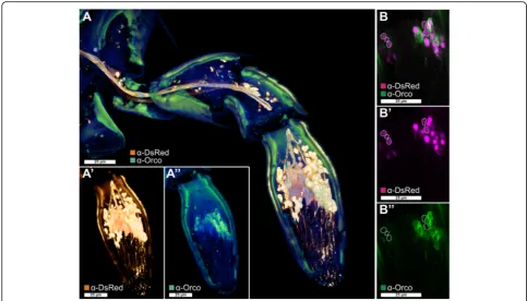
Fig. 4Orco-immunoreactive sensory neurons in the maxillary palp.aVoltex projection of a CLSM-stack showing antibody enhanced reporter expression of theEF1-B-DsRed line (a',orange) and Orco-immunoreactive cells (a'',green) in a halved maxillary palp.b–b'' Single optical section of (a) showing partial colocalization of Orco immunoreactivity and the reporter expression of theEF1-B-DsRed line (magenta). Dotted lines in (b) highlight reporter-expressing cells that are not Orco-immunoreactive.CLSMconfocal laser-scanning microscopy,Orcoodorant receptor co-receptor
statistical analysis as significantly enriched over body. All
raw values and the re-annotated gene models are
summa-rized in Additional file 8.
Tissue-specific expression of ionotropic glutamate-like
receptors
The RNAseq based revision of the 23 previously
anno-tated IRs [114] confirmed the sequences of three open
reading frames (ORFs); 17 had to be modified, two were
incompletely covered by reads, and for a single one, no
expression was detected (color coded in Additional file
8: Table S1, column B). In antennae, 16 of the IRs were
significantly
enriched
compared
to
body
(Fig.
7;
Additional file 9: Figure S5a). In the mouthparts, five IRs
are expressed, two are significantly enriched.
Comparing expression profiles of the IRs from
T.
castaneum
,
D. melanogaster
, and
Anopheles gambiae
confirmed the antennal specific expression, as well as
the high degree of phylogenetic conservation of the
antennal IRs (Fig. 8; Additional file 10: Figure S6;
highlighted in yellow) as proposed [114]. In contrast, the
divergent IRs are non-antennal specifically expressed
and are highly radiated within species clades as
previ-ously shown or predicted [114].
T. castaneum
has a
lower number of IRs compared to
D. melanogaster
and
An. gambiae
, due to lesser expansions of divergent IRs,
but maintains the basic repertoire of antennal IRs (Fig. 8;
highlighted in yellow). The homologs of IR25a, IR93a,
and IR40a, which are necessary for humidity perception
in
D. melanogaster
[119], are significantly enriched in
antennae. IR40a is exclusively expressed in antennae,
which correlates with the essential role of antennae in
T.
castaneum
hygro-perception [90]. The homolog of the
highly sensitive salt receptor and possible co-receptor IR76b
[20, 120] is significantly enriched in antennae, mouthparts,
and legs, while the co-receptors IR8a and IR25a [20] are
highly expressed in all tissues of
T. castaneum
(Fig. 7).
Tissue-specific expression of gustatory receptors
Of the 220 previously annotated GRs [81], only 207
genes had available gene models [82, 83]. Our
transcrip-tome analysis verified the ORFs of 58 GRs, showed slight
differences for 20 GRs, but did not or only incompletely
cover 129 GRs (Additional file 8: Table S1, column B).
In the antennae, 62 GRs are expressed, with 34 being
significantly enriched and 10 being antennal-specific. Of
the 69 mouthpart-expressed GRs, 36 are significantly
enriched and 19 exclusive. Seventeen GRs are
signifi-cantly enriched in both antenna and mouthparts. In legs,
18 GRs are expressed with three being significantly
enriched (Fig. 9; Additional file 11: Figure S7a).
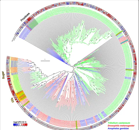
The phylogenetic comparison of the GRs in
T.
castaneum
,
D. melanogaster
, and
An. gambiae
(Fig. 10;
Additional file 12: Figure S8) confirmed that only the
CO
2receptors (highlighted in orange) are highly
con-served [121]. The other GRs seem to have undergone
inde-pendent radiation during the transition to
T. castaneum
:
e.g. the sugar receptor-related branch (highlighted in light
yellow) contains 16 genes [122], twice the number
com-pared to the two chosen dipterans. In addition, the single
fructose receptor (highlighted in grey) found in
D.
melano-gaster
and
An. gambiae
is represented by eight homologs in
T. castaneum
. The remaining 180 GRs belong to several
T.
castaneum
-specific expansion groups. Specific orthologs to
the known bitter receptors of
D. melanogaster
[13] as well
as to the thermo-sensitive
Dmel
GR28bD [123] cannot be
predicted based on our phylogenetic analysis.
Like other insects [121, 124, 125],
T. castaneum
has three
CO
2receptors (
Tcas
GR1,
Tcas
GR2, and
Tcas
GR3), while
D.
melanogaster
has only two that form functional heteromers
[126, 127]. In
T. castaneum
, the expression of the CO
2re-ceptors is not restricted to one of the chemosensory organs
with
Tcas
GR2 and
Tcas
GR3 being significantly enriched in
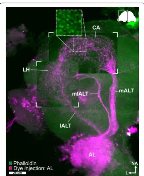
Fig. 5Antennal lobe tracts. Maximum intensity projection of a CLSM image stack after dye injection into theAL(magenta) revealed three antennal lobe tracts–the medial (mALT), mediolateral (mlALT), and the lateral antennal lobe tract (lALT)–as well as the calyx (CA) and the lateral horn (LH). In theCA, most fibers from themALTform microglomeruli (insetobtained from another preparation). The staining in the optical lobe is an artifact caused by diffusion of the dye during application. Phalloidin counterstaining ingreen. AL
antennae but also being expressed together with
Tcas
GR1
in the mouthparts (Fig. 9; highlighted in orange). This dual
input is in contrast to but combines both the expression of
the three
An. gambiae
CO
2receptors that are restricted to
the maxillary palps [128, 129], as well as the two
D.
melano-gaster
CO
2receptors that are mainly expressed in the
an-tennae [126, 127, 130].
The presence of GRs on insect antenna had
previ-ously been postulated based on physiological response
to sugars [95, 131
–
133] and was identified by
anten-nal expression aanten-nalysis [124, 127, 128, 134
–
136]. Our
interspecies comparison (Fig. 10) confirms the
anten-nal enrichment of several GRs in the two aanten-nalyzed
dipterans. However, the high number of 34
signifi-cantly enriched GRs in the antenna of
T. castaneum
is unusual, but reflects the increased total number of
GRs in this species. Interestingly, the GRs of
T.
casta-neum
are present in both antenna and mouthparts at
similar
numbers
and
expression
levels
(Fig.
9;
Additional file 11: Figure S7a).
Tissue-specific expression of odorant receptors
Of the 341 previously annotated OR sequences [115], we
could analyze 337 based on our RNAseq data. This
re-vision confirmed 97, and 22 were re-annotated reviving
eight previously indicated pseudogenes [115], namely
Tcas
OR2,
Tcas
OR18,
Tcas
OR19,
Tcas
OR22,
Tcas
OR85,
Tcas
OR99,
Tcas
OR104, and
Tcas
OR122. Moreover, 219
genes were not or only partially covered by our
transcrip-tome data (color coded in Additional file 8: Table S1,
col-umn B). Over all samples, 170 ORs are expressed (Fig. 11;
Additional file 13: Figure S9a). In antennae, 129 ORs are
expressed, with 92 being significantly enriched and 99
ex-clusive. In the mouthparts, 49 ORs are expressed, with 28
being significantly enriched and 27 exclusive. In addition,
16 of the significantly mouthpart-enriched ORs are not
enriched in the antenna (Fig. 11). The expression of
typical ORs in the mouthparts is consistent with the high
expression of Orco in this tissue (Figs. 1a and 4) and with
observations in other insect species [128, 129, 137, 138].
In legs, ten ORs are expressed (Additional file 13: Figure
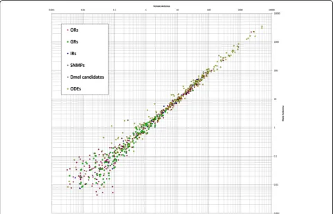
Fig. 6Comparison of expression levels in male and female antenna. Comparison of expression levels of odorant receptors (ORs,magenta), gustatory receptors (GRs,green), ionotropic glutamate-like receptors (IRs,blue), sensory neuron membrane proteins (SNMPs,orange), orthologous of candidates obtained fromD. melanogaster(Dmel candidates,grey) and potential odorant degrading enzymes (ODEs,yellow) in male and female antennae. Average values based on two male and three female antennal samples. Scatter plot of the RPKM values.Dmel D. melanogaster,GRsgustatory receptors,IRs
ionotropic glutamate-like receptors,ODEsodorant degrading enzymes,ORsodorant receptors,RPKMreads per kilobase per million,SNMPssensory neuron membrane proteins
S9a) but only one, namely
Tcas
OR127, is statistically
enriched (Fig. 9).
The phylogenetic comparison of OR expression
pat-terns in
T. castaneum
,
D. melanogaster
, and
An. gambiae
(Fig. 12; Additional file 14: Figure S10) revealed that the
atypical odorant co-receptor Orco (in
T. castaneum
pre-viously called
Tc
OR1 [115]) is the highest expressed OR
in all tissues of all three species. In
T. castaneum
, Orco
is expressed highest in antenna, followed by mouthparts.
Orco is the only OR of
T. castaneum
with clear
ortho-logs in dipterans [115, 139]. The high expression levels,
the distribution, and the evolutionary conservation of
Orco are consistent with its ancestral origin [24] and its
outstanding role as a chaperone and co-receptor,
forming functional heteromers with all typical ORs
[140, 141].
The exceptional high number of typical ORs (Fig. 12)
in
T. castaneum
is the result of large gene radiations
within the coleopteran and tenebrionid lineages [136],
which were previously subdivided into six expansion
groups (Fig. 12) [115]. Expansion groups 1, 2, and 3 are
conserved in other coleopterans [136] and are mainly
expressed in antennae. The ORs of the expansion groups
4, 5, and 6 are highly derived, have no described
homo-logs in other insects, and their expression is unusually
often mouthpart-enriched (Fig. 11; grey lettering). This
is consistent with the elaborated role of the mouthparts
in
T. castaneum
olfaction. Specific orthologs to
deorpha-nized ORs of
D. melanogaster
[142] cannot be predicted
based on our phylogenetic analysis.
Identification and expression of potential odorant
degrading enzymes
The genome of
T. castaneum
contains 15 aldehyde
dehy-drogenases (ALDHs) (Fig. 13 and Additional file 15) with
two of them being significantly enriched, but not
exclu-sively expressed in antenna. We found four predicted
genes encoding aldehyde oxidases (ALOXs) with one
be-ing highly enriched in antennae and mouthparts, which,
in contrast to ALOX ODEs from lepidopterans [143
–
145],
does not encode a signal peptide (Fig. 13). Five of the 54
identified
carboxylesterases
(CESs)
are
significantly
enriched in antenna, with two of them also in the
mouth-parts. Two other CESs are significantly enriched
exclu-sively in the mouthparts. Five of these seven candidates
show a predicted signal peptide for secretion (Fig. 13).
Tcas
CESXA shares sequence similarities with
D.
melano-gaster
Est6, and
Tcas
CES7J with
Dmel
JHEdup, with both
D. melanogaster
homologs having previously
been
identified as ODE candidates [54, 55].
TcasCES10C
is
expressed highest in antennae and related to a pheromone
degrading enzyme from the Japanese beetle,
Popillia
japonica
[146]. We identified six epoxide hydrolases
(EHs), which are supposed to be membrane bound ODEs
[147], with one being significantly enriched in antennae
and having a predicted signal peptide (Fig. 13). The
gluta-thione S-transferases (GSTs) of
T. castaneum
had already
been annotated [148]. The revision confirmed most gene
models, only
TcasGSTd2
and
TcasMGST2
had to be
modified (available in Additional file 8: Table S1). Eight of
the 41 GSTs are significantly enriched in antennae, with
Fig. 7Expression ofT. castaneumionotropic glutamate-like receptors (IRs). Heat map showing the expression level of the 23IRsas a log2[RPKM +
1] value in different tissues [adult antennae, head (missing antennae but including mouthparts), mouthparts, legs, and body]. The candidates are ordered according to their chromosomal localization (Additional file 9: Figure S5b).Horizontal bracketsabove indicate clustering in the genome. The
three also in the mouthparts (Fig. 13). One of these three,
TcasGSTd2
represents a member of the GST delta
sub-family such as GST-msolf1 from
Manduca sexta
, which is
an olfactory-specific GST expressed specifically in the
sex-pheromone-detecting sensilla [149]. Analysis of the 141
previously described cytochrome P450s (CYPs) [150]
revealed that two predicted gene models (
CYP347A4
and
CYP351B1
) were fusions of two separate genes (now
termed
CYP347A4A
and
CYP347A4B
,
as
well
as
CYP351B1A
and
CYP351B1B
, respectively). Seven other
predictions had to be adjusted based on RNAseq data
(sequences available in Additional file 8: Table S1). The
expression analysis of these 141 genes showed that 26 are
significantly enriched in the antenna, with 11 also in the
Fig. 8Phylogenetic tree of IRs. Based on protein sequences fromT. castaneum(green branches),D. melanogaster(red branches), andAn. gambiae
(blue branches). The tree was rooted using the IR8/IR25 clade, according to [114]. Robustness of the tree topology was evaluated by 100 rapid bootstrap replications.Outer ringsrepresent the expression in antennae and mouthparts (T. castaneum: palps, mandible, labrum, and labium;D. melanogaster: palp and proboscis;An. gambiae: maxillary palp) as log2-fold change compared to body corresponding to the scale in theleft lower corner. Thescale barswithin the trees represent one amino acid substitution per site. Antennal IRs are highlighted inyellow. Basically the same figure is available with absolute values instead of fold changes to get an impression of the tissue-specific abundance of the transcripts as Additional file 10: Figure S6.IRionotropic glutamate-like receptor
mouthparts (Fig. 13). In addition, six CYPs are
signifi-cantly enriched in mouthparts, but not in antennae. For
the coleopteran
Phyllopertha diversa
, CYPs have been
shown to be involved in pheromone degradation in a
membrane-bound manner [51].
Expression of potential olfaction signal transduction
pathway components
The orthologs of genes encoding signal transduction
pathway components known to be involved in olfaction
of
D. melanogaster
[46] were identified by BLAST and
manually curated. The expression analysis revealed that
four of them (
rdgB
,
itpr
,
dgkd
, and
dgkt
) are significantly
enriched in the antennae (Fig. 14). However, there is no
chemosensory-specific candidate exclusively expressed
in antennae or mouthparts. Our data, therefore, do not
indicate a chemosensory-specific metabotropic signal
transduction pathway.
Expression and distribution of sensory neuron membrane
proteins
The transcriptome analysis revealed that one of the
seven previously identified
Tcas
SNMPs [116, 117],
namely XP_969729 [116], was incorrectly annotated and
does not encode for a CD36-related protein. Moreover, the
gene model previously named SNMP1c (XM_001816389)
was a fusion of two SNMPs and overlaps with SNMP1d
(XM_001816391) [117]. In our re-annotation, we removed
XP_969729 and separated
Tcas
SNMP1c and
Tcas
SNMP1d.
In addition, the gene models of
Tcas
SNMP2,
Tcas
SNMP1a,
and XP_975606 [116] had to be modified based on
tran-scriptome and RACE-PCR data. For XP_975606, we
propose the name
Tcas
SNMP3, to reflect its unclear
phylo-genic relationship. Despite the more SNMP1-like
expres-sion pattern (Fig. 15) and chromosomal localization
(Additional file 9: Figure S5b) of
Tcas
SNMP3, the
compari-son of the amino acid composition revealed no clear
affili-ation to either the SNMP1 or the SNMP2 subgroup [151].
Interspecies comparison revealed no clear orthology of
Tcas
SNMP3 to SNMPs from other species, including the
so-called SNMP3 of
Calliphora stygia
[152], which, based
on phylogeny, clearly represents an SNMP1 homolog. All
six
Tcas
SNMPs are expressed in antennae (Fig. 15), which
was also confirmed by rapid amplification of cDNA-ends
PCR (RACE-PCR) based on an antennae cDNA pool, but
only
Tcas
SNMP1a-d and
Tcas
SNMP3 are significantly
enriched in antennal tissue. Moreover, three of the
Tcas
SNMP1, as well as
Tcas
SNMP3, are also enriched in
mouthparts (Fig. 15), further supporting the importance of
the mouthparts for olfaction in
T. castaneum
. In contrast,
Fig. 9Expression ofT. castaneumgustatory receptors (GRs). Heat map showing the expression level of the 207 analyzedGRsas a log2[RPKM + 1]
value in different tissues [adult antennae, head (missing antennae but including mouthparts), mouthparts, legs, and body]. The candidates are ordered according to their chromosomal localization (Additional file 11: Figure S7b).Horizontal bracketsabove indicate clustering in the genome. Thearrowheadsrepresent the orientation of the open reading frame. The expression levels are represented by agreyscalewith the highest shown expression levels labeledblack. Theasterisksmark statistically significantly differentially expressed genes compared to body (based on biological replicates of five antennal, two head, three mouthpart, two leg, and two body samples). Thered asterisksrepresent up- and thebluedown-regulation (pvalues adjusted are * < 0.05, ** < 0.01, and *** < 0.001). CO2receptors are highlighted inorange, fructose receptor related genes ingrey, and sugar receptors inyellow. GR
Tcas
SNMP2 is expressed highest in body and significantly
underrepresented
in
antennae
and
mouthparts
(Fig. 15), which is similar to its ortholog in
D.
mela-nogaster
[49]. Despite the observation that in most
in-sects with a fully sequenced genome only two SNMPs
were found [116, 117], the relatively high amount of
six
Tcas
SNMPs of
T. castaneum
is not unique, since
transcriptome analysis, e.g., of other beetles, revealed
four
SNMPs
in
Dendroctonus
valens
[153]
and
Dastarcus helophoroides
[154], as well as three in
Ips
typographus
and
Dendroctonus
ponderosae
[136].
However,
T. castaneum
is currently only exceeded by
the hessian fly (
Mayetiola destructor
) with seven
expressed SNMPs [155].
Fig. 10Phylogenetic tree of gustatory receptors (GRs). Mid-point rooted tree based on protein sequences fromT. castaneum(green branches),D. melanogaster(red branches), andAn. gambiae(blue branches). Robustness of the tree topology was evaluated by 100 rapid bootstrap replications.
Outer ringsrepresent the expression in antennae and mouthparts (T. castaneum: palps, mandible, labrum, and labium;D. melanogaster: palp and proboscis;An. gambiae: maxillary palp) as log2-fold change compared to body corresponding to the scale in theleft lower corner. Thescale bars
within the trees represent one amino acid substitution per site. Potential sugar receptors (highlighted inyellow), fructose receptors (highlighted in
grey), and CO2receptors (highlighted inorange) are labeled. Known bitter receptors fromD. melanogasterare highlighted ingreen, and the
thermos-sensitive GR28bD in light blue. Basically the same figure is available with absolute values instead of fold changes to get an impression of the tissue-specific abundance of the transcripts as Additional file 12: Figure S8.GRgustatory receptor
Discussion
Independent integration centers for antennal and palpal
olfactory perception
In
T. castaneum
, odorants are mainly perceived with the
last three segments of the antenna, which carries three
types of chemoreceptive sensilla (SBas, cSTri, and SCoe),
as well as with the maxillary and labial palps (Fig. 16).
Accordingly, expression analysis revealed that ORs are
mostly expressed in antennae, but also in the
mouth-parts (Figs. 11 and 12; Additional file 13: Figure S9a) as
previously shown for several dipteran species [128, 129,
137, 156
–
158]. In contrast to the Diptera, where the
Fig. 11Expression ofT. castaneumodorant receptors (ORs). Heat map showing the expression levels of the 337 analyzed ORs as log2[RPKM + 1]
with a maximum of 8.1 (Orco has a value of 11.1 in antenna) in different tissues [adult antennae, head (missing antennae but including mouthparts), mouthparts, legs, and body]. The candidates are ordered according to their chromosomal localization (Additional file 13: Figure S9c).Horizontal bracketsabove indicate clustering in the genome, and thearrowheadsrepresent the orientation of the open reading frame. ORs that are member of clades four, five, and six [115] are written ingrey letters. The line labeled withAdultandLarvarefers to data from [115]. The characterH(respectivelyB) indicates that the correspondingORwas detected in head or body cDNA samples by reverse PCR of the labeled developmental stage. Ablack letter
palps are chemosensory appendages with limited odor
coding complexity, the relatively high number of
Orco-immunoreactive CSNs (Fig. 4) as well as the high
num-ber of expressed ORs, SNMPs, potential ODEs, and
OBPs [89] in
T. castaneum
mouthparts (Figs. 11, 13, 15,
and 16) imply a more prominent role of the palps in
olfaction. The palpal ORs are possibly involved in the
evaluation of the quality of food sources, like the ORs
on the proboscis of
Manduca sexta
[159].
Moreover, in addition to the differences on the
percep-tion level, major dissimilarities to the Diptera occur on
the level of odor processing. The data from the partial
Fig. 12Phylogenetic tree of odorant receptors (ORs). Protein sequences (>300 amino acids) fromT. castaneum(green branches),D. melanogaster
(red branches), andAn. gambiae(blue branches). The tree was rooted using the Orco clade, according to [24]. Robustness of the tree topology was evaluated by 100 rapid bootstrap replications.Outer ringsrepresent the expression in antennae and mouthparts (T. castaneum: palps, mandible, labrum, and labium;D. melanogaster: palp and proboscis;An. gambiae: maxillary palp) as log2-fold change compared to body corresponding to
the scale in theleft upper corner. The surrounding numbers on theouter thin lineindicate the expansion groups 1 to 6 [115].TcasOR71and
TcasOR72PSEwere previously assigned to expansion group 1. Thescale barwithin the tree represents one amino acid substitution per site. Basically the same figure is available with absolute values instead of fold changes to get an impression of the tissue-specific abundance of the transcripts as Additional file 14: Figure S10.ORodorant receptor
Orco
-Gal4 line as well as the backfills from the antenna
and the mouthparts indicate that processing olfactory
in-formation at least at the level of the first central relay
station occurs independently of each other (Fig. 16).
This is surprising, as many of the ORs expressed on the
mouthparts are also expressed on the antennae. In
contrast, typical OR expression is mutually exclusive
between antenna and palps in
D. melanogaster
and
An.
gambiae
[128, 137], where in addition, projections from
the palps innervate several AL glomeruli [63, 160, 161].
In
T. castaneum
, the olfactory input stemming from the
antenna seems to be processed exclusively in the AL
(Fig. 3a; Additional file 5: Movie S2; Additional file 6:
Figure S4), whereas the palpal-derived olfactory
informa-tion is essentially processed outside the AL, in the LG
(Fig. 3e, f; Additional file 5: Movie S2) and the GOC, an
unpaired and glomerularly organized first olfactory
cen-ter in the GNG (Fig. 3d, f; Additional file 7: Movie S3).
The LG had, as far as we know, previously been
de-scribed only in hemimetabolous insects [57, 64
–
66, 105].
A glomerularly organized olfactory center in the GNG
such as the GOC has, to our knowledge, not been
de-scribed in any insect so far. The number of 49 ORs (with
28 being significantly enriched compared to body) that
are expressed in the mouthparts is roughly consistent
with the estimated 30 to 40 glomeruli in the GOC. This
Fig. 13Expression ofT. castaneumpotential odorant degrading enzymes (ODEs). Heat map showing the expression level of the 263 potential ODEs as a log2[RPKM + 1] value in different tissues [adult antennae, head (missing antennae but including mouthparts), mouthparts, legs, and
body]. The candidates are ordered according to their protein family and chromosomal localization.Horizontal bracketsabove indicate clustering in the genome (Additional file 15: Figure S11), and thearrowheadsrepresent the orientation of the open reading frame. Underlined genes were previously found on the protein level in antennae by [89]. The expression levels are represented by agreyscalewith the highest shown expression levels labeledblack. Theasterisksmark statistically significantly differentially expressed genes compared to body (based on biological replicates of five antennal, two head, three mouthpart, two leg, and two body samples). Thered asterisksrepresent up- and thebluedown-regulation (pvalues adjusted are * < 0.05, ** < 0.01, and *** < 0.001). Ablack dotin the lowest line indicates a predicted signal peptide according to a SignalP 4.0 [211] prediction.ODE
suggests that the wiring in the GOC may resemble the
situation in the ALs with the difference being
conver-gence into an unpaired medial structure. The only palpal
projection into the AL is a mutually exclusive
innerv-ation of a single ipsilateral glomerulus (Fig. 3a;
Additional file 5), which may be involved in CO
2percep-tion, as described in several moth species [99] and
proposed for some mosquitoes [161, 162].
Antennae serve also as key organs for gustatory
perception
In
T. castaneum
, antennae and mouthparts express
simi-lar high numbers and levels of GRs, which indicates the
antenna as a key gustatory organ besides the mouthparts
(Fig. 9; Additional file 11: Figure S7a). This finding may
reflect the beetles
’
ground-dwelling life style and
indi-cates that the scanning behavior with the antennae not
Fig. 14Expression ofT. castaneumhomologs of genes described to be involved in olfaction ofD. melanogaster. Heat map showing the expression level of the several genes supposed to be involved inD. melanogasterolfaction, as a log2[RPKM + 1] value in different tissues [adult antennae, head
(missing antennae but including mouthparts), mouthparts, legs, and body]. The expression levels are represented by agreyscalewith highest shown expression levels labeledblack. Theasterisksmark statistically significantly differentially expressed genes compared to body (based on biological replicates of five antennal, two head, three mouthpart, two leg, and two body samples). Thered asterisksrepresent up-regulation (pvalues adjusted are * < 0.05).RPKMreads per kilobase per million
Fig. 15Expression ofT. castaneumsensory neuron membrane proteins (SNMPs). Heat map showing the expression level of the six sensory neuron membrane proteins ofT. castaneum, as a log2[RPKM + 1] value in different tissues [adult antennae, head (missing antennae but including
mouthparts), mouthparts, legs, body, as well as larval head and body]. The candidates are ordered according to their chromosomal localization (Additional file 9: Figure S5b).Horizontal bracketsabove indicate clustering in the genome, and thearrowheadsrepresent the orientation of the open reading frame. The expression levels are represented by agreyscalewith highest shown expression levels labeledblack. Theasterisksmark statistically significantly differentially expressed genes compared to body (based on biological replicates of five antennal, two head, three mouthpart, two leg, and two body samples). Thered asterisksrepresent up- and thebluedown-regulation (pvalues adjusted are * < 0.05, ** < 0.01, and *** < 0.001).
SNMPsensory neuron membrane protein,RPKMreads per kilobase per million
only gathers tactile but also chemical stimuli. This is in
contrast to the Diptera, where the labellum is the main
gustatory organ [163
–
165].
Postulation of exceptions to the central dogma
The number of 129 ORs that we found to be expressed
in
T. castaneum
antennae (Fig. 11; Additional file 13:
Figure S9a) exceed the numbere of about 90 glomeruli
in the AL. Moreover, some glomeruli are likely to get
exclusive innervation by OSNs that express IRs, as
de-scribed in
D. melanogaster
[20]. These observations do
not conciliate with the central dogma postulating that
OSNs express only one typical OR and all OSNs carrying
this same OR converge into one and the same glomerulus,
which was hypothesized to be the typical situation for
insects [61, 137, 166]. However, for
D. melanogaster
, both
co-expression of more than one typical OR per OSN as well
as co-convergence due to innervation of one AL
glomeru-lus by more than one OSN sub-type have been already
described as exceptions [137, 167]. For
T. castaneum
, we
propose that such exceptions are much more frequent.
Large repertoire of potentially functional odorant
receptor genes and possible environmental regulation
The genome of
T. castaneum
harbors 341 OR genes
[81, 115], of which 270 seem to encode for functional
ORs. Of the 337 ORs with available full sequence
infor-mation [115], we find in our RNAseq data 161 ORs to
be expressed in adult antennae, mouthparts, and head by
a threshold of 0.5 RPKM (Additional file 13: Figure S9a).
Fig. 16T. castaneumhead scheme depicting the major olfactory pathway components.aHead section (dorsal view) showing the brain and the
CSNsfrom the antenna (blue) and the mouthparts (green).bHead section (ventral view) showing theGNG. Section orientation is indicated at the
upper right corner(lateral view of the head;V, ventral).Double-headed arrowsindicate body (A, anterior↔P, posterior;black) and neuro-axis (NA, n-anterior↔NP, n-posterior;red). Chemical signals are sensed by about 720CSNslocated in 56SBas, 87cSTri, and 11SCoeon the last three antennal segments. TheseCSNsexpress 16IRs, 62GRs, 129ORs, and sixSNMPs. Chemosensory information is also perceived in the palps by fiveIRs, 69GRs, 49
ORs, and sixSMNPs(number in brackets indicates significantly enriched members compared to body). The antennal nerve (AN) projects into the ipsilateralAL, where all except one (light green) of the about 90GL(dark blue) are innervated. A separate antennal tract (*1) descends into theGNG(b, blue), where presumably gustatory and mechanosensory information is processed. Incoming olfactory information is processed by a complex network of local interneurons (LNs) in theALand further relayed by projection neurons forming threeALTs. The medialALT(mALT) projects to and arborizes in the calyx of theMBformed by about 2700KCs(orange) to eventually innervate theLH(light blue). The mediolateral ALT (mlALT) and lateralALT(lALT) directly innervate theLH. From the mouthparts,CSNsproject via the maxillary (*3) and labial palp nerves (*4) into theGNG, where the gustatory information is processed in thePGC. The olfactory sensory input from the palps is processed in an unpaired glomerularly organizedGNGstructure, the
GOC, as well as in theLG, which receives input from some palpal OSNs via ascending neurons (*2) passing through theGOC. Some of the palp-derived chemosensory information is processed in the singleALglomerulus, which lacks antennal innervation and is, therefore, exclusively innervated by projections from the mouthparts (light green).ALantennal lobe,ALTantennal lobe tracts,ANantennal nerve,CSNchemosensory neuron,cSTrichemosensilla trichoidea,GLantennal lobe glomeruli,GNGgnathal ganglia,GOCgnathal olfactory center,GRgustatory receptor,IRionotropic glutamate-like receptor,KC
![Fig. 1 Sensilla types and distribution onlongitudinally corrugated, connected to a neuron at the socket (sensilla squamiformium [95]are small, smooth, and dome-shaped sensilla restricted to segment 11 (Additional file 3: Figure S3a);enhanced Tribolium cast](https://thumb-us.123doks.com/thumbv2/123dok_us/8910809.1836523/4.595.58.537.85.486/sensilla-distribution-onlongitudinally-corrugated-squamiformium-restricted-additional-tribolium.webp)


![Fig. 7 Expression of T. castaneum ionotropic glutamate-like receptors (IRs). Heat map showing the expression level of the 23 IRs as a log2[RPKM +1] value in different tissues [adult antennae, head (missing antennae but including mouthparts), mouthparts, le](https://thumb-us.123doks.com/thumbv2/123dok_us/8910809.1836523/10.595.57.542.86.259/expression-castaneum-ionotropic-glutamate-receptors-expression-mouthparts-mouthparts.webp)

![Fig. 9 Expression of T. castaneum gustatory receptors (GRs). Heat map showing the expression level of the 207 analyzed GRs as a log2[RPKM + 1]value in different tissues [adult antennae, head (missing antennae but including mouthparts), mouthparts, legs, an](https://thumb-us.123doks.com/thumbv2/123dok_us/8910809.1836523/12.595.56.540.88.321/expression-castaneum-gustatory-receptors-expression-including-mouthparts-mouthparts.webp)