R E S E A R C H A R T I C L E
Open Access
The effects of different shapes of
capsulorrhexis on postoperative refractive
outcomes and the effective position of the
intraocular lens in cataract surgery
Shixu Li, Yiping Hu, Ran Guo, Yushuang Shao, Jiangyue Zhao, Jinsong Zhang and Jing Wang
*Abstract
Background:To evaluate the effects of anterior capsular opening size on deviation from predicted refraction and
the effective position of the intraocular lens (ELP) in cataract surgery.
Methods:Nonrandomized clinical trial. Eighty patients (80 eyes) with simple age-related cataracts were treated
from May 2018 to September 2018 at the Fourth Affiliated Hospital of China Medical University. All patients undergoing phacoemulsification received intraocular lens based on the voluntary principle. Forty eyes were implanted with the C-loop haptic intraocular lens (AMO Tecnis ZCB00) while the other 40 eyes were implanted with the plate haptic intraocular lens (CT ASPHINA 509 M). Follow-up visits were conducted postoperatively at 1 week, 1 month, and 3 months during which patients underwent refraction and data collection after pupil dilation, which included anterior segment photography and Scheimpflug imaging by Pentacam. The area, horizontal and vertical diameter of the capsulorrhexis, circularity, decentration, and package were analysed using the image analysis software Image-Pro-Plus 6.0,then evaluated the relationship between the different shapes of capsulorrhexis with deviation from predicted refraction and ELP in cataract surgery.
Results:Deviation from predicted refraction and all of the parameters of capsulorrhexis were not correlative in the
509 M IOL group, however, in the Tecnis IOL group, while the deviation from predicted refraction and all of the capsulorrhexis parameters were not correlative at 1 week, the deviation from predicted refraction did correlate with capsulorrhexis area, horizontal diameter at 1 month (P= 0.029,P= 0.048), and with capsulorrhexis area, vertical diameter at 3 months (P= 0.03,P= 0.017). The ELP correlated with package in both groups postoperatively (r > 0,P< 0.05), but there is no other capsulorrhexis parameters correlated with ELP in the 509 M IOL group (allP> 0.05). For the Tecnis IOL group, the ELP and capsulorrhexis area were correlated at 1 week and 1 month, while the ELP and horizontal diameter, the ELP and vertical diameter were correlated at 1 week, but did not correlate with the other capsulorrhexis parameters in the Tecnis IOL group (allP> 0.05).
Conclusions:The shape of the capsulorrhexis has an effect on postoperative refractive outcomes and the effective
position of the intraocular lens in cataract surgery, and plate haptic intraocular lenses have better refractive stability than C-loop haptic intraocular lenses.
Trial registration:ChiCTR1800015638,2018-04-12.
Keywords:Effective intraocular lens position, Capsulorrhexis, Postoperative refractive outcomes
* Correspondence:[email protected]
Department of Ophthalmology, the Fourth Affiliated Hospital of China Medical University, Eye Hospital of China Medical University, the Key Lenticular Laboratory of Liaoning Province, Shenyang 110005, China
© The Author(s). 2019Open AccessThis article is distributed under the terms of the Creative Commons Attribution 4.0 International License (http://creativecommons.org/licenses/by/4.0/), which permits unrestricted use, distribution, and reproduction in any medium, provided you give appropriate credit to the original author(s) and the source, provide a link to the Creative Commons license, and indicate if changes were made. The Creative Commons Public Domain Dedication waiver (http://creativecommons.org/publicdomain/zero/1.0/) applies to the data made available in this article, unless otherwise stated. Liet al. BMC Ophthalmology (2019) 19:59
Background
Currently, phacoemulsification and intraocular lens im-plantation are the most effective treatments for cataract. With improvements in surgical techniques and the de-velopment of the refractive field, patients have increas-ingly high precision requirements for long-term and stable optical quality following an operation, and this has become a common pursuit for both doctors and pa-tients. Continuous circular capsulorrhexis (CCC) is a common method in cataract surgery. This method is broadly popular and has an irreplaceable special status in the cataract field. This method allows the capsule to remain relatively intact to ensure accurate implantation of the intraocular lens, effectively preventing the optical centre of the intraocular lens from moving or tilting. A capsule that is irregularly shaped, eccentric, or that does not fully cover the optic portion of the intraocular lens may lose these advantages and may lead to the postoper-ative refraction that does not match the preoperpostoper-ative predicted refraction, which will ultimately reduce the
pa-tient’s visual quality and influence the outcome of
cata-ract surgery.
The effective intraocular lens position is defined as the vertical distance from the posterior corneal apex to the optical plane of the intraocular lens on the visual axis. It reflects the longitudinal position of the intraocular lens in the eye, and the fusion and fibrosis processes of the lens capsule produce the forward and backward forces, while the ELP reflects the unbalanced result of those
forces [1]. When the intraocular lens moves forward it
causes a myopic shift, and when it moves backward it causes a hyperopic shift, therefore, the ELP determines
the refractive condition after cataract surgery [2]. This
paper intends to explore the effects of the morphological parameters of the capsulorrhexis on postoperative re-fractive outcomes and the effective position of the intra-ocular lens.
Methods
Patients selection
This study was approved by the Fourth Affiliated Hos-pital of China Medical University. All research and data collection practices adhered to the tenets of the Declar-ation of Helsinki and good clinical practices, and all pa-tients provided a signed informed consent to participate in a clinical research study.
Eighty patients (80 eyes) with simple age-related cata-racts were treated from May 2018 to September 2018 in the Fourth Affiliated Hospital of China Medical Univer-sity. We specifically included patients who were bodily healthy and underwent a successful implantation of an IOL. The exclusion criteria for this study were irregular astigmatism, corneal opacity, glaucoma, retinal disease, a history of ocular inflammation, a history of ocular trauma,
any other previous intraocular surgery, and intraoperative complications including anterior or posterior capsular tears or postoperative macular edema. The patients were divided into 2 groups according to the different intraocu-lar lenses implanted: 40 eyes were implanted with the C-loop haptic intraocular lens (AMO Tecnis ZCB00) while the other 40 eyes were implanted with plate haptic intraocular lenses (CT ASPHINA 509 M).
Surgery
All phacoemulsification and IOL implantations were performed by one experienced surgeon under topical an-aesthesia with 0.5% proparacaine hydrochloride. The complete anterior capsular opening was made approxi-mately 5.5 mm in diameter using capsulorrhexis forceps. After phacoemulsification of the nucleus and aspiration of the cortex, the Tecnis IOL or 509 M IOL was injected into the capsular bag, and the IOL centration was con-firmed again after viscoelastic removal. The Haigis for-mula was used for the lens power selection. All patients received an anti-inflammatory treatment consisting of Tobramycin and Dexamethasone eye drops (5 ml (15 mg/5 mg)) (TobraDex®, Alcon) that were used every 6 h for 2 months along with 0.1% Bromfenac Sodium Hy-drate eye drops (BRONUCK®, SENJU) that were used every 12 h for 1 month.
Postoperative evaluation
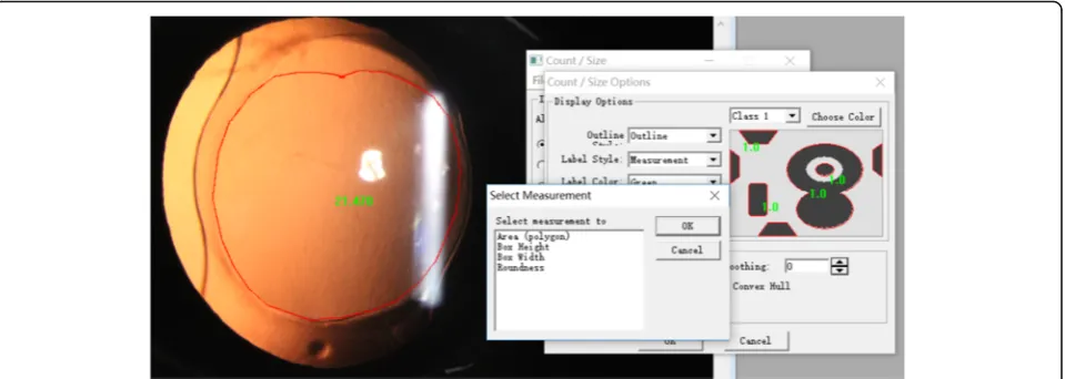
Postoperative examinations were performed at 1 week, 1 month, and 3 months after the surgery. All patients were measured for subjective refraction that was converted into spherical equivalent (SE), and the deviation from predicted refraction was the difference between postoperative SE of the subjective refraction and the predicted refraction. The patients then underwent slit-lamp digital photography measurements and Pentacam measurements, and the cap-sulorrhexis were documented using digital retroillumina-tion photographs through dilated pupils. All photographs were imported into Image-Pro-Plus 6.0 to analyse the cap-sulorrhexis parameters including the capcap-sulorrhexis area, horizontal diameter, vertical diameter, circularity,
decen-tration, and package (Fig.1). Circularity is defined by the
formula: Circularity = 4π× area/perimeter2, an index of
1.0 denotes a theoretically perfect circle, and the lower the
index, the more irregular the capsulorrhexis [3].
Decentra-tion is the vector between the centre of the pupil and the centre of the capsulorrhexis outline. Package is expressed as the ratio of the minimum distance to the maximum distance of the capsule edge from the optical plane edge (package = minimum distance/maximum distance).
Statistical analyses
of all data distributions was confirmed using the Kolmogorov-Smirnov test. Paired t-tests were used to com-pare normally distributed data, while nonparametric data were analysed by a one-way ANOVA. Spearman correl-ation tests were used to determine the correlcorrel-ations between capsulorrhexis parameters and postoperative re-fractive outcomes or correlations between capsulorrhexis parameters and ELP. Statistical significance was defined as
aPvalue that was less than 0.05.
Results
Patient characteristics
Our study protocol included 80 eyes from 80 patients who were aged between 62 and 74 years. Patient recruit-ment took place from May 2018 to September 2018: Forty eyes were implanted with the C-loop haptic intra-ocular lens (AMO Tecnis ZCB00) and the other forty eyes were implanted with plate haptic intraocular lenses (CT ASPHINA 509 M). The patient demographics and IOL models for the two groups are summarized in
Table 1. There was no significant (P> 0.05) difference
between the two groups (Table1).
Capsulorrhexis morphological features
The capsulorrhexis area, horizontal diameter, vertical diameter, circularity, decentration, and package were not
significantly different in the 509 M IOL group at 1 week,
1 month, or 3 months after the operation (all P> 0.05).
However, the capsulorrhexis area, particularly the hori-zontal diameter, was significantly different in the Tecnis IOL group at 1 week to 1 month, and at 1 week to 3
months (allP< 0.05) (Table2).
Morphological features of Capsulorrhexis
One-way ANOVA was performed in the 509 M IOL group and the Tecnis IOL group and the results showed that the Tecnis IOL group had a significantly higher de-gree of package than the 509 M IOL group at 1 month
(P< 0.001), but there were no other differences in the
morphological features of capsulorrhexis (all P> 0.05)
(Table3).
Postoperative refractive outcomes
There was no significant difference in postoperative re-fractive outcomes between two groups at 1 week, 1 month,
and 3 months. (509 M group: t =−0.891P= 0.381, Tecnis
group: t = 0.325 P= 0.750, P all> 0.05). This finding
indi-cates that the postoperative dioptre is stable across 3
months and does not show differences over time (Fig.2).
Effective position of the intraocular Lens
A non-parametric test for the effective position of the intraocular lens indicated that the effective positions of the intraocular lens in the 509 M IOL group at 1 month and 3 months were significantly larger than that of
post-operative 1 week (P= 0.023, P= 0.038); however, since
the ELP tends towards stability after 1 month, there was no significant difference between 1 month and 3 months
(P= 0.961). For the Tecnis IOL group, no statistical
dif-ferences were found at 1 week, 1 month, or 3 months
after the operation (allP> 0.05) (Fig.3).
Fig. 1Using Image-Pro-Plus 6.0 to analyse the capsulorrhexis parameters including the capsulorrhexis area, horizontal diameter, vertical diameter, circularity, decentration, and package
Table 1Demographic and clinical information for patients
included in this study
Groups Age(years) Eyes(n) Sex Laterality
Male Female Right Left
509 M 62.54 ± 11.81 40 24 16 20 20
Tecnis 63.00 ± 9.08 40 18 23 21 19
t 0.376 0.841 2.875
Pvalue 0.71 0.365 0.098
The effect of Capsulorrhexis morphological features on the deviation from predicted refraction
The deviation from predicted refraction and all capsulor-rhexis parameters were not correlated in the 509 M IOL group. However, for the Tecnis IOL group, while the de-viation from predicted refraction and all capsulorrhexis parameters were not correlated at 1 week, the deviation from predicted refraction did correlate with the
capsulor-rhexis area and horizontal diameter at 1 month (P= 0.029,
P= 0.048),and the capsulorrhexis area and vertical diameter
at 3 months (P= 0.03, P= 0.017) (Table 4). The deviation
from predicted refraction showed positive correlations with the change in the capsulorrhexis area, the horizontal
diameter, and the vertical diameter (Figs.4,5and6).
How-ever, for the 509 M IOL, the morphological features of the
capsulorrhexis showed no correlation with the deviation
from the predicted refraction (allP> 0.05).
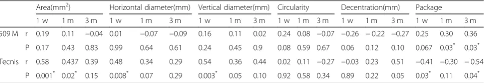
The effect of Capsulorrhexis morphological features on the ELP
The ELP correlated with package in both groups
postop-eratively (r > 0, P< 0.05), but there is no other
capsulor-rhexis parameters correlated with ELP in the 509 M IOL
group (all P> 0.05). For the Tecnis IOL group, the ELP
and capsulorrhexis area were correlated at 1 week and 1 month, while the ELP and horizontal diameter, the ELP and vertical diameter were correlated at 1 week, but did not correlate with the other capsulorrhexis parameters
in the Tecnis IOL group (allP> 0.05) (Table5).
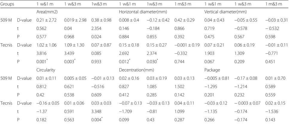
Table 2The two groups’difference value on capsulorrhexis morphological features at different time points
Groups 1 w&1 m 1 w&3 m 1w&3 m 1 w&1 m 1w&3 m 1 m&3 m 1 w&1 m 1 w&3 m 1 m&3 m
Area(mm2) Horizontal diameter(mm) Vertical diameter(mm)
509 M D-value 0.21 ± 2.72 0.019 ± 2.98 0.38 ± 0.98 0.008 ± 0.4 −0.12 ± 0.42 0.42 ± 0.29 0.04 ± 0.43 −0.05 ± 0.55 −0.03 ± 0.31
t 0.562 0.04 2.354 0.146 −0.184 0.866 0.719 −0.578 −0.532
P 0.577 0.968 0.024 0.884 0.855 0.392 0.475 0.567 0.598
Tecnis D-value 1.02 ± 1.06 1.09 ± 1.30 0.07 ± 0.87 0.15 ± 0.18 0.15 ± 0.27 −0.001 ± 0.19 0.07 ± 0.21 0.06 ± 0.19 −0.01 ± 0.11
t 3.816 3.439 0.085 2.692 2.374 −0.332 1.903 1.309 −0.771
P 0.001* 0.003* 0.933 0.012* 0.030* 0.744 0.067 0.209 0.451
Circularity Decentration(mm) Package
509 M D-value 0.01 ± 0.11 0.005 ± 0.05 −0.01 ± 0.13 0.02 ± 0.16 0.03 ± 0.19 0.03 ± 0.13 −0.005 ± 0.81 −0.17 ± 0.08 0.01 ± 0.70
t 0.812 0.621 −0.516 0.827 1.085 1.502 −1.295 −1.214 0.589
P 0.42 0.538 0.609 0.412 0.285 0.142 0.201 0.232 0.559
Tecnis D-value −0.16 ± 0.05 0.01 ± 0.06 0.03 ± 0.03 −0.07 ± 0.13 −0.03 ± 0.13 0.04 ± 0.11 −0.03 ± 0.12 −0.003 ± 0.07 0.02 ± 0.15
t −1.37 0.591 3.348 −1.709 −0.81 1.099 −1.135 −0.174 −1.536
P 0.182 0.563 0.004* 0.099 0.43 0.287 0.266 −0.174 0.143
*P < 0.05
Table 3Comparison of the capsulorrhexis parameters in the 509 M IOL group and the Tecnis IOL group at 1 week, 1 month, and 3
months postoperatively
Area (mm2) Horizontal diameter (mm) Vertical diameter (mm) Circularity Decentration (mm) Package
1 w 509 M 21.96 ± 4.76 5.39 ± .55 5.19 ± .63 .85 ± .060 0.34 ± .18 .072 ± .12
Tecnis 21.15 ± 3.39 5.23 ± .40 5.17 ± .49 0.84 ± .050 0.28 ± .16 0.11 ± .14
F 0.673 1.951 0.043 0.034 2.221 1.544
P 0.414 0.166 0.836 0.854 0.140 0.218
1 m 509 M 21.74 ± 4.68 5.89 ± .59 5.15 ± .71 .84 ± .12 .32 ± .18 .087 ± .12
Tecnis 20.31 ± 3.17 5.12 ± .35 5.11 ± .48 .86 ± .03 0.32 ± .20 0.14 ± .17
F 2.774 3.615 0.363 0.186 0.036 97.533
P 0.101 0.062 0.549 0.668 0.85 0.000*
3 m 509 M 21.46 ± 4.79 5.34 ± .58 5.18 ± .82 0.84 ± .05 0.30 ± .19 .10 ± .15
Tecnis 20.17 ± 3.23 5.10 ± .44 5.12 ± .44 0.84 ± .03 0.34 ± .16 0.12 ± .13
F 0.259 1.118 0.020 0.200 1.297 0.004
Fig. 2The deviation from predicted refraction of the 509 M IOL group and the Tecnis IOL group at 1 week, 1 month, and 3 months postoperatively
Fig. 3The ELP of the 509 M IOL group and the Tecnis IOL group at 1 week, 1 month, and 3 months postoperatively
Discussion
Currently, with the development of refractive cataract technology, the requirement for accurate postopera-tive refraction is increasingly high, and surgeons are required to design the best operation plan periopera-tively. There are many factors that affect the
refract-ive outcomes of cataract surgery, including the
influence of small surgical incisions, functional intra-ocular lens, advanced instruments and the surgical navigation system, these factors have been gradually solved, but irregular anterior capsular openings also affect the refractive outcomes, especially the size and shape of the capsular bag and the effective intraocular
lens position [4, 5]. Therefore, we designed a
pro-spective cohort study to investigate the relationships between the morphological parameters of capsulor-rhexis and deviation from predicted refraction and the effective position of the intraocular lens.
Two types of intraocular lenses were included in this study. The Tecnis ZCB00 IOL is designed by C-loop haptic and the 509 M IOL is designed by plate haptic. The Tecnis IOL material is hydrophobic acrylate, de-signed by OptiEdge edge. This design reduces the inci-dence of posterior capsule opacification and improves the stability of the postoperative intraocular lens. The 509 M IOL is a single-piece hydrophilic acrylic intraocu-lar lens and is more compatible with human tissue, while the plate haptic design makes the intraocular lens more stable and centred. Additionally, the analysis showed that there was no significant difference in the total-eye predicted spherical aberration between the two groups (P > 0.05),therefore, we chose these two different designs of intraocular lens to represent the two commonly used aspherical monofocal IOLs, which are typical and repre-sentative. The morphological features of capsulorrhexis
(including the area of capsulorrhexis, horizontal
Fig. 4For every 1-mm2 increase in area, the deviation from predicted refraction increases by 0.0152 D
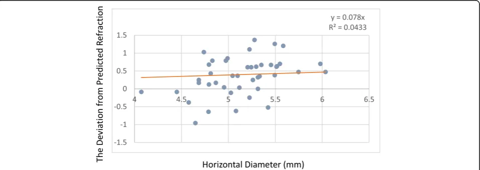
Fig. 5For every 1-mm increase in the horizontal diameter, the deviation from predicted refraction increases by 0.07 D
diameter, vertical diameter, circularity, decentration, and package) that were measured at 1 week, 1 month, and 3 months showed that the 509 M IOL does not have sig-nificant differences over time. However, the area of cap-sulorrhexis and the horizontal diameter of the Tecnis IOL showed significant differences at 1 week to 1 month and at 1 week to 3 months. These results show that the
Tecnis IOL’s capsulorrhexis had the largest area and
horizontal diameter at 1 week after the operation, and with the passage of time, the area and horizontal diam-eter gradually decreased up to 1 month and remained stable to 3 months. These findings indicate that the area and the horizontal diameter gradually stabilize at 1 month after the operation. Further, the circularity showed significant differences at 1 month to 3 months postoperatively. These differences compared to the 509 M IOL, are likely due to the design of the IOL loops. The contractive force around the C-loop haptic IOL is not equivalent on every side of the capsule bag, and the parts of the IOL loops that do not completely and tightly contact the capsule bag can result in poor stability. Therefore, in terms of stability, the C-loop haptic IOL is
not as good as the 509 M IOL [6]. In addition, capsule
contraction syndrome occurs at 1 month after surgery, which leads to decreases in the area and horizontal diameter of the capsulorrhexis. As a result, we believe that the stability of the intraocular lens with the plate
haptic design is better than that of the C-loop intraocu-lar lens, and they have lower requirements for capsulor-rhexis. Meanwhile, some studies show that the material of the optical surface also affects the contraction of the anterior capsule.
The effective position of the intraocular lens can re-flect the longitudinal position of the intraocular lens in the eye. It has been shown that changes in anterior
chamber depth of approximately 720μm lead to a 1.00
D refractive deviation [7]. Moving forward to the retina
leads to a myopic deviation while moving backward to the retina leads to a hyperopic deviation. Therefore, the ELP is particularly important for cataract surgery, espe-cially for patients with refractive intraocular lens
im-plantation [8,9].
The effective position of the intraocular lens is related to the fibre shrinkage, capsular opacity, and the material
of lens [10,11]. If the opening of the anterior capsular is
too small, it can cause contraction of the capsular bag that leads to the displacement of the ELP. Therefore, when the size of the anterior capsular is only wrapped around the edge of the optic surface of the intraocular lens (5.5 mm in diameter), the contraction of the capsu-lar bag can be avoided and the influence on the ELP can
be minimized [12].
These results showed that the effective position of the intraocular lens increased gradually at 1 week, 1 month, Fig. 6For every 1-mm increase in the vertical diameter, the deviation from predicted refraction increases by 0.05 D
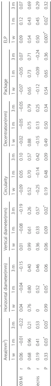
Table 5Correlations of the morphological features of capsulorrhexis and the ELP
Area(mm2) Horizontal diameter(mm) Vertical diameter(mm) Circularity Decentration(mm) Package
1 w 1 m 3 m 1 w 1 m 3 m 1 w 1 m 3 m 1 w 1 m 3 m 1 w 1 m 3 m 1 w 1 m 3 m
509 M r 0.19 0.11 −0.04 0.01 −0.07 −0.09 0.16 0.11 0.02 0.24 0.08 −0.07 −0.26 −0.22 −0.27 0.25 0.30 0.36
P 0.17 0.43 0.83 0.99 0.64 0.61 0.24 0.45 0.9 0.08 0.59 0.67 0.06 0.12 0.10 0.067 0.03* 0.03*
Tecnis r 0.58 0.437 0.39 0.48 0.34 0.29 0.54 0.36 0.44 0.02 0.11 −0.27 −0.03 0.23 0.51 −0.41 −0.30 −0.54
P 0.001* 0.02* 0.15 0.008* 0.07 0.29 0.003* 0.05 0.10 0.92 0.58 0.34 0.89 0.22 0.05 0.03* 0.11 0.04*
and 3 months in the 509 M IOL group, but there was no significant difference of the ELP in the Tecnis IOL at 1 week, 1 month, and 3 months after the operation. There are reports that this difference is related to the
mechanical properties of the IOL [13, 14]. Different
IOL edges have different effects on the axial motion of the intraocular lens. This finding may be because the 509 M IOL is a plate haptic design, and the four points of the intraocular lens all move forward dur-ing the contraction of the anterior capsule. This movement can result in the central axial force of the IOL moving backward, which leads to an increase in the effective position of the intraocular lens. For the C-loop haptic IOL, the small gap between the IOL loops and the optical zone may reduce the influence on the ELP during from the contraction of the anter-ior capsule.
The effective position of the intraocular lens may also be related to the material and design of the intraocular lens, which can affect the incidence of posterior cataract. The hydrophobic acrylate material lens is designed with a square edge, which can better block the movement of lens epithelial cells in the equatorial region and reduce the oc-currence of posterior cataract. The hydrophilic acrylate material lens provides a better matrix structure for cell migration, leading to an increase in the rate of posterior
cataract [15]. The axial movement of the lens depends on
the forward and backward forces generated during the contraction of the capsule. The postoperative increase of ELP in the 509 M group and the stability of the ELP in the Tecnis group showed that the axial stability of the hydro-phobic acrylate material was relatively better.
A precisely positioned, predictably sized CCC not only is an important guarantee for the surgeon to complete the operation safely but also ensures a lasting curative effect for the patient after surgery. In this study, the ef-fects of the capsulorrhexis morphological features on the deviation from predicted refraction and ELP were ob-served by using the relevant parameters of area, horizon-tal diameter, vertical diameter, circularity, decentration, and package. We found that there was a positive correl-ation between the area of capsulorrhexis and the devi-ation from predicted refraction at 1 month and 3 months in the Tecnis IOL group, as well as horizontal diameter at 1 month and vertical diameter at 3 months. For every
1-mm2
increase in size, the deviation from predicted re-fraction will increase by 0.0152 D. For every 1-mm in-crease in horizontal diameter, the deviation from predicted refraction will increase by 0.07 D. In addition, for every 1-mm increase in vertical diameter, the devi-ation from predicted refraction will increase by 0.05 D. For the 509 M IOL, there were no significant correla-tions between the morphological parameters and the
de-viation from predicted refraction (all P> 0.05). This
single-piece hydrophilic acrylic intraocular lens has good flexibility and the loop can adapt to different sizes of the capsule bag due to the unique design of the bending loops so that asymmetric contraction force in different directions can be balanced and maintain stability after
IOL implantation. Yu Fang et al. [16] compared the
sta-bility of a single-piece and a three-piece aspheric intra-ocular lens from the same company and found that the stability of the single-piece IOL is better than that of the three-piece IOL.
Simultaneously, in the correlation analysis between the capsulorrhexis morphological features and the ELP, it was found that the area, horizontal diameter, and vertical diameter of the capsulorrhexis were correlated with the ELP in the Tecnis IOL group, while the package was correlated with ELP in both groups. Studies have shown that in all capsulorrhexis parameters, the package affects the horizontal and vertical shift of the intraocular lens. Improvements to the package of capsulorrhexis leads to a smaller shift of the intraocular lens, a more stable IOL position, a central positon and a smaller refractive
devi-ation after surgery [17]. Dick et al. proposed that the
centre of the capsulorrhexis should be located on the optic axis, that the capsulorrhexis should be perfectly round and that the best diameter is 5.25 mm, which could be the most effective way to prevent the aberra-tion, coma, and ametropia caused by the contraction of the capsular bag, the posterior cataract, and the tilt of
the intraocular lens [18]. Kim et al. [19] proposed that
the posterior continuous circular capsulorrhexis has a more-stable refractive outcome and prevents the effect of posterior capsular opacity on the stability of the IOL. In recent years, the emergence of electronic capsulor-rhexis and femtosecond-assisted capsulorcapsulor-rhexis has en-abled the capsulorrhexis to develop in a more precise direction. It was found that the excellent rate of the con-ventional artificial capsulorrhexis group was only 20%, the failure rate was 60%,while the excellent rate of the electronic capsulorrhexis group was 100%, and the dif-ference between the two groups was statistically
signifi-cant [20].
In summary, the morphological factors of the capsu-lorrhexis can affect ELP value, ultimately affecting the postoperative diopter. At the time of implantation of C-loop haptic IOLs, the surgeon should pay attention to attention the size of the capsulorrhexis and the package. These factors have influence on ELP and postoperative diopter. For plate haptic IOLs, the requirements for the capsulorrhexis can be slightly fewer, the package of the capsulorrhexis is the most important factor for the intra-ocular lens, which can make the position of the intraoc-ular lens more stable postoperatively. The four-point force is symmetrical, which is effective at reducing and offsetting each other. The buffering capacity prevents it
from being affected by external forces, and reduces the tilt and decentration of the IOL.
However, the effective position of the intraocular lens is affected not only by the capsulorrhexis, but also by the tension of capsular, the supporting force and the lens zonules. The intraocular lens positions the visual axis under the combined action of these three forces. The 509 M IOL group showed an increase in ELP at 1 week and 1 month after surgery, We suspected that after the lens capsule lost its original support, the lens zonules were relaxed by the support of the intraocular lens. The postoperative intraocular lens position moves backwards, which increases the ELP, but this change in ELP has little effect on the change of the postoperative diopter. We suspected that as the ELP increases, the interactions among hydrophilic material IOL, ciliary muscle, lens zonules and capsules can change the postoperative di-opter and maintain the stability of the didi-opter. In the Tecnis IOL group, the ELP was relatively stable, and there is no significant change in postoperative diopter, but if ELP changes, it affects postoperative diopter. We consider that this hydrophobic acrylate IOL can increase adhesion to capsules. Compared to 509 M IOLs, Tecnis IOLs may be more closely connected to the capsule bag. Although the intraocular lens is relatively stable, once the intraocular force is given, the ELP will increase and affect postoperative diopter, the intraocular lens may not have enough flexibility to keep the diopter stable as that of hydrophilic IOLs. Therefore, each type of intraocular lens has advantages and disadvantages, and various fac-tors must be considered in the selection of IOLs.
In the future, we will increase the sample size, increase the different types of intraocular lenses, and extend the follow-up time to supplement and improve these data and provide a reference and clinical basis for the size and shape of the capsulorrhexis for different intraocular lenses.
Conclusions
In summary, the current study compared the postopera-tive refracpostopera-tive outcomes and the effecpostopera-tive position of 509 M IOLs and Tecnis IOLs. For Tecnis IOLs, the size of the capsulorrhexis and the package are important fac-tors influencing the ELP and the postoperative refractive outcomes, while for 509 M IOLs, the package is the only factor of capsulorrhexis influencing the ELP. Only the full mastery of CCC can minimize the postoperative re-fractive shift and the ELP, which will enable the patients to achieve satisfactory visual effects after surgery.
Abbreviations
ANOVA:Analysis of Variance; CCC: Continuous circular capsulorrhexis; D: Diopters; ELP: Effective position of the intraocular lens; IOL: Intraocular lens; SE: Spherical equivalent
Acknowledgements
Not applicable.
Funding
This clinical study was supported by the National Natural Science Foundation of China (81870644). The funding offered support in the design of the study and collection, analysis, and interpretation of data.
Availability of data and materials
The datasets used and analysed during the current study are available from the corresponding author on reasonable request.
Authors’contributions
SXL was a major contributor in writing the manuscript. SXL, YPH, RG and YSS involved in the collection, management, analysis, and interpretation of the data. JW, JYZ and JSZ were responsible for the clinical management of the patients and the design of the study. JSZ and JW provided critical revision. All authors read and approved the final manuscript.
Ethics approval and consent to participate
This study was approved by the Institutional Ethical Committee of the Fourth Affiliated Hospital of China Medical University, Shenyang (No.: 2018–0212). We confirmed that all written consents were obtained from participants.
Consent for publication
Not applicable.
Competing interests
The authors declare that they have no competing interests.
Publisher’s Note
Springer Nature remains neutral with regard to jurisdictional claims in published maps and institutional affiliations.
Received: 6 December 2018 Accepted: 8 February 2019
References
1. McDonnell PJ, Zarbin MA, Green WR. Posterior capsule opacification in pseudophakic eyes. Ophthalmology. 1983;90:1548–53.
2. Kuszak JR. A re-examination of primate lens epithelial cell size, density and structure as a function of development, growth and age [J]. Nova Acta Leopoldina. 1997;75:45–66.
3. Okada M, Hersh D, Pau E, et al. Effect of centration and circularity of manual capsulorrhexis on cataract surgery refractive outcomes. Ophthalmology. 2014;121(3):763–670.https://doi.org/10.1016/j.ophtha.2013.09.049. 4. Liu YZ, Cheng B, Liu YH, et al. Changes of lens capsule after
phacoemulsification. Zhonghua Yan Ke Za Zhi [Chin J Ophthalmol]. 2003; 39(5):283–5.
5. Cai N, Zhang J, Zhang Y. Current status of research on prevention and treatment of posterior capsule opacification after cataract surgery. Chin J Pract Ophthalmol. 1996;14(8):454–8.
6. Zhang F, Zhang J, Zhou L. Relative factors analysis on the stability of intraocular lens after cataract surgery. Guoji Yanke Zazhi (Int Eye Sci). 2017; 17(10):1859–63.
7. Haigis W, Trier HG. Linsenberechnungsformeln[M]//Ophthalmologische Ultraschalldiagnostik. Berlin: Springer-Verlag; 1989. p. 75–80.
8. Eom Y, Kang SY, Song JS, et al. Effect of effective lens position on cylinder power of toric intraocular lenses. Can J Ophthalmol. 2015;50(1):26–32. 9. Savini G, Naeser K. An analysis of the factors influencing the residual
refractive astigmatism after cataract surgery with toric intraocular lenses. Invest Ophthalmol Vis Sci. 2015;56:827–35.
10. Stifter E, Menapace R, Luksch A, et al. Anterior chamber depth and change in axial intraocular lens position after cataract surgery with primary posterior capsulorhexis and posterior optic buttonholing[J]. J Cataract Refract Surg. 2008;34(5):749–54.
12. Çekiç O, Batman C. The relationship between capsulorhexis size and anterior chamber depth relation. Ophthalmic Surg Lasers Imag Retina. 1999; 30(3):185–90.
13. Seland JH. Ultrastructural changes in the normal human lens capsule from birth to old age. Acta Ophthalmol(Copenh). 1974;52:688–706.
14. Fisher RF. The influence of age on some ocular basement membranes. Eye(Lond). 1987;1(Pt 2):184–9.
15. Hu P-C, Yuan F. Material and design of intraocular lens and it’s relationship with visual quality. Adv Ophthalmol. 2006;26(10):785–7.
16. Yu F, Chang P, Li J, et al. Comparative study of the tilt, decentration and higher-order aberrations (HOA) of single-piece and 3-piece tecnis aspheric intraocular lenses. Zhonghua Yan Ke Za Zhi [Chin J Ophthalmol]. 2015;51(4):270–5. 17. Han LI, Wang Y, Cao D, et al. Effects of femtosecond laser-assisted anterior
capsulotomy versus manual continuous curvilinear capsulorhexis on intraocular lens centration. Recent Adv Ophthalmol. 2017;37(8):747–50. 18. Dick HB, Pena-Aceves A, Manns M, et a1. New technology for sizing the
continuous eurvilinear capsulorhexis:prospective trial. Cataract Refract Surg, 2008, 34(7):1136—1144. doiI:https://doi.org/10.10161/j.jers.2008.03.025. 19. Kim KH, Kim WS. Intraocularlensstabilityandrefractiveoutcomes after cataract
surgery using primary posterior continuous curvilinear capsulorrhexis. Ophthalmology. 2010;117(12):2278–86.
20. Cheng-Hai W, Liang-Cheng WU, Ting-Ting XU, et al. Application of electrical capsulotomy tip in phacoemulsification for white cataract. Recent Adv Ophthalmol. 2016.