R E S E A R C H A R T I C L E
Open Access
Comparison of postoperative pulmonary function
and air leakage between pleural closure vs.
mesh-cover for intersegmental plane in
segmentectomy
Kentaro Yoshimoto
1†, Hiroaki Nomori
1,2*†, Takeshi Mori
1†, Yasuomi Ohba
1†, Kenji Shiraishi
1†and Koei Ikeda
1†Abstract
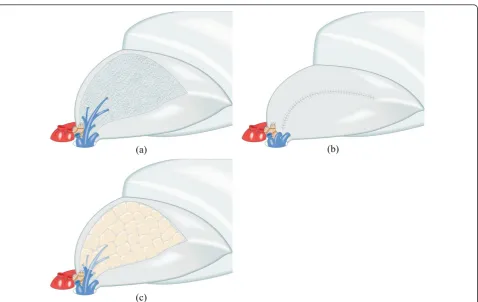
Background:To prevent postoperative air leakage after lung segmentectomy, we used two methods for the intersegmental plane: closing it by suturing the pleural edge (pleural closure), or opening it with coverage using polyglycolic acid mesh and fibrin glue (mesh-cover). The preserved forced expiratory volume in one second (FEV1) of each lobe and the postoperative air leakage were compared between the two groups.
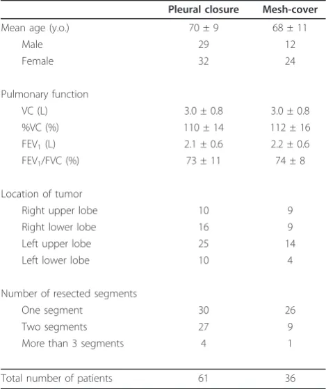
Methods:For 61 patients who underwent pleural closure and 36 patients who underwent mesh-cover, FEV1 of the lobe before and after segmentectomy was measured using lung-perfusion single-photon-emission computed tomography and CT (SPECT/CT). The groups’ results were compared, revealing differences of the preserved FEV1of the lobe for several segmentectomy procedures and postoperative duration of chest tube drainage.
Results:Although left upper division segmentectomy showed higher preserved FEV1 of the lobe in the mesh-cover group than in the pleural closure one (p = 0.06), the other segmentectomy procedures showed no
differences between the groups. The durations of postoperative chest drainage in the two groups (2.0 ± 2.5 vs. 2.3 ± 2.2 days) were not different.
Conclusions:Mesh-cover preserved the pulmonary function of remaining segments better than the pleural closure method in left upper division segmentectomy, although no superiority was found in the other segmentectomy procedures. However, the data include no results obtained using a stapler, which cuts the segment without recognizing even the intersegmental plane and the intersegmental vein. Mesh-cover prevented postoperative air leakage as well as the pleural closure method did.
Background
Advances in high-resolution CT scanning have led to frequent detection of peripheral T1N0M0 non-small cell lung cancers (NSCLCs). Although a randomized trial of lobectomy vs. limited resection for T1N0M0 NSCLC by the Lung Cancer Study Group in 1995 demonstrated that limited resection showed inferiority for prognosis and no advantage for postoperative pulmonary function compared to lobectomy [1], several studies conducted in Japan have demonstrated that segmentectomy is
superior to lobectomy for preserving pulmonary func-tion without worsening prognosis [2-7]. To preserve the pulmonary function of residual segments after segmen-tectomy, two techniques are considered important [8]: (1) sparing the intersegmental vein to preserve the venous drainage of residual segments, and (2) opening the intersegmental plane without closing it for sufficient re-expansion of the residual segments. However, open-ing the intersegmental plane causes postoperative air leakage. To prevent air leakage from the intersegmental plane, closing the pleural edge of preserved segments would be useful, but it would shrink the preserved seg-ments, resulting in insufficient re-expansion. As another method to prevent air leakage, coverage of the opened
* Correspondence: [email protected]
†Contributed equally
1
Department of Thoracic Surgery, Faculty of Life Sciences, Kumamoto University, 1-1-1 Honjo, Kumamoto 860-8556, Japan
Full list of author information is available at the end of the article
segments to prevent postoperative air leakage. During the second term, January 2008 - March 2009, we opened the intersegmental plane with coverage by a PGA mesh and fibrin glue, not only to maintain re-expansion of the preserved segments but also to prevent air leakage. To evaluate the effectiveness of using PGA mesh and fibrin glue on the intersegmental plane for preserving pulmon-ary function and for preventing air leakage, we mea-sured the preserved forced expiratory volume of lobes in one second (FEV1) using lung-perfusion
single-photon-emission computed tomography and CT (SPECT/CT) and the postoperative duration of chest tube drainage. Subsequently, we compared data obtained from patients of the two groups.
Methods
Eligibility
The Ethics Committees of Kumamoto University Hospi-tal approved the study protocol for sublobar resection in patients with c-T1N0M0 NSCLC. Informed consent was obtained from all patients after a comprehensive discus-sion of the risks and benefits of the proposed procedures.
Patients
Between April 2005 and March 2009, 198 patients with c-T1N0M0 NSCLC were treated with segmentectomy. Of the 198 patients, 166 patients underwent the conven-tional segmentectomy and 32 underwent the combined subsegmentectomy. Of the 166 patients who underwent conventional segmentectomy, 92 patients underwent both the pulmonary function test and lung-perfusion SPECT/CT before and after surgery. In addition to them, four patients with metastatic lung tumor and one with benign lung tumor were enrolled in the present study, constituting 97 patients in total.
Treatment for Intersegmental Plane
During segmentectomy, the intersegmental plane was identified using the procedure reported by Tsubota et al. as follows [12]: (1) After the segmental bronchus was iso-lated, the whole lung was temporarily inflated; (2) The segmental bronchus was first ligated to retain the air
the following two methods. (1) During the first term of April 2005 - December 2007, the intersegmental plane was closed by continuous suturing the pleural edge of preserved segments (pleural closure) (Figure 1b). (2) Dur-ing the second term of January 2008 - March 2009, the intersegmental plane was kept opened with coverage by PGA mesh and fibrin glue (mesh-cover) (Figure 1c). The pleural closure and mesh-cover groups respectively included 61 and 36 patients (Table 1).
Pulmonary Function Tests
Vital capacity (VC), forced vital capacity (FVC), and
FEV1 were measured before and more than 6 months
after surgery with a patient in a seated position using a dry rolling-seal spirometer (CHESTAC-9800DN; Chest Inc. Tokyo, Japan) according to American Thoracic Society standards [13].
Measurement of Pulmonary Function of Lobes
Lung-perfusion SPECT/CT was conducted both before and more than 6 months after surgery, at the same day with pulmonary function test. Preoperative and
post-operative FEV1 of the lobe underwent segmentectomy
was measured from pulmonary function test and lung-perfusion SPECT/CT, as previously reported [14-16]. Briefly, images of the lobe before segmentectomy and of the remained lobe after segmentectomy were traced on the CT image with a region of interest, of which the radioisotope (RI) was counted on the SPECT image (Figure 2).
The FEV1of the lobe before (A) and after (B)
segmen-tectomy was calculated from the preoperative or post-operative SPECT/CT according to the following formulae.
A = Preoperative FEV1×[RI counts of the lobe/RI counts of the whole lung] B = Postoperative FEV1×[RI counts of the lobe/RI counts of the whole lung]
The percentage of preserved FEV1of the lobe (C) was
calculated according to the following formula:
C = B/A
Resected sites compared between the two groups
The percentage of preserved FEV1 of each lobe was
groups in several resected sites of segmentectomy, i.e., resections of one segment of the right upper lobe, one segment of the left upper lobe, apical segment of the right lower lobe, apical segment of the left lower lobe, and the left upper division.
Statistical Analysis
Student’st-test was used to compare the preoperative VC, %VC, FEV1, FEV1/FVC, preserved FEV1of the lobe,
percentage of preserved FEV1 of the lobe and the
post-operative duration of chest tube drainage between the pleural closure and mesh-cover groups. Differences in mean percentage of preserved FEV1of each lobe in each
resected sites were analyzed by using multivariate analy-sis. Software (SPSS; SPSS Inc., Chicago, Illinois) was used for these analyses. Values ofp< 0.05 were inferred as significant. All values in the text and table are given as mean ± SD.
Results
No difference in preoperative pulmonary function was found between the pleural closure and mesh-cover groups, as shown in Table 1. In the pleural closure
group, the respective mean values of FEV1 before and
after surgery were 2.1 ± 0.6 and 1.9 ± 0.5 l, of which the
mean percentage of postoperative FEV1 was 89 ± 9%. In
the mesh-cover group, the respective mean values of FEV1before and after surgery were 2.2 ± 0.6 and 2.0 ±
0.6 l, of which the mean percentage of postoperative
FEV1was 92 ± 8%. The mean percentage of
postopera-tive FEV1 in the mesh-cover group was higher than that
in the pleural closure group, with marginal significance (p= 0.09).
In the pleural closure group, the preoperative and
postoperative FEV1 of each lobe that had undergone
segmentectomy were 0.51 ± 0.20 and 0.22 ± 0.15 l, respectively, of which the mean percentage of preserved
FEV1 of the lobe was 40 ± 20%. In the mesh-cover
group, the preoperative and postoperative values were 0.52 ± 0.20 and 0.23 ± 0.12 l, respectively, of which the
mean percentage of preserved FEV1 of the lobe was 46
± 24%. The mean percentage of postoperative FEV1 of
the lobe was not different between the two groups. Table 2 presents the mean percentages of preserved
FEV1 of the lobe in each resected site of the two
groups. The pleural closure group showed a lower
per-centage of preserved FEV1 than the mesh-cover group
for left upper division segmentectomy, with marginal
(a) (b)
(c)
significance (21 ± 10 vs. 35 ± 15%,p= 0.06). However, no significant difference in the values was found between the two groups at any other resected site, i.e., resections of one segment of the right or left upper lobe, or of an apical segment of the right or left lower lobe. The FEV1 values of the lobe before and after the
upper division segmentectomy in the pleural closure group were 0.59 ± 0.21 and 0.13 ± 0.10 l, respectively, whereas the values in the mesh-cover group were, respectively, 0.47 ± 0.18 and 0.17 ± 0.10 l. Multivariate
analysis of the mean percentages of preserved FEV1 of
the lobe in each resected site of the two groups also showed no significant difference (p= 0.38).
No significant difference was found in the respective durations of chest drainage, which were 2.0 ± 2.5 and 2.3 ± 2.2 days in the pleural closure and mesh-cover groups.
Discussion
The results of this study elucidated the following points. (1) Mesh-cover is useful to preserve the pulmonary function of the residual lingular segment after the left upper division segmentectomy, although no difference was found between the mesh-cover and pleural closure methods at other resected sites. (2) Covering the inter-segmental plane with PGA mesh and fibrin glue can
ment has only four. This study showed that pleural clo-sure in the upper division segmentectomy was
associated with lower FEV1 of the remaining lingular
segment more than the mesh-cover method, although no difference between the two methods was found at other segmentectomy sites. The following reasons might explain this outcome. (1) The remaining left lingular segment after left upper division segmentectomy has lit-tle lung volume, similar to the corresponding right mid-dle lobe. (2) The functional volume of the lingular segment is likely to be decreased after left upper division segmentectomy because of the excessive upward bend-ing and rotation of the lbend-ingular bronchus, similar to the occurrence of right middle lobe syndrome after right upper lobectomy [17]. (3) For these two reasons, pleural closure of the remained lingular segment shrink it and further decrease of the pulmonary function of the criti-cally preserved lingular segment. The left upper division segmentectomy is a popular procedure for segmentect-omy. Therefore, we must keep in mind that pleural clo-sure in the left upper division segmentectomy preserves little pulmonary function of the remaining lingular ment. Furthermore, because the left upper division seg-mentectomy decreases the postoperative pulmonary function to a greater degree than segmentectomy of other kinds [12], left upper division segmentectomy should be examined separately in a controlled study of postoperative pulmonary function between the lobect-omy and segmentectlobect-omy.
Recent development of stapling devices has added a new dimension to the technique for dissecting interseg-mental plane. However, the present data include none related to closure of the intersegmental plane using a stapler. Although the pleural closure method in this study cut the lung tissue along the inflated-deflated line and spared intersegmental veins, the stapling method do not only cut the lung tissue without recognizing the intersegmental plane but also injure the intersegmental veins, which are instrumental for venous return of the residual segments. Therefore, segmental resection using a stapler will further decrease the pulmonary function of the remaining lobe, even compared to the pleural clo-sure method described in this report. The use of staple
FEV1/FVC (%) 73 ± 11 74 ± 8
Location of tumor
Right upper lobe 10 9 Right lower lobe 16 9 Left upper lobe 25 14 Left lower lobe 10 4 Number of resected segments
One segment 30 26 Two segments 27 9 More than 3 segments 4 1 Total number of patients 61 36
VC: vital capacity, FVC: forced vital capacity, FEV1: forced expiratory volume in
devices in the dissection of intersegmental plane for pre-serving pulmonary function should be further evaluated in a separate study.
Results reported herein demonstrate that pleural clo-sure does not decrease pulmonary function of the
pre-served segments compared to the mesh-cover
procedure, except for left upper division segmentectomy. For left upper division segmentectomy, the intersegmen-tal plane should be opened to preserve the pulmonary function of the residual lingular segment. Furthermore, results showed that coverage of the opened interseg-mental plane using the PGA mesh and fibrin glue can prevent postoperative air leakage with the same degree of beneficial effect as pleural closure.
Abbreviations
NSCLCs: non-small cell lung cancers; PGA: polyglycolic acid; FEV1: forced
expiratory volume in 1 second; SPECT/CT: lung-perfusion
single-photon-(a)
(b)
(c)
Figure 2Images of before and after segmentectomy. (a) Axial image of CT before surgery, showing lung cancer in posterior apical segment of the left upper lobe. (b) Sagittal image of the lung-perfusion single-photon-emission computed tomography and CT (SPECT/CT) of the left upper lobe before operation. (c) Sagittal image of the lung-perfusion SPECT/CT of the remaining lingular segment after resection of upper division segmentectomy.
Table 2 Mean percentage of preserved FEV1of each lobe
in each resected sites
Percentage of FEV1of each lobe (%)
Resected site Pleural closure Mesh-cover Difference
One segment of right upper lobe
38 ± 18 (n = 9) 35 ± 27 (n = 6) p = 0.77
One segment of left upper lobe
46 ± 13 (n = 9) 52 ± 15 (n = 8) p = 0.37
Apical segment of right lower lobe
59 ± 13 (n = 5) 63 ± 8 (n = 4) p = 0.62
Apical segment of left lower lobe
46 ± 11 (n = 3) 44 ± 8 (n = 3) p = 0.81
Upper division of left upper lobe
21 ± 10 (n = 10) 35 ± 15 (n = 4) p = 0.06
coordination. All authors have read and approved the final manuscript.
Competing interests
The authors declare that they have no competing interests.
Received: 5 January 2011 Accepted: 25 April 2011 Published: 25 April 2011
References
1. Lung Cancer Study Group, Ginsberg RH, Rubinstein LV:Randomized trial of lobectomy versus limited resection for T1N0 non-small cell lung cancer. Ann Thorac Surg1995,60:615-23.
2. Okada M, Koike T, Higashiyama M, Yamato Y, Kodama K, Tsubota N:Radical sublobar resection for small-sized non-small cell lung cancer: a multicenter study.J Thorac Cardiovasc Surg2006,132:769-75. 3. Yoshikawa K, Tsubota N, Kodama K, Ayabe H, Taki T, Mori T:Prospective
study of extended segmentectomy for small lung tumors: the final report.Ann Thorac Surg2002,73:1055-9.
4. Kodama K, Doi O, Higashiyama M, Yokouchi H:Intentional limited resection for selected patients with T1 N0 M0 non-small-cell lung cancer: a single-institution study.J Thorac Cardiovasc Surg1997,
114:347-53.
5. Sienel W, Dango S, Kirschbaum A, Cucuruz B, Horth W, Stremmel C, Passlick B:Sublobar resections in stage IA non-small cell lung cancer: segmentectomies result in significantly better cancer-related survival than wedge resections.Eur J Cardiothorac Surg2008,33:728-34. 6. Koike T, Yamato Y, Yoshiya K, Shimoyama T, Suzuki R:Intentional limited
pulmonary resection for peripheral T1 N0 M0 small-sized lung cancer.J Thorac Cardiovasc Surg2003,125:924-8.
7. Harada H, Okada M, Sakamoto T, Matsuoka H, Tsubota N:Functional advantage after radical segmentectomy versus lobectomy for lung cancer.Ann Thorac Surg2005,80:2041-5.
8. Fell SC:Segmental resection.Chest Surg Clin N Am1995,5:205-21. 9. Matsumura Y, Okada Y, Shimada K, Endo C, Chida M, Sakurada A, Sato M,
Kondo T:New surgical technique of pulmonary segmentectomy by ultrasonic scalpel and absorbable sealing materials.Kyobu Geka2004,
57:31-7.
10. Nomori H, Ikeda K, Mori T, Kobayashi H, Iwatani K, Kawanaka K, Shiraishi S, Kobayashi T:Sentinel node navigation segmentectomy for clinical stage IA non-small cell lung cancer.J Thorac Cardiovasc Surg2007,133:780-5. 11. Nomori H, Ohba Y, Shibata H, Shiraishi K, Mori T, Shiraishi S:Required area
of lymph node sampling during segmentectomy for clinical stage IA non-small cell lung cancer.J Thorac Cardiovasc Surg2010,139:38-42. 12. Tsubota N:An improved method for distinguishing the intersegmental
plane of the lung.Surg Today2000,30:963-4.
13. American Thoracic Society:Standardization of spirometry - 1987 update. Am Rev Respir Dis1987,136:1285-98.
14. Yoshimoto K, Nomori H, Mori T, Kobayashi H, Ohba Y, Shibata H, Tashiro K, Shiraishi S, Kobayashi T:Quantification of the impact of segmentectomy on pulmonary function by perfusion single-photon-emission computed tomography and multidetector computed tomography.J Thorac Cardiovasc Surg2009,137:1200-5.
15. Yoshimoto K, Nomori H, Mori T, Ohba Y, Shiraishi K, Tashiro K, Shiraishi S:
Postoperative change in pulmonary function of the ipsilateral preserved lung after segmentectomy compared with that after lobectomy.Eur J Cardiothorac Surg2010,37:36-9.
Submit your next manuscript to BioMed Central and take full advantage of:
• Convenient online submission
• Thorough peer review
• No space constraints or color figure charges
• Immediate publication on acceptance
• Inclusion in PubMed, CAS, Scopus and Google Scholar
• Research which is freely available for redistribution