generally has negative effects on the host genome. To counteract this threat, host cells have evolved genetic and epigenetic mechanisms that keep TEs silenced. One such mechanism involves the Piwi-piRNA complex, which represses TEs in animal gonads either by cleaving TE transcripts in the cytoplasm or by directing specific chromatin modifications at TE loci in the nucleus. Most Piwi-interacting RNAs (piRNAs) are derived from genomic piRNA clusters. There has been remarkable progress in our understanding of the mechanisms underlying piRNA biogenesis. However, little is known about how a specific locus in the genome is converted into a piRNA-producing site. In this review, we will discuss a possible link between chromatin boundaries and piRNA cluster formation.
Keywords:Transposable elements, Piwi, piRNA, piRNA cluster, Chromatin boundary
Review
Background
Large fractions of eukaryotic genomes comprise trans-posable elements (TEs), which are repetitive DNA ele-ments that can mobilize to take up new chromosomal locations within a genome. TEs act as insertional muta-gens that can alter gene expression or rearrange chro-mosomes. Therefore, they can cause disease and may even drive evolution [1-4]. TEs are diverse in sequence and in the way they transpose [5,6]. They possess a lim-ited gene set of their own, but use the gene expression machinery of their host to thrive in the genome. DNA transposons move by a “cut-and-paste” mechanism, in which they are excised from one genomic site and inserted into a new location using their own transposase. Therefore, generally, the copy number of DNA transpo-sons in a genome does not expand. By contrast, retrotran-sposons use a “copy-and-paste” mechanism to propagate their copies through RNA intermediates. Retrotranspo-sons are transcribed from the genome, reverse transcribed and integrated into a new location, in a process mediated by a transposon-encoded reverse transcriptase. Retrotran-sposons are distinguished by their DNA sequence top-ology and mechanism of transposition: those that possess long terminal repeats (LTRs), such asgypsy, and those that
do not (non-LTRs), such as long interspersed repetitive el-ements (LINEs) and short interspersed repetitive elel-ements (SINEs). Both DNA transposons and retrotransposons have non-autonomous subtypes and defective copies, which require the reverse transcriptase and endonucle-ase supplied by the autonomous type to jump around the genome.
As an example,Drosophilaharbors around 100 differ-ent TEs, and the only conserved and universal property shared by them is the ability of transposition [7]. Thus, the requirements for host cells for repression of TEs are at least two-fold: 1) a mechanism that recognizes such a diverse set of TE types, and 2) a mechanism that distin-guishes them from other cellular genes and selectively targets them for silencing. Recent studies have postu-lated that host cells have evolved an elaborate silencing mechanism to meet these two requirements. Host cells may have taken advantage of the only universal property of TEs, their transposition ability to trap them in specific genomic locations and subject them to a silencing pro-gram, which employs small RNA-based immunity to se-lectively silence homologous elements [8-10]. In animal gonads, small non-coding RNAs (ncRNAs), termed Piwi-interacting RNAs (piRNAs), mediate TE silencing to en-sure genome integrity during germ cell development [11,12]. Most piRNAs are derived from particular genomic sites termed piRNA clusters, which contain a large num-ber and various types of TEs. Thus, the sequences of piR-NAs derived from these clusters are homologous not only * Correspondence:[email protected]
1
Department of Molecular Biology, Keio University School of Medicine, 35 Shinanomachi Shinjuku-ku, Tokyo 160-8582, Japan
Full list of author information is available at the end of the article
to TEs in the clusters, but also to related TEs located else-where in the genome and can therefore act as guide mole-cules to repress TEs in trans. Thus, piRNA clusters are genetic elements that regulate the activity of TEs. How-ever, relatively little is known about how piRNA clusters are formed. In this review, we emphasize the role of chro-matin boundaries in piRNA cluster formation. To this end, we briefly review our current knowledge of piRNAs and piRNA clusters. We then discuss a possible link be-tween chromatin boundaries and piRNA clusters, and propose some models as to how piRNA clusters are formed in chromatin boundaries.
TE silencing mediated by piRNAs
RNA interference (RNAi) and related pathways are cellu-lar pathways in which small ncRNAs of 20 to 35 nucleo-tides (nt) guide Argonaute-containing effector complexes, termed RNA-induced silencing complexes (RISCs), to RNA targets by means of base-pairing, and promote the inactivation of homologous sequences [13-16]. They have been shown to suppress the activity of TEs in plants and animals. In animal germline cells, piRNAs of 24 to 35 nt are produced and loaded onto germline-specific Argo-naute proteins (termed PIWI proteins) to form piRNA-induced silencing complexes (piRISCs). Genetic analyses ofDrosophila PIWIgenes (ago3,aubergine/aub, andpiwi) have revealed that mutations in these genes affect germ-line development [17-20]. TEs are deregulated in mutant ovaries defective in these genes, suggesting a model in which TE overexpression and mobilization triggers DNA damage signaling-dependent defects in an early step in the germ cell patterning cascade [21].
Unlike other small silencing RNAs such as microRNAs (miRNAs) and small interfering RNAs (siRNAs), piRNAs in most animals are processed in a Dicer-independent manner from single-stranded precursors, which are tran-scribed mostly from genomic piRNA clusters [22,23]. A large number of genes have been identified to function in piRNA biogenesis [24]. In the Drosophila genome,
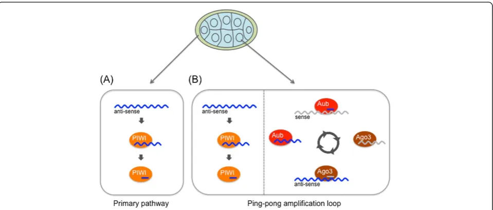
142 regions have been identified as piRNA clusters [22]. Although these sites account for less than 5% of the as-sembled genome, over 90% of all sequenced piRNAs can be derived from these regions [25]. The piRNA clusters cover chromosomal regions of several to hundreds of ki-lobases, and they contain TEs that are mostly inactive copies or truncated fragments, arranged in a nested fashion [22]. Among all the piRNA clusters in Drosoph-ila, theflamencolocus produces a major fraction of piR-NAs in somatic support cells in the ovary [25]. This locus was originally discovered as a regulator of the ac-tivity of thegypsy,idefix, andZAMTEs [26,27]. piRNAs from this cluster, which spans about 150 kb, are derived from one DNA strand only, most likely through unidir-ectional transcription oriented in the anti-sense direc-tion to most TEs in the locus (Figure 1). This provides a molecular basis of why Piwi, the only PIWI protein expressed in ovarian somatic cells, loads with piRNAs that are anti-sense-oriented relative to TEs. Mutants of flamencoin which theP-elementis inserted in the 5′ re-gion and those lacking flamenco partial genomic se-quence lose the ability to regulate TEs [22,26,28,29]. These data indicate that the single long transcripts from theflamencolocus are processed into piRNAs. This lin-ear biogenesis of piRNAs from precursor transcripts has been called the ‘primary piRNA processing path-way’ (Figure 2). piRNA maturation and Piwi-piRNA complex (Piwi-piRISC) formation occur in the cyto-plasm [30]. Piwi-piRISCs are then imported into the nucleus where they repress TEs in trans at transcrip-tional level by directing specific histone modifications to TE loci [31-34]. This suggests that Piwi-piRISCs re-cruit the relevant enzymes to modify histones at TE loci. Because depletion ofpiwiactivity rapidly results in derepression of TEs, the TE silencing state requires the continual activities of Piwi-piRISCs [30,35,36]. There-fore, Piwi-piRISCs are genetic elements that mediate and maintain epigenetic chromatin modifications of target TE loci.
Compared with the situation in somatic support cells, the piRNA biogenesis in germline cells in the fly ovary is more complex. In contrast to the unidirectional fla-mencopiRNA cluster, many piRNA clusters in the Dros-ophila germline are transcribed from both strands, and both precursor transcripts are processed into piRNAs [22,25]. Therefore, both sense and anti-sense piRNAs relative to the TE sequences are produced from the clus-ters. All three PIWI proteins are expressed in the germ-line, but Piwi is nuclear, and both Aub and Ago3 are cytoplasmic [22,37,38]. Anti-sense precursor transcripts from dual-stranded piRNA clusters are processed into anti-sense piRNAs that are loaded onto Aub and Piwi (“primary piRNA processing pathway”). Piwi-piRISCs then move into the nucleus where they repress TEs, probably by a mechanism similar to that observed in somatic support cells. Aub-piRISCs, by contrast, remain in the cytoplasm and cleave both sense precursor scripts from dual-stranded piRNA clusters and tran-scripts from active TEs, using the small RNA-directed endonuclease or Slicer activity exhibited by PIWI pro-teins [37]. This cleavage results in the production of sense piRNAs, which in turn are loaded onto Ago3. This process initiates a feed-forward amplification loop of piRNA production, the so-called “ping-pong cycle”, in which sense and anti-sense transcripts of dual-stranded piRNA clusters and active TEs are reciprocally cleaved by the Slicer activity of Ago3 and Aub [22,37] (Figure 2). Both Ago3-piRISCs and Aub-piRISCs act catalytically,
and thus the cycle leads to repeated rounds of piRNA production by consuming both cluster transcripts and TE transcripts, thereby silencing TEs at posttranscrip-tional levels in the cytoplasm.
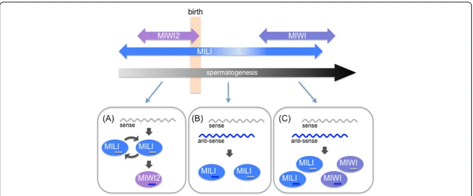
studies have shown that the Slicer activity of MILI is re-quired for the secondary piRNA production, which ampli-fies MILI-bound piRNAs through an intra-MILI ping-pong loop, and generates all MIWI2-bound secondary piRNAs [45] (Figure 3). In contrast to the cytoplasmic localization of MILI and MIWI, MIWI2-piRISCs are imported into the nucleus where they direct specific DNA methylation of TE loci, thereby establishing TE silencing at the transcriptional level [39,45,47]. However, the Slicer activity of both MIWI and MILI is still required to main-tain TE silencing in the mouse testis after birth, suggesting that continuous cleavage of TE transcripts by the Slicer activity is essential to repress TEs in mouse testes [44,45].
piRNA clusters in diverse organisms
TE insertions inDrosophilaare mostly located in hetero-chromatin and proximal heterohetero-chromatin-euhetero-chromatin boundary zones [22]. Of 142 piRNA clusters identified in Drosophila, only 7 are in presumed euchromatic re-gions, while the rest reside within cytologically defined pericentromeric and telomeric heterochromatin regions. Within these heterochromatin regions, the piRNA clus-ters tend to be located near the boundary region between heterochromatin and euchromatin. Heterochromatin re-gions in theDrosophilagenome can be found at the peri-centromeric and subtelomeric regions, and are megabases in size [48-50]. Their constituent sequences fall into roughly three categories: tandemly repeated short se-quences (satellite DNAs), moderately repetitive elements
(such as TEs), and some single-copy genes [48-50]. In the Drosophilagenome, intact and potentially active TEs pre-vail across the genome, while fragmented or inactive copies of TEs are strongly enriched in the transition zones between heterochromatin and euchromatin near to the centromere, and constitute piRNA clusters [22,50] (Figure 4).
Because most piRNAs are derived from piRNA clus-ters that genetically control the activity of TEs and largely comprise various types of defective TEs, a model in which piRNA clusters act as“TE traps”has been pro-posed [8,51-53]. This model relies on the transposition ability of TEs for piRNA clusters to passively acquire new content by chance transposition. TEs that happen to jump into piRNA clusters can then become fixed by evolutionary selection, and produce corresponding piR-NAs and regulate other homologous elements expressed from different genomic positions in germ cells.
As mentioned above, two types of piRNA clusters exist in the Drosophila gonads: unidirectional clusters and dual-stranded clusters. Most piRNA clusters in somatic support cells are unidirectional, while the predominant fraction of germline piRNA clusters is dual-stranded [22,25] (Figure 5).
An example of a unidirectional piRNA cluster is the flamenco locus, which is located near the pericentro-meric heterochromatin boundary of the X chromosome, and contains a large number of truncated or inactivated TEs. Most of these TEs belong to the gypsy family and Figure 3The Piwi-interacting RNA (piRNA) biogenesis pathway in mouse testis.The piRNA biogenesis pathway in mouse can be
are anti-sense-oriented with regard to the polarity of transcription. This requires the transcription factor Cu-bitus interruptus, a segment polarity gene that controls a number of genes, including Hedgehog genes [22,54]. The molecular mechanism that restricts the directional-ity of transposition into a unistrand piRNA cluster is not well understood.
A representative dual-stranded cluster is the 42AB cluster, which spans around 240 kb, near the pericentro-meric heterochromatin boundary. However, the orienta-tion of truncated TEs in this cluster is random rather than uniform, and piRNAs are produced from both sense and anti-sense strands.
Although many factors that are required for piRNA production are shared between these two types of clus-ters, there are some differences between them. Rhino (a variant of heterochromatin protein 1; HP1), Cutoff (a homolog of the yeast decapping nuclease and transcrip-tion terminatranscrip-tion factor Rai1), and Deadlock (which acts as a linker between Rhino and Cutoff ), are all required for piRNA production only in germline cells of the oo-cyte [22,55-57]. Interestingly, most piRNA clusters in Drosophila are within cytologically defined heterochro-matic regions. A recent chromatin immunoprecipitation (ChIP)-sequencing analysis of H3K9me3, the most estab-lished marker for heterochromatic regions, revealed that the promoter and its surrounding region of flamenco, a unistrand piRNA cluster, is fairly devoid of this repressive
histone mark, which may explain the active transcription of the locus by RNA polymerase II [34]. By contrast, the germline cell-specific dual-strand piRNA clusters, such as 42AB, are coated with H3K9me3, but are still transcrip-tionally active [55] (see also below).
In the Bombyx moritissue cultured cell line BmN4, a portion of piRNAs are derived from TEs [58]. piRNA clusters in BmN4 cells have been shown to have a high level of H3K4me3 mark, which is a hallmark of active transcription [59], suggesting the open nature of silk-worm piRNA clusters.
These findings suggest that piRNA clusters are highly transcribed units within heterochromatic regions, and raise the question of how this kind of special location in the genome has been selected for piRNA clusters to pro-duce piRNAs.
In the mouse, over 90% of piRNA reads have been mapped to roughly 100 genomic regions, ranging from a few kb to over 100 kb in length. Most mouse clusters show profound strand asymmetry, with reads arising from only one strand within a cluster (unidirectional cluster). When piRNAs map to both strands within one piRNA cluster, the transcription units are arranged in a divergent manner (bidirectional cluster) [42,43] and the piRNA-producing region on one strand does not overlap with that on the other strand. In prenatal mouse testes, piRNAs are produced from both strands in the same region (dual-strand cluster) [39] (Figure 5). Recent comprehensive
Figure 5Three types of Piwi-interacting RNA (piRNA) cluster. (A)Unistrand piRNA cluster; piRNAs are produced from only one genomic DNA strand.(B)Dual-strand piRNA cluster; piRNAs are produced from both strands of the same genomic region.(C)Bidirectional piRNA cluster; two unistrand piRNA clusters are arranged in a divergent manner.
deep sequencing analysis of postnatal mouse testes reveals that the transcription factor A-MYB drives pachytene piRNA production, suggesting a model in which a specific transcription factor engages in transcription of most piRNA clusters [60,61]. It should be noted that A-MYB is not spe-cific for piRNA clusters, but rather has a number of target genes, suggesting that A-MYB has been co-opted to drive transcription of piRNA clusters. This also raises the question of what might be the difference between the A-MYB bind-ing sites that direct piRNA production and the A-MYB binding sites that produce mRNAs but not piRNAs. piRNA clusters have been identified in other mammals including primates [62]. Synteny analysis has revealed conservation in the genomic location of piRNA clusters among mammals, although the precise sequence of each piRNA shows no ap-parent similarity [42,43,62]. This indicates that the relative chromosomal position has some marked features with re-gard to production of piRNAs, and such special features are maintained across mammals.
Caenorhabditis elegans has two PIWI proteins, PRG-1 and PRG-2. PRG-1 is required in germline maintenance, and interacts with a class of small RNAs, called 21U-RNAs [63,64]. By definition, 21U-21U-RNAs are the pi21U-RNAs ofC. elegans. As their name implies, they are character-ized by a first U bias, and their length is exclusively 21 nt, which is shorter than that of piRNA species in other organisms [65]. The vast majority of the 21U-RNAs are derived from thousands of individual loci broadly scattered in two large clusters on chromosome IV [65]. These regions are gene-poor compared with other regions of the genome. A marked feature of 21U-RNAs is the existence of a clearcismotif located around 40 bp upstream of the 21U-RNA encoding site [65]. The consensus motif is CTGTTTCA and is flanked by an AT-rich sequence, which is specifically recognized by Forkhead family transcription factors [65,66]. In addition, ChIP-on-chip experiments have shown a low level of histone H3 across the two piRNA clusters, which corre-lates well with DNase-sensitive sites [66,67]. Moreover, it was also revealed that each upstream consensus motif corresponds with the nucleosome-depleted region (NDR) [66]. These findings strongly suggest that each piRNA in C. elegans is produced from an independent trans-cription unit.
Tetrahymena thermophila has a unique genome pro-cessing mechanism, called ‘programmed DNA elimin-ation’. Most ciliated protozoans, includingT. thermophila, exhibit nuclear dimorphism, with a germline micronu-cleus (Mic) and somatic macronumicronu-cleus (Mac) [68]. The genomic sequence of this organism is processed during the course of meiosis. Mic has an unprocessed genome, and Mac has a processed one, but has a much larger gen-ome size due to polyploidy. In contrast to the role of Mic as a reservoir of genetic information, gene expression for
maintaining the organism takes place in Mac. The smaller genome size of Mac compared with Mic is attributable to DNA elimination induced by scan RNA (scnRNA). In-ternal eliminated sequences (IESs) are specific regions that are selectively eliminated from the developing Mac gen-ome, and there are over 6,000 IESs within the Mic genome. scnRNA are loaded onto Twi, one of the Tetrahymena PIWI proteins and are, therefore,T. thermophila piRNAs [69]. Twi1-scnRNA complexes are then transported to the developing Mac, which has an unprocessed genome, and they recognize and eliminate IESs through base-pairing be-tween IESs and scnRNAs [70]. Strikingly, scnRNA produc-tion requires a Dicer-like protein, which is in clear contrast to piRNA production in other animals [71]. scnRNAs map predominantly to IESs, therefore, it can be said that IESs are piRNA clusters in Tetrahymena [72]. Recent high throughput analysis has uncovered biased transcription of IESs in Mic; that is, IESs are destined to have high tran-scription activity [72]. Because of the lack of clear consen-sus sequence between different IESs, IESs are thought to be epigenetically marked as piRNA clusters.
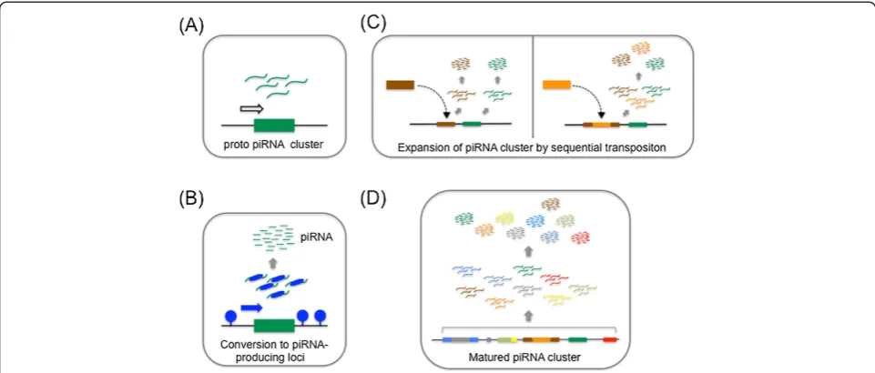
These findings in various animals suggest possible re-quirements to establish piRNA clusters, which are as follows (in random order): 1) an ability to recruit chromatin-modifying enzymes that contribute to the main-tenance of open chromatin so as to attract and trap TEs, 2) an ability to recruit DNA specific factors (for example, spe-cific transcriptional factors) to drive transcription of that region, and 3) an ability to distinguish transcripts from that region from other cellular transcripts and to specifically process them into small RNAs (Figure 6B).
Transposition and chromatin boundaries
A prerequisite for genomic regions to act as TE traps is that they must be frequent and non-deleterious sites for TE insertion. TEs jump around the genome by transpos-ition, but this appears to occur in a non-random manner [73]. The P-elementis a DNA transposon that has been used for insertional mutagenesis to isolate specific alleles inDrosophila[74,75]. Because of this, a large body of data has accumulated concerning the preferential P-element insertion sites in the genome. Analysis of 100,000 transposition events identified thatP-elementinsertion preferentially occurs immediately 5′to genes or within 5′ exons [76].piggyBac, another TE that is also often used for mutagenesis, also shows a high preference of insertion at or near promoter regions of genes [77]. These results indicate that these TEs tend to target genomic regions that presumably contain open chro-matin and/or are actively transcribed at the time of transposition.
regions known to have relatively open chromatin [78,79]. These studies clearly argue for the relationships between open chromatin and preferential transposition sites. How-ever, it should be noted that these TE insertions at or near promoters alter the transcriptional activity of genes and are, therefore, often highly deleterious to the host. Thus, individual genomes with these insertions tend to be elimi-nated from the population. So are there any genomic re-gions where TE insertions are tolerated?
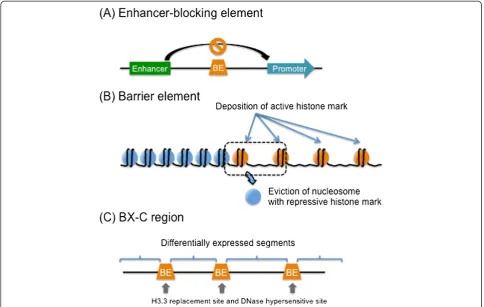
In addition to gene promoters and their neighboring re-gions, chromatin boundaries are also known to have rela-tively open chromatin structures. A chromatin boundary can act as a buffer between two functional chromatin do-mains by resisting the proliferation of epigenetic changes that are characteristic of each, thus genes present in one domain are not affected by regulatory sequences present in a different domain [80-84] (Figure 7).Cis-regulatory el-ements are located at chromatin boundaries, and have dif-ferent compositions of trans-acting proteins. They limit the spreading of heterochromatin domains into regions of actively transcribed genes (andvice versa) and prevent ad-ventitious interactions between enhancers and promoters when located between them (acting as “insulators”) [83,84] (Figure 7A). However, chromatin boundaries, especially those in Drosophila, between constitutive heterochromatin and euchromatin are not fixed but stochastic, as evident in position effect variegation (PEV), in which the heritable inactivating influence of the heterochromatin on a neighboring gene can spread in some, but not all, cells of the same cell type [85].
In fission yeast, tRNA gene clusters near to the site of constitutive heterochromatin, such as those around centromere, serve as strong boundary elements that in-hibit the encroachment of heterochromatin into the eu-chromatic region [86,87] (Figure 7B). One explanation of this phenomenon is that the high transcriptional activity from tRNA genes creates a discontinuity in arrayed nu-cleosomes that serves as a barrier to the propagation of heterochromatin [88,89]. This high transcriptional activ-ity might also function by promoting the activactiv-ity of histone-modifying enzymes that contribute to the main-tenance of open chromatin conformation [90]. A num-ber of chromatin boundaries are associated with active promoters. In addition, the recruitment of histone ace-tyltransferase activity correlates well with barrier activity in multiple organisms [82]. These results suggest the possibility that some promoters or transcription units with specific characteristics may determine their own chromosomal environment to ensure their activity, thereby allowing them to effectively resist and even counteract het-erochromatin formation, probably by manipulating histone modifications.
enhancers, and chromatin boundaries [17,92-95]. These H2A.Z–rich regions are common NDRs, and are there-fore DNase-hypersensitive. H2A.Z, together with H3.3, a histone H3 variant, forms histone octamers, which con-stitute the most labile nucleosome state in human cells. This leads to the dissociation of nucleosomes from chro-matin, thereby forming NDRs [93,96]. Mapping the pref-erential H3.3 deposition sites in Drosophila S2 cells revealed that there are specific sites at which H3.3 is heav-ily deposited [97,98]. The bithorax complex (BX-C) regu-lates the identity of each of the segments that contributes to the posterior two-thirds of the fly [99]. The region en-codes three genes, Ultrabithorax (Ubx), abdominal A (abd-A), and Abdominal B (Abd-B). It has been shown that nine body segments are defined by the combination of expression level of the three genes. Boundary elements demarcate the BX-C region into nine parts, making pos-sible the differential expression pattern of the three genes. Interestingly, the preferential deposition sites of H3.3 match well with the BX-C boundary elements, such as Fab-7, Fab-8, and Mcp [98]. Moreover, those sites are in-dependently identified as DNase-hypersensitive sites [100] (Figure 7C). Therefore, both H2A.Z and H3.3 serve as
molecular indicators of open chromatin conformation. Interestingly, both H2A.Z and H3.3 have been recovered from genome-wide RNAi screening to identify factors re-quired for transposon silencing in Drosophila[35]. Thus, it is tempting to speculate that both histone variants are involved in piRNA production, possibly through maintain-ing the boundary nature of piRNA clusters (see below).
Of note, certain types of TEs themselves also show high rates of H3.3 deposition [97], implying that a TE it-self can be a good recipient of a transposon. In addition, it is known that transposition of retrotransposons tends to occur within even older retrotransposons. For example, nearly all retrotransposon insertions in the Arabidopsis genome are into older retrotransposons [101,102]. The recent ENCODE project has also revealed that DNase I hypersensitive sites are strongly enriched at specific LTR retrotransposons in the human genome in some cultured cells, suggesting the possibility that TEs can transpose into certain types of TE [95]. This would explain the reason why TEs in piRNA clusters, such asflamenco, tend to be arranged in a nested fashion.
blocking insulator, a type of boundary element, when inserted between promoter and enhancer [104]. There-fore, this gypsy insulator locus could be a prototype for TE transposition landing pads. The aforementioned find-ings in Drosophila, mouse and other animals also imply that special chromatin status with accompanying tran-scriptional factors and/or epigenetic factors at the chro-matin boundary can give transcriptional license to that region [22,61,66,72]. There is increasing evidence that TEs often carry with them an array of transcription fac-tor binding sites that, when integrated into the genome, can become either alternative promoters or new en-hancers [105]. Thus, transposition to a chromatin bound-ary of a TE that has a specific transcription factor binding site, the transcription factor for which is already expressed in gonads, may make that region transcriptionally active and put it under the control of the transcription factor. In this way, boundary-specific elements may drive transcrip-tion of that boundary region to produce transcripts in go-nads. A study describing the relationships between TE insertion and de novopiRNA production shows that not all TE insertions drivede novo piRNA production [106]. The transcriptional status at the insertion site might affect whether the TE transcript is processed into piRNA [106]. This is consistent with the view we have discussed. The chromatin boundaries are gene-poor regions, and there-fore TE transposition at those regions is likely to be neutral to the host, thereby allowing not only TE accu-mulation at those regions, but accuaccu-mulation of nucleotide changes in those accumulated TEs. Repeated transposition events at the same boundary region would expand the size of clusters. Thus, it is possible that special transcriptional units in the boundary regions are primitive piRNA pro-duction sites.
What makes the piRNA cluster so special?
When thinking about the process by which piRNA clus-ters are formed, the biggest outstanding question is how does a specific locus turn into a piRNA-producing site? In other words, what is the prerequisite for certain loci to produce piRNAs? We propose two scenarios based on the data described so far.
Another model is that transcripts from piRNA clusters have some special property allowing them to be proc-essed into piRNA, and this property is used by the piRNA-producing machinery to distinguish piRNA tran-scripts from the vast majority of other trantran-scripts. This special property can be either altered splicing, character-istic 3′-end processing, or specific ciselements that dir-ect recognition by specialtransfactors. Recently, Madhani and colleagues showed that stalled spliceosomes are a sig-nal for an RNAi response in a human pathogenic yeast, Cryptococcus neoformans [108]. These authors proposed that splicing intermediates are a preferred substrate for small interfering RNA biogenesis. This work explains how specific transcripts are differentially recognized by the small RNA biogenesis machinery. It was recently reported that Rhino can suppress normal splicing in theDrosophila germ line with the aid of Uap56, making the piRNA pre-cursor transcript different from other pol II transcripts [55,107,109]. However, inDrosophilafollicle cells, splicing of a long single-stranded transcript (more than 150 kb) produced from theflamlocus was reported [54]. Further-more, the first intron of flam was found to be constitu-tively spliced [54]. In addition, there are numerous 3′-end processing signals of TEs located in theflamlocus. There-fore, there could be a certain mechanism that suppresses transcription termination and poly(A) addition for the flamtranscripts. Therefore, the transcript itself is sending some message that it is different from other transcript.
Conclusions
the transcript from piRNA cluster. Taking advantage of specific DNA-protein interactions, such as LexA with LexA-binding sites, LacI with LacO repeats and modified transcription activator-like effector (TALE), recent studies have successfully immunopurified a chromatin locus of interest and identified associated proteins [110-113]. A combination of RNA-binding proteins and their specific binding sites, such as MS2 and BoxB sites, can be applied to identify the proteins that bind to piRNA transcripts. These types of strategy will allow us to identify the hidden triggers for piRNA production.
Abbreviations
ChIP:Chromatin immunoprecipitation; CLIP: Cross-linking immunoprecipitation; IES: Internal eliminated sequence; LINE: Long interspersed repetitive element; miRNA: microRNA; NDR: Nucleosome-depleted region; Nt: Nucleotide; PEV: Position effect variegation; piRNA: Piwi-interacting RNA; RISC: RNA-induced silencing complex; scnRNA: Scan RNA; SINE: Short interspersed repetitive element; siRNA: Small interfering RNA;
TALE: Transcription activator-like effector; TE: Transposable element; tRNA: Transfer RNA.
Competing interests
The authors declare that they have no competing interests.
Authors' contributions
SY and HS designed the structure of the review, and wrote the paper along with MCS. All authors commented on the manuscript, and read and approved the final manuscript.
Acknowledgements
We thank Yasunori Aizawa, Kojiro Ishii, and Yota Murakami for critical reading of the manuscript. We are grateful to the members of the Siomi Laboratory for discussions. This work was supported by MEXT grants to HS, MCS, and SY, and a Keio University Grant-in-Aid for Encouragement of Young Medical Scientists to SY.
Author details
1
Department of Molecular Biology, Keio University School of Medicine, 35 Shinanomachi Shinjuku-ku, Tokyo 160-8582, Japan.2Department of Biological Sciences, Graduate School of Science, The University of Tokyo, Tokyo 113-0032, Japan.
Received: 18 June 2014 Accepted: 9 July 2014 Published: 1 August 2014
References
1. Cordaux R, Batzer MA:The impact of retrotransposons on human genome evolution.Nat Rev Genet2009,10:691–703.
2. Fedoroff NV:Presidential address. Transposable elements, epigenetics, and genome evolution.Science2012,338:758–767.
3. Feschotte C:Transposable elements and the evolution of regulatory networks.Nat Rev Genet2008,9:397–405.
4. Han JS, Boeke JD:LINE-1 retrotransposons: modulators of quantity and quality of mammalian gene expression?Bioessays2005,27:775–784. 5. Kazazian HH Jr:Mobile elements: drivers of genome evolution.Science
2004,303:1626–1632.
6. Wicker T, Sabot F, Hua-Van A, Bennetzen JL, Capy P, Chalhoub B, Flavell A, Leroy P, Morgante M, Panaud O, Paux E, SanMiguel P, Schulman AH: A unified classification system for eukaryotic transposable elements. Nat Rev Genet2007,8:973–982.
7. Kaminker JS, Bergman CM, Kronmiller B, Carlson J, Svirskas R, Patel S, Frise E, Wheeler DA, Lewis SE, Rubin GM, Ashburner M, Celniker SE:The
transposable elements of the Drosophila melanogaster euchromatin: a genomics perspective.Genome Biol2002,3:RESEARCH0084.
8. Malone CD, Hannon GJ:Small RNAs as guardians of the genome.Cell 2009,136:656–668.
9. Matranga C, Zamore PD:Small silencing RNAs.Curr Biol2007,17:R789–R793.
10. Saito K, Siomi MC:Small RNA-mediated quiescence of transposable elements in animals.Dev Cell2010,19:687–697.
11. Pillai RS, Chuma S:piRNAs and their involvement in male germline development in mice.Dev Growth Differ2012,54:78–92.
12. Siomi MC, Sato K, Pezic D, Aravin AA:PIWI-interacting small RNAs: the vanguard of genome defence.Nat Rev Mol Cell Biol2011,12:246–258. 13. Siomi H, Siomi MC:On the road to reading the RNA-interference code.
Nature2009,457:396–404.
14. Carthew RW, Sontheimer EJ:Origins and mechanisms of miRNAs and siRNAs.Cell2009,136:642–655.
15. Ghildiyal M, Zamore PD:Small silencing RNAs: an expanding universe.Nat Rev Genet2009,10:94–108.
16. Kim VN, Han J, Siomi MC:Biogenesis of small RNAs in animals.Nat Rev Mol Cell Biol2009,10:126–139.
17. Cox DN, Chao A, Baker J, Chang L, Qiao D, Lin H:A novel class of evolutionarily conserved genes defined by piwi are essential for stem cell self-renewal.Genes Dev1998,12:3715–3727.
18. Harris AN, Macdonald PM:Aubergine encodes a Drosophila polar granule component required for pole cell formation and related to eIF2C. Development2001,128:2823–2832.
19. Li C, Vagin VV, Lee S, Xu J, Ma S, Xi H, Seitz H, Horwich MD, Syrzycka M, Honda BM, Kittler EL, Zapp ML, Klattenhoff C, Schulz N, Theurkauf WE, Weng Z, Zamore PD:Collapse of germline piRNAs in the absence of Argonaute3 reveals somatic piRNAs in flies.Cell2009,137:509–521. 20. Lin H, Spradling AC:A novel group of pumilio mutations affects the
asymmetric division of germline stem cells in the Drosophila ovary. Development1997,124:2463–2476.
21. Khurana JS, Theurkauf W:piRNAs, transposon silencing, and Drosophila germline development.J Cell Biol2010,191:905–913.
22. Brennecke J, Aravin AA, Stark A, Dus M, Kellis M, Sachidanandam R, Hannon GJ: Discrete small RNA-generating loci as master regulators of transposon activity in Drosophila.Cell2007,128:1089–1103.
23. Vagin VV, Sigova A, Li C, Seitz H, Gvozdev V, Zamore PD:A distinct small RNA pathway silences selfish genetic elements in the germline.Science 2006,313:320–324.
24. Ishizu H, Siomi H, Siomi MC:Biology of PIWI-interacting RNAs: new insights into biogenesis and function inside and outside of germlines.Genes Dev 2012,26:2361–2373.
25. Malone CD, Brennecke J, Dus M, Stark A, McCombie WR, Sachidanandam R, Hannon GJ:Specialized piRNA pathways act in germline and somatic tissues of the Drosophila ovary.Cell2009,137:522–535.
26. Desset S, Meignin C, Dastugue B, Vaury C:COM, a heterochromatic locus governing the control of independent endogenous retroviruses from Drosophila melanogaster.Genetics2003,164:501–509.
27. Pelisson A, Song SU, Prud'homme N, Smith PA, Bucheton A, Corces VG: Gypsy transposition correlates with the production of a retroviral envelope-like protein under the tissue-specific control of the Drosophila flamenco gene.EMBO J1994,13:4401–4411.
28. Mevel-Ninio M, Pelisson A, Kinder J, Campos AR, Bucheton A:The flamenco locus controls the gypsy and ZAM retroviruses and is required for Drosophila oogenesis.Genetics2007,175:1615–1624.
29. Prud'homme N, Gans M, Masson M, Terzian C, Bucheton A:Flamenco, a gene controlling the gypsy retrovirus of Drosophila melanogaster. Genetics1995,139:697–711.
30. Saito K, Inagaki S, Mituyama T, Kawamura Y, Ono Y, Sakota E, Kotani H, Asai K, Siomi H, Siomi MC:A regulatory circuit for piwi by the large Maf gene traffic jam in Drosophila.Nature2009,461:1296–1299.
31. Huang XA, Yin H, Sweeney S, Raha D, Snyder M, Lin H:A major epigenetic programming mechanism guided by piRNAs.Dev Cell2013,24:502–516. 32. Le Thomas A, Rogers AK, Webster A, Marinov GK, Liao SE, Perkins EM, Hur JK, Aravin AA, Toth KF:Piwi induces piRNA-guided transcriptional silencing and establishment of a repressive chromatin state.Genes Dev2013, 27:390–399.
33. Rozhkov NV, Hammell M, Hannon GJ:Multiple roles for Piwi in silencing Drosophila transposons.Genes Dev2013,27:400–412.
34. Sienski G, Donertas D, Brennecke J:Transcriptional silencing of transposons by Piwi and maelstrom and its impact on chromatin state and gene expression.Cell2012,151:964–980.
40. Deng W, Lin H:miwi, a murine homolog of piwi, encodes a cytoplasmic protein essential for spermatogenesis.Dev Cell2002,2:819–830. 41. Kuramochi-Miyagawa S, Kimura T, Yomogida K, Kuroiwa A, Tadokoro Y,
Fujita Y, Sato M, Matsuda Y, Nakano T:Two mouse piwi-related genes: miwi and mili.Mech Dev2001,108:121–133.
42. Aravin A, Gaidatzis D, Pfeffer S, Lagos-Quintana M, Landgraf P, Iovino N, Morris P, Brownstein MJ, Kuramochi-Miyagawa S, Nakano T, Chien M, Russo JJ, Ju J, Sheridan R, Sander C, Zavolan M, Tuschl T:A novel class of small RNAs bind to MILI protein in mouse testes.Nature2006,442:203–207. 43. Girard A, Sachidanandam R, Hannon GJ, Carmell MA:A germline-specific
class of small RNAs binds mammalian Piwi proteins.Nature2006, 442:199–202.
44. Reuter M, Berninger P, Chuma S, Shah H, Hosokawa M, Funaya C, Antony C, Sachidanandam R, Pillai RS:Miwi catalysis is required for piRNA amplification-independent LINE1 transposon silencing.Nature2011,480:264–267. 45. De Fazio S, Bartonicek N, Di Giacomo M, Abreu-Goodger C, Sankar A,
Funaya C, Antony C, Moreira PN, Enright AJ, O'Carroll D:The endonuclease activity of Mili fuels piRNA amplification that silences LINE1 elements. Nature2011,480:259–263.
46. Aravin AA, Sachidanandam R, Girard A, Fejes-Toth K, Hannon GJ:Developmentally regulated piRNA clusters implicate MILI in transposon control.Science2007, 316:744–747.
47. Kuramochi-Miyagawa S, Watanabe T, Gotoh K, Totoki Y, Toyoda A, Ikawa M, Asada N, Kojima K, Yamaguchi Y, Ijiri TW, Hata K, Li E, Matsuda Y, Kimura T, Okabe M, Sakaki Y, Sasaki H, Nakano T:DNA methylation of
retrotransposon genes is regulated by Piwi family members MILI and MIWI2 in murine fetal testes.Genes Dev2008,22:908–917.
48. Adams MD, Celniker SE, Holt RA, Evans CA, Gocayne JD, Amanatides PG, Scherer SE, Li PW, Hoskins RA, Galle RF, George RA, Lewis SE, Richards S, Ashburner M, Henderson SN, Sutton GG, Wortman JR, Yandell MD, Zhang Q, Chen LX, Brandon RC, Rogers YH, Blazej RG, Champe M, Pfeiffer BD, Wan KH, Doyle C, Baxter EG, Helt G, Nelson CR,et al:The genome sequence of Drosophila melanogaster.Science2000,287:2185–2195.
49. Hoskins RA, Carlson JW, Kennedy C, Acevedo D, Evans-Holm M, Frise E, Wan KH, Park S, Mendez-Lago M, Rossi F, Villasante A, Dimitri P, Karpen GH, Celniker SE:Sequence finishing and mapping of Drosophila melanogaster heterochromatin.Science2007,316:1625–1628.
50. Hoskins RA, Smith CD, Carlson JW, Carvalho AB, Halpern A, Kaminker JS, Kennedy C, Mungall CJ, Sullivan BA, Sutton GG, Yasuhara JC, Wakimoto BT, Myers EW, Celniker SE, Rubin GM, Karpen GH:Heterochromatic sequences in a Drosophila whole-genome shotgun assembly.Genome Biol2002, 3:RESEARCH0085.
51. Bergman CM, Quesneville H, Anxolabehere D, Ashburner M:Recurrent insertion and duplication generate networks of transposable element sequences in the Drosophila melanogaster genome.Genome Biol2006,7:R112. 52. Karginov FV, Hannon GJ:The CRISPR system: small RNA-guided defense
in bacteria and archaea.Mol Cell2010,37:7–19.
53. Zanni V, Eymery A, Coiffet M, Zytnicki M, Luyten I, Quesneville H, Vaury C, Jensen S:Distribution, evolution, and diversity of retrotransposons at the flamenco locus reflect the regulatory properties of piRNA clusters.Proc Natl Acad Sci U S A2013,110:19842–19847.
54. Goriaux C, Desset S, Renaud Y, Vaury C, Brasset E:Transcriptional properties and splicing of the flamenco piRNA cluster.EMBO Rep2014, 15:411–418.
55. Mohn F, Sienski G, Handler D, Brennecke J:The rhino-deadlock-cutoff complex licenses noncanonical transcription of dual-strand pirna clusters in drosophila.Cell2014,157:1364–1379.
Handel MA, Shen L, Schimenti JC:A-MYB (MYBL1) transcription factor is a master regulator of male meiosis.Development2011,138:3319–3330. 61. Li XZ, Roy CK, Dong X, Bolcun-Filas E, Wang J, Han BW, Xu J, Moore MJ,
Schimenti JC, Weng Z, Zamore PD:An ancient transcription factor initiates the burst of piRNA production during early meiosis in mouse testes.Mol Cell2013,50:67–81.
62. Hirano T, Iwasaki Y, Lin ZY, Imamura M, Seki NM, Sasaki E, Saito K, Okano H, Siomi MC, Siomi H:Small RNA profiling and characterization of piRNA clusters in the adult testes of the common marmoset, a model primate. RNA2014,20:1223–1237.
63. Batista PJ, Ruby JG, Claycomb JM, Chiang R, Fahlgren N, Kasschau KD, Chaves DA, Gu W, Vasale JJ, Duan S, Conte D Jr, Luo S, Schroth GP, Carrington JC, Bartel DP, Mello CC:PRG-1 and 21U-RNAs interact to form the piRNA complex required for fertility in C. elegans.Mol Cell2008, 31:67–78.
64. Wang G, Reinke V:A C. elegans Piwi, PRG-1, regulates 21U-RNAs during spermatogenesis.Curr Biol2008,18:861–867.
65. Ruby JG, Jan C, Player C, Axtell MJ, Lee W, Nusbaum C, Ge H, Bartel DP: Large-scale sequencing reveals 21U-RNAs and additional microRNAs and endogenous siRNAs in C. elegans.Cell2006,127:1193–1207.
66. Cecere G, Zheng GX, Mansisidor AR, Klymko KE, Grishok A:Promoters recognized by forkhead proteins exist for individual 21U-RNAs.Mol Cell 2012,47:734–745.
67. Valouev A, Ichikawa J, Tonthat T, Stuart J, Ranade S, Peckham H, Zeng K, Malek JA, Costa G, McKernan K, Sidow A, Fire A, Johnson SM:A high-resolution, nucleosome position map of C. elegans reveals a lack of universal sequence-dictated positioning.Genome Res2008,18:1051–1063. 68. Chalker DL, Meyer E, Mochizuki K:Epigenetics of ciliates.Cold Spring Harb
Perspect Biol2013,5:a017764.
69. Mochizuki K, Kurth HM:Loading and pre-loading processes generate a distinct siRNA population in Tetrahymena.Biochem Biophys Res Commun 2013,436:497–502.
70. Mochizuki K, Fine NA, Fujisawa T, Gorovsky MA:Analysis of a piwi-related gene implicates small RNAs in genome rearrangement in tetrahymena. Cell2002,110:689–699.
71. Mochizuki K, Gorovsky MA:A Dicer-like protein in Tetrahymena has distinct functions in genome rearrangement, chromosome segregation, and meiotic prophase.Genes Dev2005,19:77–89.
72. Schoeberl UE, Kurth HM, Noto T, Mochizuki K:Biased transcription and selective degradation of small RNAs shape the pattern of DNA elimination in Tetrahymena.Genes Dev2012,26:1729–1742.
73. Huang CR, Burns KH, Boeke JD:Active transposition in genomes.Annu Rev Genet2012,46:651–675.
74. Bellen HJ, Levis RW, He Y, Carlson JW, Evans-Holm M, Bae E, Kim J, Metaxakis A, Savakis C, Schulze KL, Hoskins RA, Spradling AC:The Drosophila gene disruption project: progress using transposons with distinctive site specificities.Genetics2011,188:731–743.
75. Bellen HJ, Levis RW, Liao G, He Y, Carlson JW, Tsang G, Evans-Holm M, Hiesinger PR, Schulze KL, Rubin GM, Hoskins RA, Spradling AC:The BDGP gene disruption project: single transposon insertions associated with 40% of Drosophila genes.Genetics2004,167:761–781.
76. Spradling AC, Bellen HJ, Hoskins RA:Drosophila P elements preferentially transpose to replication origins.Proc Natl Acad Sci U S A2011, 108:15948–15953.
Joo D, Killpack K, Laufer A, Mazzotta J, Smith RD, Stevens LM, Stuber C, Tan LR, Ventura R, Woo A, Zakrajsek I,et al:A complementary transposon tool kit for Drosophila melanogaster using P and piggyBac.Nat Genet2004, 36:283–287.
78. Bowen NJ, Jordan IK, Epstein JA, Wood V, Levin HL:Retrotransposons and their recognition of pol II promoters: a comprehensive survey of the transposable elements from the complete genome sequence of Schizosaccharomyces pombe.Genome Res2003,13:1984–1997. 79. Levin HL, Weaver DC, Boeke JD:Two related families of retrotransposons
from Schizosaccharomyces pombe.Mol Cell Biol1990,10:6791–6798. 80. Barkess G, West AG:Chromatin insulator elements: establishing barriers to
set heterochromatin boundaries.Epigenomics2012,4:67–80. 81. Labrador M, Corces VG:Setting the boundaries of chromatin domains
and nuclear organization.Cell2002,111:151–154.
82. Lunyak VV:Boundaries. Boundaries…Boundaries???Curr Opin Cell Biol 2008,20:281–287.
83. Gaszner M, Felsenfeld G:Insulators: exploiting transcriptional and epigenetic mechanisms.Nat Rev Genet2006,7:703–713.
84. Valenzuela L, Kamakaka RT:Chromatin insulators.Annu Rev Genet2006, 40:107–138.
85. Elgin SC, Reuter G:Position-effect variegation, heterochromatin formation, and gene silencing in Drosophila.Cold Spring Harb Perspect Biol 2013,5:a017780.
86. Donze D, Kamakaka RT:RNA polymerase III and RNA polymerase II promoter complexes are heterochromatin barriers in Saccharomyces cerevisiae.EMBO J2001,20:520–531.
87. Noma K, Cam HP, Maraia RJ, Grewal SI:A role for TFIIIC transcription factor complex in genome organization.Cell2006,125:859–872.
88. Bi X, Yu Q, Sandmeier JJ, Zou Y:Formation of boundaries of
transcriptionally silent chromatin by nucleosome-excluding structures. Mol Cell Biol2004,24:2118–2131.
89. Dion MF, Kaplan T, Kim M, Buratowski S, Friedman N, Rando OJ:Dynamics of replication-independent histone turnover in budding yeast.Science 2007,315:1405–1408.
90. Oki M, Valenzuela L, Chiba T, Ito T, Kamakaka RT:Barrier proteins remodel and modify chromatin to restrict silenced domains.Mol Cell Biol2004, 24:1956–1967.
91. Guillemette B, Gaudreau L:Reuniting the contrasting functions of H2A.Z. Biochem Cell Biol2006,84:528–535.
92. Barski A, Cuddapah S, Cui K, Roh TY, Schones DE, Wang Z, Wei G, Chepelev I, Zhao K:High-resolution profiling of histone methylations in the human genome.Cell2007,129:823–837.
93. Jin C, Zang C, Wei G, Cui K, Peng W, Zhao K, Felsenfeld G:H3.3/H2A.Z double variant-containing nucleosomes mark 'nucleosome-free regions' of active promoters and other regulatory regions.Nat Genet2009, 41:941–945.
94. Luk E, Ranjan A, Fitzgerald PC, Mizuguchi G, Huang Y, Wei D, Wu C: Stepwise histone replacement by SWR1 requires dual activation with histone H2A.Z and canonical nucleosome.Cell2010,143:725–736. 95. Thurman RE, Rynes E, Humbert R, Vierstra J, Maurano MT, Haugen E,
Sheffield NC, Stergachis AB, Wang H, Vernot B, Garg K, John S, Sandstrom R, Bates D, Boatman L, Canfield TK, Diegel M, Dunn D, Ebersol AK, Frum T, Giste E, Johnson AK, Johnson EM, Kutyavin T, Lajoie B, Lee BK, Lee K, London D, Lotakis D, Neph S,et al:The accessible chromatin landscape of the human genome.Nature2012,489:75–82.
96. Jin C, Felsenfeld G:Nucleosome stability mediated by histone variants H3.3 and H2A.Z.Genes Dev2007,21:1519–1529.
97. Mito Y, Henikoff JG, Henikoff S:Genome-scale profiling of histone H3.3 replacement patterns.Nat Genet2005,37:1090–1097.
98. Mito Y, Henikoff JG, Henikoff S:Histone replacement marks the boundaries of cis-regulatory domains.Science2007,315:1408–1411. 99. Maeda RK, Karch F:The ABC of the BX-C: the bithorax complex explained.
Development2006,133:1413–1422.
100. Karch F, Galloni M, Sipos L, Gausz J, Gyurkovics H, Schedl P:Mcp and Fab-7: molecular analysis of putative boundaries of cis-regulatory domains in the bithorax complex of Drosophila melanogaster.Nucleic Acids Res1994, 22:3138–3146.
101. Gaut BS, Le Thierry D'Ennequin M, Peek AS, Sawkins MC:Maize as a model for the evolution of plant nuclear genomes.Proc Natl Acad Sci U S A2000, 97:7008–7015.
102. SanMiguel P, Gaut BS, Tikhonov A, Nakajima Y, Bennetzen JL:The paleontology of intergene retrotransposons of maize.Nat Genet1998, 20:43–45.
103. Walbot V, Petrov DA:Gene galaxies in the maize genome.Proc Natl Acad Sci U S A2001,98:8163–8164.
104. Geyer PK, Corces VG:DNA position-specific repression of transcription by a Drosophila zinc finger protein.Genes Dev1992,6:1865–1873. 105. Wagner GP, Lynch VJ:Evolutionary novelties.Curr Biol2010,20:R48–R52. 106. Shpiz S, Ryazansky S, Olovnikov I, Abramov Y, Kalmykova A:Euchromatic
transposon insertions trigger production of novel Pi- and endo-siRNAs at the target sites in the drosophila germline.PLoS Genet2014,10:e1004138. 107. Zhang Z, Wang J, Schultz N, Zhang F, Parhad SS, Tu S, Vreven T, Zamore PD, Weng Z, Theurkauf WE:The HP1 homolog rhino anchors a nuclear complex that suppresses piRNA precursor splicing.Cell2014,157:1353–1363. 108. Dumesic PA, Natarajan P, Chen C, Drinnenberg IA, Schiller BJ, Thompson J,
Moresco JJ, Yates JR 3rd, Bartel DP, Madhani HD:Stalled spliceosomes are a signal for RNAi-mediated genome defense.Cell2013,152:957–968. 109. Muerdter F, Olovnikov I, Molaro A, Rozhkov NV, Czech B, Gordon A, Hannon
GJ, Aravin AA:Production of artificial piRNAs in flies and mice.RNA2012, 18:42–52.
110. Akiyoshi B, Nelson CR, Ranish JA, Biggins S:Quantitative proteomic analysis of purified yeast kinetochores identifies a PP1 regulatory subunit.Genes Dev2009,23:2887–2899.
111. Byrum SD, Raman A, Taverna SD, Tackett AJ:ChAP-MS: a method for identification of proteins and histone posttranslational modifications at a single genomic locus.Cell Rep2012,2:198–205.
112. Byrum SD, Taverna SD, Tackett AJ:Purification of a specific native genomic locus for proteomic analysis.Nucleic Acids Res2013,41:e195. 113. Unnikrishnan A, Gafken PR, Tsukiyama T:Dynamic changes in histone
acetylation regulate origins of DNA replication.Nat Struct Mol Biol2010, 17:430–437.
doi:10.1186/1759-8753-5-22
Cite this article as:Yamanakaet al.:piRNA clusters and open chromatin structure.Mobile DNA20145:22.
Submit your next manuscript to BioMed Central and take full advantage of:
• Convenient online submission
• Thorough peer review
• No space constraints or color figure charges
• Immediate publication on acceptance
• Inclusion in PubMed, CAS, Scopus and Google Scholar
• Research which is freely available for redistribution