Relationship between ultrasound bone parameters, lung
function, and body mass index in healthy student
population
Selma Cvijetić, Ivana Sabolić Pipinić, Veda Maria Varnai, and Jelena Macan
Institute for Medical Research and Occupational Health, Zagreb, Croatia
[Received in August 2016; Similarity Check in August 2016; Accepted in February 2016]
Low bone mineral density has been reported in paediatric and adult patients with different lung diseases, but limited data are available on the association between lung function and bone density in a healthy young population. We explored the predictors of association between bone mass and pulmonary function in healthy first-year university students, focusing on body mass index (BMI). In this cross-sectional study we measured bone density with ultrasound and lung function with spirometry in 370 university students (271 girls and 99 boys). Information on lifestyle habits, such as physical activity, smoking, and alcohol consumption were obtained with a questionnaire. All lung function and bone parameters were significantly higher in boys than in girls (P<0.001). Underweight students had a significantly lower forced vital capacity (FVC%) (P=0.001 girls; P=0.012 boys), while overweight students had a significantly higher FVC% than normal weight students (P=0.024 girls; P=0.001 boys). BMI significantly correlated with FVC% (P=0.001) and forced expiratory volume in 1 second (FEV1 %) in both genders (P=0.001 girls; P=0.018 boys) and with broadband ultrasound attenuation (BUA) in boys. There were no significant associations between any of the bone and lung function parameters either in boys or girls. The most important determinant of lung function and ultrasound bone parameters in our study population was body mass index, with no direct association between bone density and lung function.
KEY WORDS: bone mineral density; body mass; physical activity; spirometry; young adults
A number of studies (1-5) have established an association between lung function and bone mineral density (BMD) in patients with different lung diseases and suggested that a plausible cause of bone loss could be medications patients receive for lung treatment. Other factors that could stimulate bone resorption include smoking, low physical activity, malnutrition, and increased serum levels of inflammatory cytokines in chronic pulmonary patients.
However, studies in general population presented controversial results on the association between lung function and BMD. The absence of a clear association pointed to other factors like lifestyle or body mass (6-9). Speaking of the latter, malnutrition was shown to prolong the activity of inspiratory muscles during expiration, decrease the neuromuscular inspiratory drive, and reduce tidal volume (10). On the other hand, obesity was reported to reduce lung volume and expiratory flow rate (11). Deviations in body mass, especially malnutrition, are also known risk factors for low bone density (12). Therefore, obesity and malnutrition are important confounding factors in the relationship between lung function and bone density.
The association between bone quality and lung function has rarely been investigated in young healthy people. Many known risk factors for low bone density and poor lung function like smoking, prolonged physical inactivity, alcohol consumption, other diseases, and medications are either absent or of brief duration in young people. Therefore, in this study we wanted to investigate the association between lung function and bone density in a healthy student population focusing on the body mass index as a possible determinant of that association.
PARTICIPANTS AND METHODS
We aimed for first-year students across the University of Zagreb, who were informed about the aims of the study at recruitment lectures at their schools.
Each candidate was interviewed by a physician using a modified ISAAC questionnaire (13) to determine whether the participants had any respiratory and skin allergy symptoms (asthma, rhinitis, and dermatitis) in the last twelve months. The physician also took their family history of allergies. Information about menarche, current diseases, and drug therapy were also obtained. The exclusion criteria were asthma in the last twelve months, primary or secondary amenorrhea, diseases affecting bone metabolism, and
participants (271 girls and 99 boys) entered the study. The study was designed in accordance with the Helsinki Declaration and approved by the Ethics Committee of the Institute for Medical Research and Occupational Health (by a decision of 28 February 2006). All participants volunteered and signed consent form.
Anthropometric measurements
Height and weight were measured for each participant and rounded to the nearest 0.5 cm and 0.5 kg, respectively. Body mass index (BMI) was calculated as weight (kg) divided by the square of height (m2) and the participants divided in three groups: underweight (BMI <18) , normal weight (BMI 18-24.9), overweight (BMI 25-30), and obese (BMI >30) (14).
Lifestyle
Information about lifestyle, including smoking, alcohol consumption, and use of medicines were obtained with a questionnaire designed for this study. The smoking index was calculated by multiplying the number of cigarettes by the number of years of smoking (15). According to alcohol consumption the participants were divided in three groups: never drinking, occasional consumers, and regular consumers.
Physical activity was established by quantifying self-reported frequency (hours per week) and intensity (moderate/hard) and was scored on a scale from 0 (no physical activity) to 7 (intensive physical activity).
Spirometry
We measured forced vital capacity (FVC), forced expiratory volume in the first second (FEV1), and forced expiratory flow rate at 50 % of FVC (FEF50) with a Pneumoscreen II spirometer (Jaeger, Wurtzburg, Germany), taking the best of at least three measurements for analysis. The obtained values were expressed and analysed as the percentage of the European Coal and Steel Community (CECA II) reference values (16).
Quantitative bone ultrasonography
We took quantitative ultrasound (QUS) of the heel (non-dominant side) using a Sahara sonometer (Hologic, Bedford, MA, USA). The measured parameters were broadband ultrasound attenuation (BUA; dB/MHz) and the speed of sound (SOS; m/s). From these two parameters, the quantitative ultrasound index (QUI) a. k. a. ultrasound stiffness is obtained and used to evaluate bone status. It may range from 0 to 170, with higher values being associated with greater bone mass. Z scores for QUI were calculated using the Croatian QUS normative data (17) according to the following formula:
(P-AS)/SD
mean, and SD the standard deviation of reference population. Instrumental quality control was performed daily by scanning the manufacturer-provided, temperature-sensitive phantom.
Statistical analysis
The data were analysed using the Statistica software, version 12 (StatSoft Inc., Tulsa, OK, USA). The results are shown as mean±standard deviation (SD). The distribution of variables was tested with the Kolmogorov-Smirnov test. Differences between groups were tested using Student′s t-test. The relationship between two variables was tested with Pearson′s correlation. We ran separate multiple regression for QUI, SOS, and BUA as dependent variables and for all potential confounding factors (gender, BMI, smoking index, physical activity, FVC %, FEV1 %, FEF50 %) as independent variables. For the purpose of regression analysis, variables which were not distributed normally were logarithmically transformed, and residuals were tested for normality. In all tests, a P value lower than 0.05 was considered significant. Since there were only two obese subjects, they were included in the overweight group in the analyses of associations.
RESULTS
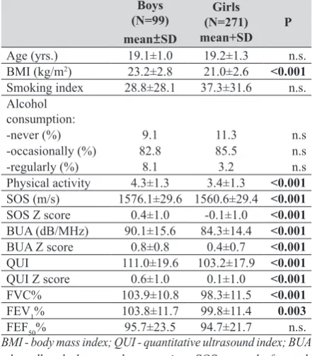
The mean age of the participants was 19.1±1.0 for boys and 19.2±1.3 for girls. Table 1 shows gender differences in the measured parameters. Boys had a significantly higher mean BMI than girls. There were 18 (18.1 %) overweight boys and 24 (8.8 %) overweight girls. Two boys (2.0 %) were obese and two boys were underweight. Thirty-three girls (12.1 %) were underweight. In addition, all bone density parameters (QUI, BUA, SOS) were significantly higher in boys than in girls (P<0.001). According to QUI Z score, low bone stiffness (Z score <-1.0) was found in 16.2 % of girls and 3.0 % of boys.
Furthermore, boys had a significantly better lung function than girls (FVC % P<0.001; FEV1 %, P=0.003). Low FVC %, FEV1 % (<80 % of the reference values), and FEF50 % (<60 % of the reference values) were found in 1.0 %, 1.0 %, and 4.0 % of boys, respectively and in 4.8 %, 4.8 % and 10.1 % of girls, respectively.
Compared to the normal weight groups, underweight boys and girls had a significantly lower FVC % (P=0.012 and P<0.001, respectively) while overweight boys and girls had a significantly higher FVC % (P=<0.001 and P=0.024, respectively) (Table 2). Underweight girls also had a significantly lower FEV1 % (P=0.005).
We found no significant association between lung function and bone density in our young, healthy participants. Our results suggest that this association in healthy, but probably also in sick people, is not a simple causal association but rather a consequence of the interaction between various factors that independently affect both parameters, like anthropometry, physical activity, or smoking. This was also proposed the authors who confirmed significant association between these two parameters, but could not establish a pathophysiological mechanism that would explain this association (6, 7).
The factor that stands out in our study as a link between ultrasound bone parameters and lung function is BMI. Underweight students (BMI <18) had significantly worse lung function test results than normal weight students. One earlier study showed that poor spirometry findings in malnourished people were the consequence of respiratory muscle weakness and lower ventilatory drive (18). Studies in young patients with anorexia showed significant worsening of lung function, including a low diffusing capacity for carbon monoxide (19), a lower peak expiratory flow (20), and a significant elevation in residual volume (21).
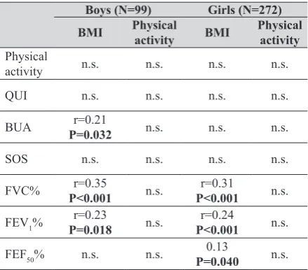
Furthermore, our overweight students had better lung function results than normal weight students. However, this finding challenges most other studies with reduced lung function in overweight children and adults (11, 22, 23-26). As a parameter of excess weight and obesity, BMI is not always reliable. Being the ratio between body mass and height, without accounting for fat tissue, BMI all too frequently categorises people as overweight even if they have higher muscle-to-fat-tissue ratio (27, 28). The mean BMI of 27.1 in our overweight students is not much higher than the upper limit of normal BMI. On the other side, our In boys BMI positively correlated with FVC %
(P<0.001), FEV1 % (P=0.018), and BUA (P=0.032). In girls BMI positively correlated with all lung function parameters (FVC % and FEV1 % - P<0.001; FEF50 % - P=0.040) (Table 3).
Table 4 shows the association between ultrasound bone density parameters and potential confounding factors. When controlled for gender, BMI significantly correlated with BUA (P=0.012).
(Student's t-test)
Boys (N=99) mean±SD
Girls (N=271)
mean+SD P
Age (yrs.) 19.1±1.0 19.2±1.3 n.s. BMI (kg/m2) 23.2±2.8 21.0±2.6 <0.001
Smoking index 28.8±28.1 37.3±31.6 n.s. Alcohol
consumption:
-never (%) 9.1 11.3 n.s -occasionally (%) 82.8 85.5 n.s -regularly (%) 8.1 3.2 n.s Physical activity 4.3±1.3 3.4±1.3 <0.001
SOS (m/s) 1576.1±29.6 1560.6±29.4 <0.001
SOS Z score 0.4±1.0 -0.1±1.0 <0.001
BUA (dB/MHz) 90.1±15.6 84.3±14.4 <0.001
BUA Z score 0.8±0.8 0.4±0.7 <0.001
QUI 111.0±19.6 103.2±17.9 <0.001
QUI Z score 0.6±1.0 0.1±1.0 <0.001
FVC% 103.9±10.8 98.3±11.5 <0.001
FEV1% 103.8±11.7 99.8±11.4 0.003
FEF50% 95.7±23.5 94.7±21.7 n.s. BMI - body mass index; QUI - quantitative ultrasound index; BUA - broadband ultrasound attenuation; SOS - speed of sound; FVC - forced vital capacity; FEV1% - forced expiratory volume
in one second; FEF50% - maximal expiratory flow at 50 %; n.s. - not significant
Table 2 Differences in physical activity, ultrasound bone density parameters, and lung function between normal weight, underweight, and overweight participants (Student's t-test)
Boys (N=99) Girls (N=272)
Normal weight Under-weight Over-weight Normal weight Under-weight Over-weight
Physical activity 4.3±1.3n.s. 2.5±1.3n.s. 4.2±1.2n.s. 3.4±1.3n.s. 3.3±1.3n.s. 3.3±1.5n.s.
QUI 112.1±17.3n.s. 97.5±0.4n.s. 111.5±17.8n.s. 103.1±16.5n.s. 103.7±22.3n.s. 103.3±16.3n.s.
BUA 89.7±15.5n.s. 69.6±1.7n.s. 93.0±16.2n.s. 83.9±13.5n.s. 82.5±19.9n.s. 86.4±14.2n.s.
SOS 1577.3±30.0n.s. 1561.0±0.5n.s. 1571.6±30.4n.s. 1560.4±28.7n.s. 1559.8±37.5n.s. 1558.2±27.1n.s.
FVC% 102.4±9.3n.s. 85.3±13.2P=0.012 111.7±11.5P<0.001 98.5±11.6n.s. 93.1±11.0P<0.001 104.1±9.3P=0.024
FEV1% 103.0±11.4n.s. 91.7±7.6n.s. 108.9±12.0n.s. 99.9±11.9n.s. 95.7±8.9P=0.005 103.7±10.5n.s.
FEF50% 95.2±23.1n.s. 82.4±0.4n.s. 101.3±26.1n.s. 94.7±21.7n.s. 92.0±22.5n.s. 99.0±21.9n.s.
normal weight subjects were relatively thin, with mean BMI of 21.3. The relative leanness in the majority of our students may be the reason why the overweight ones had a significantly better pulmonary function. Moreover, we do not have the data on fat-to-muscle ratio and muscle strength that could suggest the capacity of their respiratory muscles. Yet it is pretty safe to assume that the overweight group had a greater muscle content, considering that our overweight girls were significantly more physically active than the rest. Furthermore, muscle strength and physical activity are also well known predictors of bone density in young people (29, 30).
Body mass is a well-known predictor of bone density, with the exception of extreme obesity (32). In our participants, this correlation was clear only for the ultrasound parameter (BUA).
Our study found significant gender differences in the analysed parameters. Girls were significantly more underweight, less physically active, and had lower lung function and bone density parameters than boys. These findings highlight harmful misconceptions launched by the
aggressive promotion of healthy diet and regular physical activity in adolescent women.
Our study has two limitations: the first is that we did not measure bone density with dual-energy X-ray absorptiometry (DXA). Although many studies show a good correlation between ultrasound and DXA, others have raised concern due to differences in precision and areas being measured (31). The second limitation is the uneven number of boys and girls due to low response by boys. We tried to overcome this issue by including male dominated faculties, but did not succeed.
The merit of our study, however, remains: it found no direct relationship between BMD and lung function in a young healthy population after adjusting for confounding factors. Instead, it has singled out body mass as the most important confounder and link between the two.
Acknowledgements
The study was supported by the Croatian Ministry of Science, Education and Sports project No. 022-0222411-2410.
REFERENCES
1. Ferencz V, Meszaros S, Csupor E, Toth E, Bors K, Falus A, Horvath C. Increased bone fracture prevalence in postmenopausal women suffering from pollen-allergy. Osteoporos Int 2006;17:484-91. doi: 10.1007/s00198-005-0011-z
2. Vrieze A, de Greef MHG, Wýkstra PJ, Wempe JB. Low bone mineral density in COPD patients related to worse lung function, low weight and decreased fat-free mass. Osteoporos Int 2007;18:1197-202. doi: 10.1007/s00198-007-0355-7
3. Vondracek SF, Voelkel NF, McDermott MT, Valdez C. The relationship between adipokines, body composition, and bone density in men with chronic obstructive pulmonary disease. Int J Chron Obstruct Pulmon Dis 2009;4:267-77. PMCID: PMC2719257
4. Dam TT, Harrison S, Fink HA, Ramsdell J, Barret-Connor E; Osteoporotic Fractures in Men (MrOS) Research Group. Bone mineral density and fractures in older men with chronic obstructive pulmonary disease or asthma. Osteoporos Int 2010;21:1341-49. doi: 10.1007/s00198-009-1076-x
Boys (N=99) Girls (N=272) BMI Physical activity BMI Physical activity
Physical
activity n.s. n.s. n.s. n.s. QUI n.s. n.s. n.s. n.s.
BUA P=0.032r=0.21 n.s. n.s. n.s.
SOS n.s. n.s. n.s. n.s.
FVC% P<0.001r=0.35 n.s. P<0.001r=0.31 n.s.
FEV1% P=0.018r=0.23 n.s. P<0.001r=0.24 n.s.
FEF50% n.s. n.s. P=0.0400.13 n.s. BMI - body mass index; QUI - quantitative ultrasound index; BUA - broadband ultrasound attenuation; SOS - speed of sound; FVC - forced vital capacity; FEV1% - forced expiratory volume in one
second; FEF50% - maximal expiratory flow at 50 %; n.s. - not significant
Table 4 Regression summary for dependent variables in all participants (N=370) Dependent Variables
QUI BUA SOS
b P-value b P-value B P-value
Gender -7.64 <0.001 -0.13 0.010 -0.21 <0.001
BMI 0.30 n.s. 0.13 0.012 0.01 n.s.
Smoking index -0.03 n.s. -0.01 n.s. -0.03 n.s. Physical activity 0.04 n.s. 0.01 n.s. 0.04 n.s.
FVC% -0.03 n.s. -0.01 n.s. -0.03 n.s.
FEV1 % 0.01 n.s. -0.01 n.s. 0.01 n.s.
FEF50 % -0.03 n.s. -0.00 n.s. -0.02 n.s.
Jankovic J, Cvok T, Mitic J, Osteoporosis in COPD patients. Wien Klin Wochenschr 2012;124:484-5. doi: 10.1007/ s00508-012-0204-3
6. Lekamwasam S, Trivedi DP, Khaw KT. An association between respiratory function and bone mineral density in women from the general community: a cross sectional study. Osteoporos Int 2002;13:710-5. doi: 10.1007/s00198-004-1673-7
7. Ozen A, Ercan Saricoban H, Berber M, Sen N, Yesilyurt S, Ozdogan S, Cengizlier R. Association between respiratory function and bone mineral density in pubertal and prepubertal healthy children. J Ped Sci 2012;4:e120.
8. Jeon YK, Shin MJ, Kim WJ, Kim SS, Kim BH, Kim SJ, Kim YK, Shin YB, Kim IJ. The relationship between pulmonary function and bone mineral density in healthy nonsmoking women: the Korean National Health and Nutrition Examination Survey (KNHANES) 2010. Osteopor int 2014; 25:1571–76. doi: 10.1007/s00198-014-2627
9. Dennison EM, Dhanwal DK, Shaheen SO, Azagra R, Reading I, Jameson KA, Sayer AA, Cooper C. Is lung function associated with bone mineral density? Results from the Hertfordshire cohort study. Arch Osteoporos 2013;8:115. doi: 10.1007/s11657-012-0115-y
10. Ziora K, Ziora D, Oswiecimska J, Roczniak W, Machura E, Dworniczak S, Tomalak W, Dyduch A. Spirometric parameters in malnourished girls with anorexia nervosa. J Physiol Pharmacol 2008;59(Suppl.6):801-7. PMID:
19218707
11. Gundogdu Z, Eryilmaz N. Correlation between peak flow and body mass index in obese and non-obese children in Kocaeli, Turkey. Prim Care Respir J 2011;20:403-6. doi: 10.4104/pcrj.2011.00061
12. Johansson H, Kanis JA, Odén A, McCloskey E, Chapurlat RD, Christiansen C, Cummings SR, Diez-Perez A, Eisman JA, Fujiwara S, Glüer C-C, Goltzman D, Hans D, Khaw K-T, Krieg M-A, Kröger H, LaCroix AZ, Lau E, Leslie WD, Mellström D, Melton III LJ, O’Neill TW, Pasco JA, Prior JC, Reid DM, Rivadeneira F, van Staa T, Yoshimura N, Zillikens MC. A meta-analysis of the association of fracture risk and body mass index in women. J Bone Min Res 2014;29:223-33. doi: 10.1002/jbmr.2017
13. Asher MI, Keil U, Anderson HR, Beasley R, Crane J, Martinez F, Mitchell EA, Pearce N, Sibbald B, Stewart AW, Strachan D, Weiland SK, Williams HC. International study of asthma and allergies in childhood (ISAAC): rationale and methods. Eur Respir J 1995;8:483-91. doi: 10.1183/09031936.95.08030483
14. World Healt Ogranization (WHO). Physical status: the use and interpretation of anthropometry. Report of a WHO Expert Committee. WHO Technical Report Series 854. Geneva: WHO; 1995.
15. Brinkman GL, Coates EO Jr. The effect of bronchitis, smoking and occupation on ventilation. Am Rev Respir Dis 1963;87:684-93. doi: 10.1164/arrd.1963.87.5.684
16. Quanjer PH, Tammeling GJ, Cotes JE, Pedersen OF, Peslin R, Yernault JC. Lung volumes and forced ventilatory flows. Report working party standardization of lung function tests, European Community for Steel and Coal. Official Statement of the European Respiratory Society. Eur Respir J 1993;6(Suppl 16):5-40. doi: 10.1183/09041950.005s1693
17. Kraljevic I, Kastelan D, Kolcic I, Kardum I, Mazalin-Protulipac J, Korsic M. Calcaneal ultrasound parameters in
2007;13:MT29-33. PMID: 17767128
18. Ferrari-Baliviera E, Pierdominici S, Sarcinelli L. [Effects of the nutritional status on the respiratory system, in Italian]. Minerva Anestesiol 1989;55:443-50. PMID: 2699012
19. Gardini Gardenghi G, Boni E, Todisco P, Manara F, Borghesi A, Tantucci C. Respiratory function in patients with stable anorexia nervosa. Chest 2009;136:1356-63. doi: 10.1378/chest.08-3020
20. Kerem NC, Averin E, Riskin A, Tov N, Srugo I, Kugelman A. Respiratory functions in adolescents hospitalized for anorexia nervosa: a prospective study. Int J Eat Disorder 2012;45:415-22. doi: 10.1002/eat.20960
21. González-Moro JMR, de Miguel-Diez J, Paz-González L, Buendía-García MJ, Santacruz-Siminiani A, De Lucas-Ramos P. Abnormalities of the respiratory function and control of ventilation in patients with anorexia nervosa. Respiration 2003;70:490-5. doi: 10.1159/000074205
22. Jeon YH, Yang HY, Pyun BY. Lung function in Korean adolescent girls: in association with obesity and the menstrual cycle. J Korean Med Sci 2009;24: 20-5. DOI: 10.3346/ jkms.2009.24.1.20
23. Salome CM, King GG, Berend N. Physiology of obesity and effects on lung function. J Appl Physiol 2010;108:206-11. doi: 10.1152/japplphysiol.00694.2009
24. Li AM, Chan D, Wong E, Yin J, Nelson EA, Fok TF. The effects of obesity on pulmonary function. Arch Dis Child 2003;88:361-3. doi: 10.1136/adc.88.4.361
25. Thyagarajan B, Jacobs Jr DR, Apostol GG, Smith LJ, Jensen RL, Crapo RO, Barr RG, Lewis CE, Williams OD. Longitudinal association of body mass index with lung function: The CARDIA Study. Respir Res 2008;9:31. doi: 10.1186/1465-9921-9-31
26. Assunção SN, Daltro CH, Boa Sorte NC, da Costa Ribeiro Júnior H, Bastos ML, Queiroz CF, Moreira Lemos AC. Lung function in the absence of respiratory symptoms in overweight children and adolescents. J Bras Pneumol 2014;40:134-41. doi: 10.1590/S1806-37132014000200006
27. Going S, Davis R. Body composition. In: Roitman JL, Herridge M; American College of Sports Medicine; editors. ACSM Resource Manual for Exercise Testing and Prescription. Philadelphia: Lippincott Williams and Wilkins;
2001.
28. Reilly T, Sutton L. Methods and applications of body composition analysis. In: Bust P, editor. Proceedings of the International Conference on Contemporary Ergonomics (CE2008); 1-3 April 2008. Nottingham, UK. London: Taylor and Francis; 2008. p. 491-5.
29. Liberato SC, Bressan J, Hills AP. The role of physical activity and diet on bone mineral indices in young men: a cross-sectional study. J Int Soc Sports Nutr 2013;10:43-9. doi: 10.1186/1550-2783-10-43
30. Alghadir AM, Gabr SA, Al-Eisa E. Physical activity and lifestyle effects on bone mineral density among young adults: sociodemographic and biochemical analysis. J Phys Ther Sci 2015;27:2261-70. doi: 10.1589/jpts.27.2261
31. Flöter M, Bittar CK, Zabeu JL, Carneiro AC. Review of comparative studies between bone densitometry and quantitative ultrasound of the calcaneus in osteoporosis. Acta Reumatol Port 2011;36:327-35. PMID: 22472924
populacije
Rezultati istraživanja pokazali su da pedijatrijski i odrasli bolesnici s različitim plućnim bolestima mogu imati nisku mineralnu gustoću kostiju, međutim ograničeni podaci postoje o povezanosti plućne funkcije i gustoće kostiju u zdravoj mladoj populaciji. U ovom presječnom istraživanju analizirali smo čimbenike koji mogu utjecati na povezanost plućne funkcije i koštane gustoće u 370 zdravih studenata prve godine studija. Uz mjerenje visine i težine, ispitanicima je spirometrijski određena plućna funkcija, izmjerena mineralna gustoća kostiju ultrazvučnom metodom te su prikupljeni podaci o tjelesnoj aktivnosti, pušenju i unosu alkohola. Svi dobiveni pokazatelji plućne funkcije i mineralne gustoće kostiju bili su značajno veći u muških ispitanika u odnosu na djevojke (P<0,001). U odnosu na ispitanike s normalnom tjelesnom težinom, pothranjeni ispitanici imali su značajno manji forsirani vitalni kapacitet (FVC %) (P=0,001 djevojke; P=0,012 mladići), a ispitanici s prekomjernom težinom značajno veći FVC % (P=0,024 djevojke; P=0,001 mladići). Indeks tjelesne mase (ITM) u oba je spola značajno korelirao s FVC % (P=0,001) i s forsiranim ekspiratornim volumenom u prvoj sekundi (FEV1 %) (P=0,001 djevojke; P=0,018 mladići), kao i sa slabljenjem ultrazvučnog vala pri prolasku kroz kost (BUA) u mladića. Nije nađena značajna izravna povezanost između pokazatelja plućne funkcije i koštane gustoće. Regresijskom je analizom indeks tjelesne mase utvrđen kao najvažniji prediktor plućne funkcije i koštane gustoće u ispitanika obaju spolova.