Scholarship@Western
Scholarship@Western
Electronic Thesis and Dissertation Repository
6-20-2013 12:00 AM
Structure of C-terminal Domain of Parkin, IBR-RING2
Structure of C-terminal Domain of Parkin, IBR-RING2
Yeong Ju Noh
The University of Western Ontario
Supervisor Dr. Shaw
The University of Western Ontario
Graduate Program in Biochemistry
A thesis submitted in partial fulfillment of the requirements for the degree in Master of Science © Yeong Ju Noh 2013
Follow this and additional works at: https://ir.lib.uwo.ca/etd
Part of the Biochemistry Commons
Recommended Citation Recommended Citation
Noh, Yeong Ju, "Structure of C-terminal Domain of Parkin, IBR-RING2" (2013). Electronic Thesis and Dissertation Repository. 1324.
https://ir.lib.uwo.ca/etd/1324
This Dissertation/Thesis is brought to you for free and open access by Scholarship@Western. It has been accepted for inclusion in Electronic Thesis and Dissertation Repository by an authorized administrator of
(Thesis format: Monograph)
by
Yeong Ju Noh
Graduate Program in Biochemistry
A thesis submitted in partial fulfillment of the requirements for the degree of
Master of Science
The School of Graduate and Postdoctoral Studies The University of Western Ontario
London, Ontario, Canada
© Yeong Ju Noh, 2013
ii
Abstract
Parkin is an E3 ubiquitin ligase which degrades misfolded proteins and prevents the formation of abnormal protein aggregates often formed in Parkinson’s disease. The main goal of this thesis was to perform structural analysis on the IBR(In-Between-RING)-RING2 (Really Interesting New Gene) domain of parkin. After determining the three-dimensional solution structure of the protein by NMR spectroscopy, the RING2 domain was identified to be similar to the IBR domain, showing that it is not a canonical RING domain. The catalytic cysteine on RING2 was also shown to be solvent exposed, supporting the recently proposed RING/HECT hybrid mechanism of parkin as an RBR E3 ligase. The structure also revealed that IBR and RING2 domains do not interact. This was confirmed with two dimensional NMR experiments and split GFP system. The 14 disease-state IBR-RING2 proteins were analyzed using NMR spectroscopy to monitor the structural impact of autosomal recessive juvenile parkinsonism (ARJP) related mutations.
Keywords:
iii
Acknowledgments
I would like to thank my supervisor, Dr. Gary Shaw, for the opportunity to work in his lab and his support, guidance, encouragement, and patience throughout my project. Thank you for being the best supervisor one could ever ask for.
I would also like to thank my advisory committee, Dr. David Litchfield and Dr. James Choy for your valuable advice and feedback.
To all the past and present the members of the Shaw lab, I really appreciate your support, assistance, and advice during my graduate studies. I could not have done it without all of you, and it was an honor learning so much from many of you and discussing and working together. Especially to Dr. Pascal Mercier, I would like to show my appreciation for all of his assistance regarding structure calculation and making the structure complete at the end.
iv
Table of Contents
Abstract ... ii
Acknowledgments... iii
Table of Contents ... iv
List of Tables ... vii
List of Figures ... viii
List of Abbreviations and Acronyms ... x
List of Appendices ... xii
Chapter 1.Introduction ... 1
1.1 Parkinson’s Disease ... 1
1.2 Ubiquitin Protease System ... 2
1.3 E3 Ubiquitin Ligases ... 4
1.3.1 HECT E3 ligases ... 6
1.3.2 RING E3 ligases ... 6
1.3.3 RBR E3 ligases ... 10
1.4 Parkin ... 11
1.4.1 Interactions of parkin ... 11
1.4.2 Structures of parkin ... 13
1.4.3 ARJP mutations in parkin ... 14
1.5 Thesis Overview ... 16
1.6 References ... 18
Chapter 2.Materials and Methods ... 23
2.1 Drosophila melanogaster Parkin C-terminus, IBR-RING2 ... 23
2.1.1 Design of C-terminal domain constructs of Dm Parkin ... 23
v
2.1.3 Site-directed Mutagenesis of Parkin IBR-RING2 domain ... 24
2.2 Expression of IBR-RING2 ... 25
2.3 Purification of IBR-RING2 ... 27
2.4 NMR Spectroscopy ... 28
2.4.1 Chemical Shift Assignment of IBR-RING2 ... 28
2.4.2 Secondary Structure Prediction ... 29
2.4.3 T1/T2 experiments ... 30
2.4.4 Heteronuclear NOE ... 30
2.5 NMR Structure Determination ... 31
2.6 Solubility Test ... 31
2.7 References ... 33
Chapter 3.Results and Discussion ... 35
3.1 GST-IBR-RING2 Expression ... 35
3.2 Determination of Optimal Conditions for NMR Data Collection ... 38
3.3 Structural Determination of the Parkin IBR-RING2 Domain ... 43
3.3.1 ESI-MS shows the IBR-RING2 coordinates 4 Zinc ions ... 43
3.3.2 Backbone Chemical Shift Assignment of IBR-RING2 ... 44
3.3.3 Utilization of IBR-RING2 backbone assignment ... 47
3.3.4 Prediction of Zinc Coordinating Cysteines ... 47
3.3.5 Determination of Secondary Structure for parkin IBR-RING2 ... 49
3.3.6 Structure Calculation of IBR-RING2 ... 51
3.4 Interaction Studies of IBR and RING2 domains ... 58
3.4.1 The flexible linker of IBR-RING2 is confirmed with protein dynamics studies ... 58
3.4.2 Confirmation of non-interacting IBR and RING2 domains with 1H-15N HSQC spectra ... 63
vi
3.5 Mutation analysis ... 72
3.6 Summary ... 83
3.7 Closing Thoughts & Future direction ... 84
3.8 References ... 86
vii
List of Tables
Table 1. Forward and reverse primer sequences used in the amplification of different
domains of parkin’s C-terminus and mutants of IBR-RING2 and constructs of split GFP ... 26
Table 2. Cysteines involved in Zn2+ coordination in IBR-RING2 domain, as determined by chemical shifts of Cα and Cβ. ... 48
viii
List of Figures
Figure 1.1: Ubiquitination pathway showing ubiquitin conjugation. ... 5
Figure 1.2:HECT, RING and RBR type E3 ligases, illustrating the ubiquitin transfer step from E2 to E3. ... 7
Figure 1.3:Main features of Canonical RING E3 ligases. ... 9
Figure 1.4: Schematic diagram illustrating the domains of parkin, with three dimensional structures of domains in parkin that are present in the protein databank. ... 12
Figure 1.5: Zinc coordination of IBR, showing sequential zinc coordination. ... 15
Figure 3.1: SDS-PAGE gel of induced vs uninduced GST-IBR-RING2. ... 37
Figure 3.2: Chromatograms of GST purification ... 39
Figure 3.3: SDS-PAGE gels showing two-step purification of IBR-RING2 ... 40
Figure 3.4: Comparison of 1H-15N HSQC Spectra and NMR tubes of 300-400µM IBR-RING2 after the data collection. ... 42
Figure 3.5: Deconvoluted ESI-MS of proteins under denaturing and non-denaturing conditions, which show the mass of both the native and denatured state of IBR-RING2. ... 45
Figure 3.6: Two-dimensional 1H-15N HSQC spectrum of 15N-isotopically labeled IBR-RING2. ... 46
Figure 3.7: Chemical shift index of IBR-RING2 for secondary structure prediction. ... 50
Figure 3.8: Superposition of 20 structures overlay of IBR-RING2, on each of the IBR (blue) and RING2 (red) domains. ... 52
ix
Figure 3.10: Ribbon structure of parkin IBR domain with template of zinc binding sites. .... 56
Figure 3.11: Ribbon structure of parkin RING2 domain. ... 56
Figure 3.12: Steady-state heteronuclear NOE values for backbone amides of 15N-labeled IBR-RING2, obtained at 600 MHz. ... 59
Figure 3.13: Backbone 15N spin relaxation measurement of IBR-RING2 ... 61
Figure 3.14: Superposition of the IBR-RING2 1H-15N HSQC spectrum with each individual domain... 64
Figure 3.15: Superposition of the IBR-RING2 1H-15N HSQC spectrum with each individual domain, both IBR and RING2 displayed. ... 66
Figure 3.16: Titration of RING2 into 15N labeled IBR. ... 67
Figure 3.17: Illustration of split GFP system. ... 69
Figure 3.18: Solubility test of split GFP constructs shown as labelled at the top... 71
Figure 3.19: Soluble fraction of cell lysate split GFP fusions with RBR, IBR-RING2, and S100A8/A9. ... 71
Figure 3.20: Disease state substitutions within the parkin IBR-RING2. Ribbon drawings of different domains of parkin. ... 73
Figure 3.21:Solubility test of mutants of IBR-RING2, (missense mutations on RING2 only). ... 74
Figure 3.22: Superposition of 1H-15N HSQC spectra of wild type IBR-RING2 with mutated IBR-RING2 in IBR region (G349E). ... 77
Figure 3.23: Superposition of 1H-15N HSQC spectra of wild type IBR-RING2 with mutated IBR-RING2 in the linker region (D412N). ... 79
x
List of Abbreviations and Acronyms
PD ARJP PINK UPS HECT RING RBR UbL UPD IBR TEV GFP IPTG GST EDTA NMR TOCSY NOESY CYANA SDS-PAGE MW HSQC RMSD NOE Parkinson’s disease
Autosomal recessive juvenile parkinsonism PTEN-induced kinase I
Ubiquitin protease system
Homologous to E6-AP C-terminal domain Really interesting new gene
RING-between-RING Ubiquitin-like
Unique parkin domain In-between-RING Tobacco Etch Virus Green fluorescent protein
Isopropyl β-D thiogalactopyranoside Glutathione S-Transferase
Ethylenediaminetetraacetic acid Nuclear magnetic resonance Total Correlation Spectroscopy
Nuclear Overhauser effect spectroscopy
Combined Assignment and Dynamics Algorithm for NMR Applications Sodium dodecyl sulfate polyacrylamide gel electrophoresis
Molecular weight
Heteronuclear Single Quantum Coherence Root-mean-square deviation
xi HGMD DCDB E.Coli Dm Ala (A) Arg (R) Asn (N) Asp (D) Cys (C) Gln (Q) Glu (E) Gly (G) His (H) Ile (I) Leu (L) Lys (K) Met (M) Phe (F) Pro (P) Ser (S) Thr (T) Trp (W) Tyr (Y) Val (V)
Human gene mutation database Domain classification database
xii
List of Appendices
Appendix A: Data from Structure Calculation of IBR-RING2 ... 89
Appendix B: Data from Dynamics Studies of IBR-RING2 ... 90
Appendix C: Data from Mutational Analysis of IBR-RING2 ... 92
Chapter 1
Introduction
1.1 Parkinson’s Disease
Parkinson’s disease (PD) is the most prevalent movement disorder and second most common neurodegenerative disease, affecting more than 100,000 Canadians. Over the span of next years, the number of PD patients is expected to double aged 65 and over. PD is caused by the death of neurons responsible for dopamine production in the substantia nigra (Fearnley and Lees, 1990) and common clinical diagnosis of PD patients includes bradykinesia, rigidity, resting tremors and postural instability. Interestingly, the loss of smell is also a commonly noted symptom by PD patients in retrospect (Doty et al., 1995). PD patients start experiencing symptoms at approximately 55 years old, with these become increasingly prevalent. The molecular mechanisms involved in the pathogenesis of the disorder are not clearly understood, except for their proposed relation to aging, environment, and genetic predisposition (Mattson et al., 2002).
symptoms of sporadic PD are similar to those of the familial early-onset form of PD, including autosomal recessive juvenile Parkinsonism (ARJP), where early-onset occurs before the age of 40. A hallmark of idiopathic PD is the observation of cytoplasmic insoluble protein aggregates (Lewy bodies) containing α-synuclein encoded by PARK1/4, which also occurs in autosomal dominant forms of PD (Marin et al., 2004). Interestingly, for the ARJP, Lewy bodies are rarely present (Mori et al., 1998; Takahashi et al., 1994).
The most common form of ARJP (50%) is linked to mutations on the PARK2 gene on chromosome 6 (Kitada et al., 1998; Matsumine et al., 1998). This gene codes for a 465-residue-protein known as parkin. Other less frequent ARJP related proteins include; PTEN-induced kinase I (PINK), encoded by the PARK6 gene, and DJ-1 encoded by the PARK7 gene (Bonifati et al., 2003; Valente et al., 2004). Also, neural loss in ARJP patients is identified to be associated with dysfunction of parkin (Takahashi et al., 1994). The loss of function in parkin due to mutation has been suggested to cause problems for PD patients, leading to the potential neurotoxic accumulation (Sang et al., 2007).
Parkin belongs to the family of E3 ubiquitin-protein ligases in the ubiquitin protease system (UPS), and is responsible for the degradation of misfolded proteins by the proteasome, which prevents the formation of abnormal protein aggregates such as those formed in PD (Shimura et al., 2000).
1.2 Ubiquitin Protease System
most well-known mechanisms for this is the ubiquitination proteasome system (UPS) (Lehman, 2009). The UPS is characterized by the transfer of ubiquitin, a 76-residue protein, through a chain of several enzymes until it is covalently attached to a substrate.
Ubiquitin, one of the most highly conserved proteins present in eukaryotic cells, is involved in various cellular processes including: transcription (Pickart, 1997), cell-cycle (Imai et al., 2000), endocytosis (Hicke and Dunn, 2003), DNA repair (Barbour et al., 2006; Hershko and Ciechanover, 1998), and proteolysis (Glickman and Ciechanover, 2002). Therefore, it is not a surprise that when the ubiquitin system encounters abnormalities, many diseases can arise, including but not limited to: Angelman Syndrome, Cystic Fibrosis, and neurodegenerative diseases such as Alzheimer’s or Parkinson’s Disease (Glickman and Ciechanover, 2002).
The UPS conjugates ubiquitin through a highly conserved mechanism involving a cascade of enzymes, E1: ubiquitin activating, E2: ubiquitin conjugating, and E3: ubiquitin ligating enzymes, as shown in Figure 1.1. The transfer of ubiquitin is initiated by the ubiquitin activating enzyme (in an ATP-dependent manner) forming a thiolester bond between E1 and ubiquitin. Then, the ubiquitin is transferred to an E2 enzyme through a transthioesterification reaction, which releases E1. The process is complete when the E2-ubiquitin complex associates with the E3 enzyme in order to transfer the ubiquitin to the target protein (Huang et al., 2007).
In the ubiquitination process, E3 enzymes are far more specific and abundant than the other proteins involved (Schwartz and Ciechanover, 2009). Ubiquitination of a particular substrate is thought to recognize a particular E2:E3 combination. In the human genome of substrates, there are two E1s, over 38 E2s, and about 600 to 1000 E3s, known to date that allow ubiquitination.
1.3 E3 Ubiquitin Ligases
Figure 1.1: Ubiquitination pathway showing ubiquitin conjugation.
Ubiquitin is activated by the E1 ubiquitin-activating enzyme in an ATP-dependent manner. Ubiquitin attaches to E1forming a thiolester bond. Subsequently, it is transferred to the E2 ubiquitin-conjugating enzyme through a transthiolester reaction. Ubiquitin conjugated E2 interacts with an E3 ligase, bringing the substrate closer to the E2, in order for the ubiquitin to attach to the lysine of the substrate. Several repetitions of the
the direct transfer of ubiquitin from the E2 enzyme to the substrate (Passmore and Barford, 2004). Figure 1.2 illustrates the ubiquitin transfer models for the HECT and RING E3 ligases. Catalytic residues have not been identified for RING type ligases, and are suspected not to exist.
1.3.1 HECT E3 ligases
The HECT domain (about 350 residues long) is usually located at the C-terminus of these E3 ligases, identified based on its similarity to the E6 associated protein (E6AP). A conserved catalytic cysteine (reactive C-terminus) is contained in the protein database in the HECT E3 family, which can form a thiolester bond with ubiquitin. The N-terminus of the HECT domain serves as the E2 binding domain. There are about 30 HECT type E3 ligases, including E6AP, and Nedd4, all of which are known to play roles in protein trafficking, immune response, and regulation of cell growth through involvement in signaling (Rotin and Kumar, 2009).
1.3.2 RING E3 ligases
A B C
Figure 1.2:HECT, RING and RBR type E3 ligases, illustrating the ubiquitin transfer step from E2 to E3.
A. HECT type ubiquitin transfer to substrate, involving the catalytic cysteine of E3 ligase
(blue & red arrows), B. RING type ubiquitin transfer to substrate; E3 does not get
complexes, including Skp1-cullin-F-box (SCF), and Cullin-elongin B and C (CBC) complexes. Rbx1, the RING E3 ligase was later found to be part of the SCF complexes. RING domains can be located anywhere throughout the E3 ligase sequence and are characterized by the consensus sequence C-X2-C-X(9-39)-C-X(1-3)-H-X(2-3)-C/H-X2-C-X
(4-48)-C-X2-C, where X represents any given amino acid. The regular pattern of cysteines and histidines allows for the binding of two zinc ions, which stabilizes the protein structure. This coordination is referred to as a cross brace-like motif and the first and third pairs of Cys and second and fourth pairs of Cys/His allow for the proper coordination of zinc within the structure (Figure 1.3).
Figure 1.3:Main features of Canonical RING E3 ligases.
A. Cross-brace zinc coordination of RING E3 ligase, B. Canonical structure of RING,
represented by TRAF6 (PDB 3HCS) (Adapted from Spratt et al., 2013).
A
1.3.3 RBR E3 ligases
Some members of the RBR ligases include: human homolog of Drosophila Ariadne (HHARI), heme-oxidized-IRP2 ubiquitin ligase 1(HOIL-1), HOIL-1-interacting protein (HOIP), and parkin (Lucas et al., 2006; Marin et al., 2004). These RBR domains have been demonstrated to be involved in translation and immune signaling. Recently, it has been proposed that RBR E3 ligases might act as a hybrid of RING and HECT-type E3s (as shown in Figure 1.2).
1.4 Parkin
Parkin is the most studied RBR E3 ligase, due to its relation to Parkinson’s disease (PD). Parkin is an RBR-E3 ligase that has multiple domains containing numerous point mutations that are responsible for 50% or more of ARJP cases (Figure 1.4). The N-terminus of parkin contains an ubiquitin-like (UbL) domain and it spans from 1-76 residues. This domain has been proposed to be important in the recruitment of proteasome (Sakata et al., 2003; Tashiro et al., 2003). The UbL domain is followed by the unique parkin domain (UPD) that is only present in parkin, and is approximately 70 residues long. The C-terminus of parkin comprised the RBR domains with a RING0 domain preceding it (Beasley et al., 2007; Hristova et al., 2009; Morett and Bork, 1999). Of these domains, only the UbL and IBR domains have been successfully purified and had their three dimensional structures solved (Beasley et al., 2007; Hristova et al., 2009; Morett and Bork, 1999).
1.4.1 Interactions of parkin
Figure 1.4: Schematic diagram illustrating the domains of parkin, with three dimensional structures of domains in parkin that are present in the protein databank.
Parkin has UbL domain at the N-terminus, unique parkin domain (UPD) and RING0 domains in the middle, and RING1, IBR, and RING2 (RBR) at the C-terminus. Some of the amino acid substitutions related to ARJP are indicated with black arrows. Orange ribbon diagram shows UbL domain (PDB: 1IYF) and blue ribbon diagram shows IBR (PDB: 2JMO) (Adapted from von Coelln et al., 2004)
binding partner of RING1. More recently, a heterodimeric form of the E2 enzyme Ubc13/Uev2a was suggested as an interaction partner of parkin (Doss-Pepe et al., 2005; Matsuda et al., 2006). Based on the inconsistency of previous work, it still remains to be seen which RING domains in parkin are important for recruitment of E2 enzymes and control of ubiquitination.
1.4.2 Structures of parkin
The three-dimensional structure of the UbL domain present at the N-terminus of parkin has been solved by nuclear magnetic resonance (NMR) spectroscopy. The structure shows similarity to ubiquitin, possessing the β-grasp fold comprised of five β -sheets and two α-helices, as shown in Figure 1.4 (Sakata et al., 2003). A distinct feature of this domain of parkin is that it lacks a C-terminal glycine, so that unlike ubiquitin, it cannot conjugate to the lysine of a substrate. Instead, the UbL domain is followed by the UPD, a region postulated to be a disordered linker between the UbL domain and RING0-RBR domains with no obvious other function.
shown in Figure 1.5, and illustrates that the first and second pairs of cysteines and the third and fourth pairs of cysteines/histidines coordinate zinc. The IBR was proposed to act like a bridge that brings RING2 and RING2 domains together in the full-length parkin assembly, with an expectation that those two are interacting domains. Though the three-dimensional structure of the IBR domain of human parkin has been solved, structures of the RING domains and more importantly, the assembly of the entire C-terminal RBR motif, still remain unknown. It is expected that structural analysis of these domains can answer some questions regarding the catalytic mechanism of parkin in ubiquitination.
1.4.3 ARJP mutations in parkin
Figure 1.5: Zinc coordination of IBR, showing sequential zinc coordination.
However, other point mutations do not show whether ARJP is caused by a loss of its E3 activity. It is not known how parkin becomes dysfunctional with substitution, or how these alterations affect the protein’s structure or stability. To have a better understanding of the impact of these mutations, structure and interaction studies with parkin would be necessary.
1.5 Thesis Overview
Mutations in the PARK2 gene that codes for parkin are strongly linked to ARJP and have many different effects on RBR E3 ligase activity. However, given the structure determination of the parkin IBR domain (Beasley et al., 2007), the roles of the RING1 and RING2 domains in either a RING or RING/HECT E3 ligase mechanism have neither been supported nor opposed by structural analysis. The goal of this project was to determine the three-dimensional structure of IBR-RING2 in order to have a better understanding of parkin’s assembly and function. The proposed hypothesis is that the IBR and RING2 of parkin are likely to be interacting, even with the missing RING1 domain, to facilitate ubiquitin transfer to a substrate.
Specific experiments designed to address this hypothesis are:
1) Determine the three-dimensional solution structure of parkin IBR-RING2 using NMR spectroscopy.
utilizing NMR spectroscopy, NMR relaxation experiments, and a split-GFP system.
3) Analyze the impact on the solubility and structure of IBR-RING2 domains caused by mutations (ARJP substitutions) using NMR spectroscopy.
1.6 References
Ardley, H.C., Tan, N.G., Rose, S.A., Markham, A.F., and Robinson, P.A. (2001). Features of the parkin/ariadne-like ubiquitin ligase, HHARI, that regulate its interaction with the ubiquitin-conjugating enzyme, Ubch7. J Biol Chem 276, 19640-19647.
Barbour, L., Ball, L.G., Zhang, K., and Xiao, W. (2006). DNA damage checkpoints are involved in postreplication repair. Genetics 174, 1789-1800.
Beasley, S.A., Hristova, V.A., and Shaw, G.S. (2007). Structure of the Parkin
in-between-ring domain provides insights for E3-ligase dysfunction in autosomal recessive Parkinson's disease. Proc Natl Acad Sci U S A 104, 3095-3100.
Bedford, L., Lowe, J., Dick, L.R., Mayer, R.J., and Brownell, J.E. (2011). Ubiquitin-like protein conjugation and the ubiquitin-proteasome system as drug targets. Nat Rev Drug Discov 10, 29-46.
Bonifati, V., Rizzu, P., van Baren, M.J., Schaap, O., Breedveld, G.J., Krieger, E., Dekker, M.C., Squitieri, F., Ibanez, P., Joosse, M., et al. (2003). Mutations in the DJ-1 gene associated with autosomal recessive early-onset parkinsonism. Science 299, 256-259. Brzovic, P.S., Meza, J.E., King, M.C., and Klevit, R.E. (2001). BRCA1 RING domain cancer-predisposing mutations. Structural consequences and effects on protein-protein interactions. J Biol Chem 276, 41399-41406.
Buchwald, G., van der Stoop, P., Weichenrieder, O., Perrakis, A., van Lohuizen, M., and Sixma, T.K. (2006). Structure and E3-ligase activity of the Ring-Ring complex of polycomb proteins Bmi1 and Ring1b. EMBO J 25, 2465-2474.
Di Fiore, P.P., Polo, S., and Hofmann, K. (2003). When ubiquitin meets ubiquitin receptors: a signalling connection. Nat Rev Mol Cell Biol 4, 491-497.
Doss-Pepe, E.W., Chen, L., and Madura, K. (2005). Alpha-synuclein and parkin contribute to the assembly of ubiquitin lysine 63-linked multiubiquitin chains. J Biol Chem 280, 16619-16624.
Doty, R.L., Bromley, S.M., and Stern, M.B. (1995). Olfactory testing as an aid in the diagnosis of Parkinson's disease: development of optimal discrimination criteria. Neurodegeneration 4, 93-97.
Fearnley, J.M., and Lees, A.J. (1990). Striatonigral degeneration. A clinicopathological study. Brain 113 ( Pt 6), 1823-1842.
Glickman, M.H., and Ciechanover, A. (2002). The ubiquitin-proteasome proteolytic pathway: destruction for the sake of construction. Physiol Rev 82, 373-428.
Haglund, K., and Stenmark, H. (2006). Working out coupled monoubiquitination. Nat Cell Biol 8, 1218-1219.
Hershko, A., and Ciechanover, A. (1998). The ubiquitin system. Annu Rev Biochem 67, 425-479.
Hicke, L. (2001). Protein regulation by monoubiquitin. Nat Rev Mol Cell Biol 2, 195-201. Hicke, L., and Dunn, R. (2003). Regulation of membrane protein transport by ubiquitin and ubiquitin-binding proteins. Annu Rev Cell Dev Biol 19, 141-172.
Hristova, V.A., Beasley, S.A., Rylett, R.J., and Shaw, G.S. (2009). Identification of a novel Zn2+-binding domain in the autosomal recessive juvenile Parkinson-related E3 ligase parkin. J Biol Chem 284, 14978-14986.
Huang, D.T., Hunt, H.W., Zhuang, M., Ohi, M.D., Holton, J.M., and Schulman, B.A. (2007). Basis for a ubiquitin-like protein thioester switch toggling E1-E2 affinity. Nature
445, 394-398.
Imai, Y., Soda, M., and Takahashi, R. (2000). Parkin suppresses unfolded protein stress-induced cell death through its E3 ubiquitin-protein ligase activity. J Biol Chem 275, 35661-35664.
Kitada, T., Asakawa, S., Hattori, N., Matsumine, H., Yamamura, Y., Minoshima, S., Yokochi, M., Mizuno, Y., and Shimizu, N. (1998). Mutations in the parkin gene cause autosomal recessive juvenile parkinsonism. Nature 392, 605-608.
Kubota, H. (2009). Quality control against misfolded proteins in the cytosol: a network for cell survival. J Biochem 146, 609-616.
Lehman, N.L. (2009). The ubiquitin proteasome system in neuropathology. Acta Neuropathol 118, 329-347.
Lucas, J.I., Arnau, V., and Marin, I. (2006). Comparative genomics and protein domain graph analyses link ubiquitination and RNA metabolism. J Mol Biol 357, 9-17.
Marin, I., Lucas, J.I., Gradilla, A.C., and Ferrus, A. (2004). Parkin and relatives: the RBR family of ubiquitin ligases. Physiol Genomics 17, 253-263.
Matsuda, N., Kitami, T., Suzuki, T., Mizuno, Y., Hattori, N., and Tanaka, K. (2006). Diverse effects of pathogenic mutations of Parkin that catalyze multiple
Matsumine, H., Yamamura, Y., Hattori, N., Kobayashi, T., Kitada, T., Yoritaka, A., and Mizuno, Y. (1998). A microdeletion of D6S305 in a family of autosomal recessive juvenile parkinsonism (PARK2). Genomics 49, 143-146.
Mattson, M.P., Chan, S.L., and Duan, W. (2002). Modification of brain aging and neurodegenerative disorders by genes, diet, and behavior. Physiol Rev 82, 637-672. Metzger, M.B., Hristova, V.A., and Weissman, A.M. (2012). HECT and RING finger families of E3 ubiquitin ligases at a glance. J Cell Sci 125, 531-537.
Morett, E., and Bork, P. (1999). A novel transactivation domain in parkin. Trends Biochem Sci 24, 229-231.
Mori, H., Kondo, T., Yokochi, M., Matsumine, H., Nakagawa-Hattori, Y., Miyake, T., Suda, K., and Mizuno, Y. (1998). Pathologic and biochemical studies of juvenile parkinsonism linked to chromosome 6q. Neurology 51, 890-892.
Moynihan, T.P., Ardley, H.C., Nuber, U., Rose, S.A., Jones, P.F., Markham, A.F., Scheffner, M., and Robinson, P.A. (1999). The ubiquitin-conjugating enzymes UbcH7 and UbcH8 interact with RING finger/IBR motif-containing domains of HHARI and H7-AP1. J Biol Chem 274, 30963-30968.
Passmore, L.A., and Barford, D. (2004). Getting into position: the catalytic mechanisms of protein ubiquitylation. Biochem J 379, 513-525.
Pickart, C.M. (1997). Targeting of substrates to the 26S proteasome. FASEB J 11, 1055-1066.
Ross, C.A., and Pickart, C.M. (2004). The ubiquitin-proteasome pathway in Parkinson's disease and other neurodegenerative diseases. Trends Cell Biol 14, 703-711.
Rotin, D., and Kumar, S. (2009). Physiological functions of the HECT family of ubiquitin ligases. Nat Rev Mol Cell Biol 10, 398-409.
Sakata, E., Yamaguchi, Y., Kurimoto, E., Kikuchi, J., Yokoyama, S., Yamada, S., Kawahara, H., Yokosawa, H., Hattori, N., Mizuno, Y., et al. (2003). Parkin binds the Rpn10 subunit of 26S proteasomes through its ubiquitin-like domain. EMBO Rep 4, 301-306.
Sang, T.K., Chang, H.Y., Lawless, G.M., Ratnaparkhi, A., Mee, L., Ackerson, L.C., Maidment, N.T., Krantz, D.E., and Jackson, G.R. (2007). A Drosophila model of mutant human parkin-induced toxicity demonstrates selective loss of dopaminergic neurons and dependence on cellular dopamine. J Neurosci 27, 981-992.
Spratt, D.E., Julio Martinez-Torres, R., Noh, Y.J., Mercier, P., Manczyk, N., Barber, K.R., Aguirre, J.D., Burchell, L., Purkiss, A., Walden, H., et al. (2013). A molecular
Schwartz, A.L., and Ciechanover, A. (2009). Targeting proteins for destruction by the ubiquitin system: implications for human pathobiology. Annu Rev Pharmacol Toxicol 49, 73-96.
Shimura, H., Hattori, N., Kubo, S., Mizuno, Y., Asakawa, S., Minoshima, S., Shimizu, N., Iwai, K., Chiba, T., Tanaka, K., et al. (2000). Familial Parkinson disease gene product, parkin, is a ubiquitin-protein ligase. Nat Genet 25, 302-305.
Sriram, S.R., Li, X., Ko, H.S., Chung, K.K., Wong, E., Lim, K.L., Dawson, V.L., and Dawson, T.M. (2005). Familial-associated mutations differentially disrupt the solubility, localization, binding and ubiquitination properties of parkin. Hum Mol Genet 14, 2571-2586.
Takahashi, H., Ohama, E., Suzuki, S., Horikawa, Y., Ishikawa, A., Morita, T., Tsuji, S., and Ikuta, F. (1994). Familial juvenile parkinsonism: clinical and pathologic study in a family. Neurology 44, 437-441.
Tashiro, K., Tamada, S., Kuwabara, N., Komiya, T., Takekida, K., Asai, T., Iwao, H., Sugimura, K., Matsumura, Y., Takaoka, M., et al. (2003). Attenuation of renal fibrosis by proteasome inhibition in rat obstructive nephropathy: possible role of nuclear factor kappaB. Int J Mol Med 12, 587-592.
Valente, E.M., Abou-Sleiman, P.M., Caputo, V., Muqit, M.M., Harvey, K., Gispert, S., Ali, Z., Del Turco, D., Bentivoglio, A.R., Healy, D.G., et al. (2004). Hereditary early-onset Parkinson's disease caused by mutations in PINK1. Science 304, 1158-1160. van Duijn, C.M., Dekker, M.C., Bonifati, V., Galjaard, R.J., Houwing-Duistermaat, J.J., Snijders, P.J., Testers, L., Breedveld, G.J., Horstink, M., Sandkuijl, L.A., et al. (2001). Park7, a novel locus for autosomal recessive early-onset parkinsonism, on chromosome 1p36. Am J Hum Genet 69, 629-634.
von Coelln, R., Dawson, V.L., and Dawson, T.M. (2004). Parkin-associated Parkinson's disease. Cell Tissue Res 318, 175-184.
Wang, C., Tan, J.M., Ho, M.W., Zaiden, N., Wong, S.H., Chew, C.L., Eng, P.W., Lim, T.M., Dawson, T.M., and Lim, K.L. (2005). Alterations in the solubility and intracellular localization of parkin by several familial Parkinson's disease-linked point mutations. J Neurochem 93, 422-431.
Wenzel, D.M., Lissounov, A., Brzovic, P.S., and Klevit, R.E. (2011). UBCH7 reactivity profile reveals parkin and HHARI to be RING/HECT hybrids. Nature 474, 105-108. Zhang, Y., Gao, J., Chung, K.K., Huang, H., Dawson, V.L., and Dawson, T.M. (2000). Parkin functions as an E2-dependent ubiquitin- protein ligase and promotes the
Zimprich, A., Biskup, S., Leitner, P., Lichtner, P., Farrer, M., Lincoln, S., Kachergus, J., Hulihan, M., Uitti, R.J., Calne, D.B., et al. (2004). Mutations in LRRK2 cause
Chapter 2
Materials and Methods
2.1 Drosophila melanogaster Parkin C-terminus, IBR-RING2
The DNA encoding Drosophila melanogaster (Dm) parkin (PARK2) in pGEX vector was a gift from Dr. Brian Staveley (Memorial University of Newfoundland, Newfoundland and Labrador). This gene was previously cloned into a modified pGEX-6P-2 vector with a Tobacco Etch Virus (TEV)-cleavage site (ENLYFQ(G/S)) by Noah Manczyk (summer student, 2011). Site-directed mutagenesis was performed using primers that are complementary to each other which would bind to the end of TEV-cleavage site as well as the beginning of the IBR (starting from E342) sequence. This generated an N-terminal deletion construct, leaving only the IBR-RING2 domain from
Dm parkin (residues 342-482). The parkin IBR-RING2 construct was verified by DNA
sequencing (Robarts Research Institute).
2.1.1 Design of C-terminal domain constructs of Dm Parkin
products were digested with BamHI and EcoRI and ligated into the equivalent sites of the modified pGEX-6P-2. DNA sequencing verified the correct sequences of the constructs (Robarts Research Institute). The plasmids were then transformed into E.coli JM109 and BL21(DE3) Codon PlusRIL cell lines.
2.1.2 C-terminal Parkin into Split-GFP vector
RING1-IBR-RING2 and IBR-RING2 constructs of Dm parkin were inserted into a pETDuet dual expression plasmid containing the N- and C-terminal halves of GFP under identical T7-promoters (Ghosh et al., 2000). The cloning resulted in the creation of two constructs; (1) N-terminal GFP-parkin RING1-IBR-RING2-C-terminal GFP, and (2)
N-terminal GFP-parkin IBR-RING2-C-terminal GFP. PCR reactions were performed as
described previously (2.1.1) and the primers used for the insertion are listed in Table 1. The PCR products were then digested with BamHI and KpnI and ligated into the equivalent sites of the split-GFP vector. After sequencing, the vectors were transformed into E. coli BL21 BL21 (DE3) Codon PlusRIL expression cell line.
2.1.3 Site-directed Mutagenesis of Parkin IBR-RING2 domain
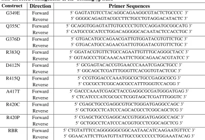
transformed into E. coli BL21(DE3) Codon PlusRIL. In total, 18 disease-related mutants were designed, and out of those, 16 mutants were successfully made. The mutants were designed for parkin IBR (G349E, Q355C, T372P, G376D, R383Q), linker (D412N, R415Q, A417T, R420C, R420P) and parkin RING2 (T433N, C436R, G447E, G448D, C449F, C459R, and M476L). Table 1 displays all the primers used in generating the mutants. Also, a RING1-IBR-RING2 construct (residues 252-482) was looped out from the PARK2 gene in the pGEX vector using restriction-free (RF) cloning (van den Ent and Lowe, 2006), using a similar protocol as outline in section 2.1.1.
2.2 Expression of IBR-RING2
The Dm parkin IBR-RING2 was overexpressed in E. coli BL21(DE3) Codon Plus RIL Escherichia coli strain, with an N-terminal GST fusion tag. The cells were streaked on Luria-Bertani (LB)–agar plates containing 100 µg/mL ampicillin (Amp) and incubated for approximately 20 hours at 37°C. Single colonies were picked for overnight cultures.
Table 1. Forward and reverse primer sequences used in the amplification of different
domains of parkin’s C-terminus and mutants of IBR-RING2 and constructs of split GFP
Cloning_pGEX
Construct Direction Primer Sequences
RING1 Forward
Reverse
5’ ATTCATATGGGATCCAATAACATCAAGAATGTTCC 3’ 5’ATACTCGAGAAGCTTGAATTCCTATTACTCCTCGGTGGCGAATCTC3’
RING1 -IBR
Forward Reverse
5’ ATTCATATGGGATCCAATAACATCAAGAATGTTCC 3’ 5’ATTCTCGAGGAATTCAAGCTTCTATTAACTAGCGCCCGTCCCCTCGG3’
IBR Forward
Reverse 5’ATTCATATGGGATCCACACGCGAGGAGTACGATC3’ 5’ATTCTCGAGGAATTCAAGCTTCTATTAACTAGCGCCCGTCCCCTCGG3’ Cloning_splitGFP RBR RBR Forward Reverse
5’ ATTGGATCCTCCGGCGGGGAGAAGG 3’ 5’ ATTGGATCCGAGTATGTCCTACAGGCAGG 3’
IBR IBR
Forward Reverse
5’ ATTGGATCCGAGTATGTCCTACAGGCAGG 3’ 5’ ATTGGATCCGAGTATGTCCTACAGGCAGG 3’
Site-directed Mutagenesis & RF Cloning _pGEX
Construct Direction Primer Sequences
G349E Forward
Reverse
5’ GAGTATGTCCTACAGGCAGAAGGCGTACTCTGCCCC 3’ 5’ GGGGCAGAGTACGCCTTCTGCCTGTAGGACATACTC 3’
Q355C Forward
Reverse
5’ GCAGGTGGAGTATTGTGCCCCTGTCCAGGATGCGGCATG 3’ 5’ CATGCCGCATCCTGGACAGGGGCACAATACTCCACCTGC 3’
G376D Forward
Reverse
5’ GTGACATGCCAGAACGATTGTGGATACGTGTTCTGC 3’ 5’ GTGACATGCCAGAACGATTGTGGATACGTGTTCTGC 3’
R383Q Forward
Reverse
5’ GGATACGTGTTCTGCCAGAATTGTTTGCAGGGCTACC 3’ 5’ GGTAGCCCTGCAAACAATTCTGGCAGAACACGTATCC 3’
D412N Forward
Reverse
5’ GCGAGTACACCGTGAACCCAAATCGAGCTGCC 3’ 5’ GGCAGCTCGATTTGGGTTCACGGTGTACTCGC 3’
R415Q Forward
Reverse
5’ CCGTGGACCCAAATGGCGCTGCCGAGGCGCG 3’ 5’ CGCGCCTCGGCAGCGCCATTTGGGTCCACGG 3’
A417T Forward
Reverse
5’ GACCCAAATCGAGCTACCGAGGCGCGATGGGATGAG 3’ 5’ CTCATCCCATCGCGCCTCGGTAGCTCGATTTGGGTC 3’
R420C Forward
Reverse
5’ CGAGCTGCCGAGGCGTGCTGGGATGAGGCCAGC 3’ 5’ GCTGGCCTCATCCCAGCACGCCTCGGCAGCTCG 3’
R420P Forward
Reverse
5’ CGAGCTGCCGAGGCACCGTGGGATGAGGCCAGC 3’ 5’ GCTGGCCTCATCCCACGGTGCCTCGGCAGCTCG 3’
RBR Forward
Reverse
5’ CTGTATTTCCAGGGGGGCGGCAATAACATCAAGAATGTTCC 3’ 5’ GGAACATTCTTGATGTTATTGCCGCCCCCCTGGAAATACAG 3’
To prepare 15N or 15N-13C labeled protein, M9 minimal media was used supplemented with 1 g/L 15N-ammonium chloride and 2% glucose (for 13C sample, 2 g/L of 13C6-D-glucose). The media also contained 2 mM MgSO4, 2 µM FeSO4, micronutrients, 100 µg/mL ampicillin and 34 µg/mL chloramphenicol. After a 20-hour expression, cells were harvested by centrifugation (6000 g, 4°C, 15min). Cell pellets were then transferred into 50mL tubes, flash frozen with liquid nitrogen and stored at -80°C until needed.
2.3 Purification of IBR-RING2
2.4 NMR Spectroscopy
All NMR spectra were obtained using Varian Unity INOVA 600 (UWO Biomolecular NMR facility) or 800 MHz NMR (NANUC) spectrometers equipped with cold probe technology. The NMR tubes used were standard 5 mm NMR tubes for 600 µL samples and Shigemi microcell NMR tubes for 300 µL samples. All of the NMR samples were prepared with 9:1 H2O/D2O in 25 mM Tris, 150 mM NaCl 5mM DTT at pH 7.5. Also, 1 µL of complete MINI EDTA free protease inhibitor (ROCHE) and 1 µM of EDTA was added to NMR sample to minimize proteolysis during data acquisition, and to prevent possible oxidation, argon gas was gently blown into the sample before closing the cap. NMR samples contained 30 µM DSS as an internal standard. The concentration of IBR-RING2 samples ranged from 300-400 µM, and all NMR spectra were collected at 25°C.
2.4.1 Chemical Shift Assignment of IBR-RING2
For the sequential backbone assignment of IBR-RING2, five different 3D experiments were used: CBCA(CO)NH (Grzesiek and Bax, 1992), HNCACB (Wittenkind and Mueller, 1993), HNCA (Kay et al., 1990), HNCO (Kay et al., 1994), and HN(CA)CO (Clubb et al., 1992). All of these experiments were collected on a 600 MHz spectrometer. The number of data points and spectral widths for the 1H(F3) and 15N(F2) dimensions were set to 1024 and 7500 Hz and 32 and1880 Hz, respectively. For CBCA(CO)NH and HNCACB, 46 increments and spectral width of 8000 Hz in were used in the 13C(F1) dimension. And for HNCO and HN(CA)CO experiments, 28 increments and 3000 Hz were used. For HNCA, 32 increments and 4521.5 Hz were used.
HC(CO)NH (Grzesiek and Bax, 1993), HCCH-TOCSY(Bax et al., 1990), 13C-NOESY (Marion et al., 1989), and 1H-13C HSQC (Kay et al., 1992) experiments. The 1H-13C HSQC experiment was collected with 1280 data points and spectral width of 7500 Hz in the 1H(F2) and 128 increments and 12000 Hz in the 13C(F1) dimension. For C(CO)NH experiment, 1280 complex data points and a spectral width of 7500Hz, 32 increments and 1880 Hz, and 52 increments and 11000 Hz in 1H(F3), 15N(F2) and 13C(F1) dimensions respectively. For the HC(CO)NH, conditions were similar to CCONH, except for F1 dimension, which was adjusted to 56 data points with 7500 Hz. For the HCCH-TOCSY experiment, the data points and spectral widths used were 1280 and 7500 Hz, 32 and 12000Hz, and 128 and 7500 for the 1H(F3), 13C(F2), 1H(F1) dimensions respectively. Aromatic side chains were assigned using (HB)CB(CGCD)HD , (HB)CB(CGCDCE)HE (Yamazaki et al., 1993), aromatic TOCSY, and aromatic 13C-HSQC(Kay et al., 1992).
All spectra were processed using the program NMRPipe (Delaglio et al., 1995) with cosine-squared function in order to minimize the artifacts in the spectra. Linear prediction was used for data processing by doubling the number of points in the F1 and F2 dimensions for the three-dimensional experiments. Manual chemical shift assignment was accomplished using the program NMRView (Johnson and Belvins, 1994) in order to analyze the three dimensional experiments.
2.4.2 Secondary Structure Prediction
chemical shifts to accurately predict secondary structures by comparing the values to those typically found in a random coil. The analysis was performed using the CSI option in the NMRViewJ program.
2.4.3 T1/T2 experiments
The backbone 15N T1 and T2 relaxation rates of IBR-RING2 were measured with the conventional series of 1H-15N-HSQC experiments with varied relaxation delays (Farrow et al., 1994). Longitudinal relaxation time (T1) and transverse relaxation time (T2) were determined from these experiments, based on the theory that relaxation times are sensitive to the intensity changes of backbone amides. The rotation correlation time (τc) was then calculated from experimental T1 and T2 experiments. Using the relationship between T1/T2 and τC, tumbling time of the IBR-RING2 was plotted on a graph by fitting them using the decay curves and determining T1 and T2.
2.4.4 Heteronuclear NOE
2.5 NMR Structure Determination
After manual assignment of chemical shifts of IBR-RING2, distance restraints for the structure calculation were extracted from signal intensities of 13C and 15N-edited 3D NOESY-HSQC spectra. Dihedral angle constraints were obtained using the TALOS+ program, based on the chemical shifts of 1Hα, 13Cα, 13C’ (Shen et al., 2009). Structures were calculated using a combination of manual and automatic NOE assignment with CYANA software (Herrmann et al., 2002). Seven cycles of combined automated NOESY assignment and structure calculation were performed followed by a final structure calculation. In this step, the knowledge of short atom-atom distances and the amino acid sequence of the protein as well as the arrangement of all atoms in space were calculated by CYANA. In the final structure calculation 100 conformers were calculated and the 20 conformers with the lowest final target function values (based upon minimal NOE, dihedral and van der Waal contact violations) were selected. The final structures were refined in water using explicit restraints for zinc coordination by Dr. Pascal Mercier.
The atomic coordinates for the structures of IBR-RING2 have been deposited in the Protein Data Bank under the accession code 2M48, and the BMRB accession code is 18990.
2.6 Solubility Test
2.7 References
Bax, A., Clore, G.M., and Gronenborn, A.M. (1990). 1H-1H correlation via isotropic mixing of 13C magnetization, a new three-dimensional approach for assigning 1H and 13C spectra of 13C-enriched proteins. Journal of magnetic resonance 88, 425-431.
Clubb, R.T., Thanabal, V., and Wagner, G. (1992). A new 3D HN(CA)HA experiment for obtaining fingerprint HN-Halpha peaks in 15N- and 13C-labeled proteins. J Biomol NMR 2, 203-210.
Delaglio, F., Grzesiek, S., Vuister, G.W., Zhu, G., Pfeifer, J., and Bax, A. (1995). NMRPipe: a multidimensional spectral processing system based on UNIX pipes. J Biomol NMR 6, 277-293.
Farrow, N.A., Muhandiram, R., Singer, A.U., Pascal, S.M., Kay, C.M., Gish, G., Shoelson, S.E., Pawson, T., Forman-Kay, J.D., and Kay, L.E. (1994). Backbone dynamics of a free and phosphopeptide-complexed Src homology 2 domain studied by 15N NMR relaxation. Biochemistry 33, 5984-6003.
Ghosh, I., Hamilton, A., and Regan, L. (2000). Antiparallel Leucine Zipper-Directed Protein Reassembly: Application to the Green Fluorescent Protein. J Am Chem Soc 122, 5658-5659.
Grzesiek, S., and Bax, A. (1992). Correlating backbone amide and side chain resonances in larger proteins by multiple relayed triple resonance NMR. J Am Chem Soc
114, 6291-6293.
Grzesiek, S., and Bax, A. (1993). Measurement of amide proton exchange rates and NOEs with water in 13C/15N-enriched calcineurin B. J Biomol NMR 3, 627-638.
Herrmann, T., Guntert, P., and Wuthrich, K. (2002). Protein NMR structure determination with automated NOE assignment using the new software CANDID and the torsion angle dynamics algorithm DYANA. J Mol Biol 319, 209-227.
Hristova, V.A., Beasley, S.A., Rylett, R.J., and Shaw, G.S. (2009). Identification of a novel Zn2+-binding domain in the autosomal recessive juvenile Parkinson-related E3 ligase parkin. J Biol Chem 284, 14978-14986.
Johnson, B.A., and Belvins, R.A. (1994). NMRView: A computer program for the visualization and analysis of NMR data. J Biomol NMR 4, 603-614.
Kay, L.E., Keifer, P., and Saarinen, T. (1992). Pure absorption gradient enhanced heteronuclear single quantum correlation spectroscopy with improved sensitivity. J Am Chem Soc 114, 10663–10665.
Kay, L.E., Torchia, D.A., and Bax, A. (1989). Backbone dynamics of proteins as studied by 15N inverse detected heteronuclear NMR spectroscopy: application to staphylococcal nuclease. Biochemistry 28, 8972-8979.
Kay, L.E., Xu, G.Y., and Yamazaki, T. (1994). Enhanced-sensitivity triple-resonance spectroscopy with minimal H2O saturation. J Mag Res 1994 109, 129–133. Marion, D., Driscoll, P.C., Kay, L.E., Wingfield, P.T., Bax, A., Gronenborn, A.M., and
Clore, G.M. (1989). Overcoming the overlap problem in the assignment of proton NMR spectra of larger proteins by use of three-dimensional heteronuclear proton-nitrogen-15 Hartmann-Hahn-multiple quantum coherence and nuclear Overhauser-multiple quantum coherence spectroscopy: application to interleukin 1. beta. . Biochemistry 28, 6150-6156.
Shen, Y., Delaglio, F., Cornilescu, G., and Bax, A. (2009). TALOS+: A hybrid method for predicting backbone torsion angles from NMR chemical shifts. J Biomol NMR 44, 213-223.
van den Ent, F., and Lowe, J. (2006). RF cloning: a restriction-free method for inserting target genes into plasmids. J Biochem Biophys Methods 67, 67-74.
Wang, G., Wylie, G.P., Twigg, P.D., Caspar, D.L.D., Murphy, J.R., and Logan, T.M. (1999). Solution structure and peptide binding studies of the C-terminal Src homology 3-like domain of the diphtheria toxin repressor protein. Proc Natl Acad Sci 96, 6119–6124.
Wishart, D.S., and Sykes, B.D. (1994). The 13C chemical-shift index: a simple method for the identification of protein secondary structure using 13C chemical-shift data. J Biomol NMR 4, 171-180.
Wittenkind, M., and Mueller, L. (1993). HNCACB, a high-sensitivity 3D NMR experiment to correlate amide-proton and nitrogen resonances with the alpha- and beba-carbon resonances in proteins. J Magn Reson B 101, 201-205.
Yamazaki, T., Forman-Kay, J.D., and Kay, L.E. (1993). Two-dimensional NMR experiments for correlating 13C.beta. and 1H.delta/epsilon chemical shifts of aromatic residues in 13C-labeled proteins via scalar couplings. J Am Chem Soc
Chapter 3
Results and Discussion
Parkin is an RBR E3 ligase involved in the degradation of proteins that are misfolded through the UPS. The most important portion of parkin is in the C-terminus that comprises the RING0, RING1, IBR, and RING2 domains. The RING2 domain is thought to be the catalytic domain/segment responsible for acquiring a ubiquitin from an E2 enzyme and transferring it to a substrate (Spratt et al., 2013). The RING1 domain is thought to be involved in the interaction with an E2 enzyme. Understanding how this ubiquitin transfer occurs is difficult because three-dimensional structures of the functional RING domains, and more importantly the assembly of the entire C-terminal RBR motif, are not known. In this chapter the structure of the IBR-RING2 region of parkin was determined.
3.1 GST-IBR-RING2 Expression
Prior to the purification of GST-IBR-RING2, expression tests were performed to determine the optimal expression time and temperature for the protein. Figure 3.1 shows an SDS-PAGE gel of an expression test of GST-IBR-RING2 in E. coli strain BL21 DE3 CodonPlus-RIL, at 37°C and 16°C. Protein expression was induced for 17 hours, after addition of 1 mM IPTG. As a negative control, comparison was made against an uninduced expression culture.
Figure 3.1: SDS-PAGE gel of induced vs uninduced GST-IBR-RING2.
MW refers to the molecular weight ladder. U represents an uninduced sample, and I represents an induced sample; samples are observed at two different temperatures (37°C and 16°C) for expression, and at different time-points (3.5, 7, and 17 hours).
GST-IBR -RING2 GST
MW 3.5Hr 7Hr 17Hr_ 3.5Hr 7Hr 17Hr (kDa) U U I U I U I U U I U I U I
37°C 16°C
1
75 50 37
25 20 15
3.2 Purification of IBR-RING2
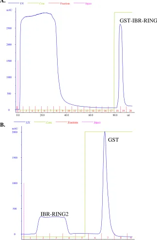
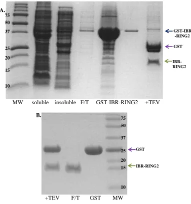
After the cell lysate was centrifuged, GST affinity chromatography was used to purify GST-IBR-RING2. The GST fusion protein was then cleaved with TEV protease and dialyzed extensively to remove excess glutathione. Thorough dialysis was necessary to ensure all glutathione was removed so that the cleaved GST tag could be separated from the IBR-RING2. A second GST purification was subsequently performed and the IBR-RING2 protein eluted in the flow-through fractions while the GST tag remained bound to the column. Figure 3.2 shows typical chromatograms of IBR-RING2 protein purification prior to and following cleavage of the GST tag from the protein. The purity of the protein throughout the process was checked by SDS-PAGE. Figure 3.3 shows segments of SDS-PAGE gels of the complete purification scheme of IBR-RING2. As stated in Chapter 2, all proteins that are GST-tagged were expressed and purified in the same manner. At the end of the purification, the amount of protein in solution was approximately 4-5 mg (from 2L of growth).
3.2 Determination of Optimal Conditions for NMR Data Collection
UV Conc Fractions Inject 0 500 1000 1500 2000 mAU
0.0 5.0 10.0 15.0 20.0 25.0 ml
1 2 3 4 5 6 7 8 9
A.
B.
Figure 3.2: Chromatograms of GST purification
A. Chromatogram of a typical GST affinity purification. The absorbance at 280 nm is
shown in blue. GST-IBR-RING2 is eluted from the column by the addition of 100% elution buffer (shown in green, 20mM Tris, 10mM GSH, at pH 8.0). B. Chromatogram of a second GST purification after TEV cleavage. Sharp and intense elution peak is an indication that thorough dialysis of glutathione was achieved, allowing separation of GST and IBR-RING2.
GST-IBR-RING2
GST
IBR-RING2
UV Conc Fractions Inject
0 500 1000 1500 2000 2500 mAU
0.0 20.0 40.0 60.0 80.0 ml
Figure 3.3: SDS-PAGE gels showing two-step purification of IBR-RING2
A. First, GST purification and TEV cleavage reaction. B. TEV-cleaved protein through the GSTrap HP column. Abbreviations used for each lanes are, MW: molecular weight marker, soluble: soluble fraction, insoluble: insoluble fraction, F/T: flow-through, and +TEV: TEV cleaved GST-IBR-RING2.
MW soluble insoluble F/T GST-IBR-RING2 +TEV
+TEV F/T GST MW
collect accurate data with degraded or precipitated protein in NMR studies. Therefore, it was important to find out what conditions would ensure that the IBR-RING2 was homogeneous, intact, well folded, and soluble.
As mentioned above, a major concern was the possible oxidation of 17 cysteine residues found in IBR-RING2. In order to avoid oxidation and/or disulphide bond formation, the sample was purged with Argon gas to displace oxygen in the NMR tube. In addition, Shigemi NMR tubes were used which limit the amount of oxygen exposure experienced by the sample due to the insertion of a plunger. Finally, addition of 5 mM DTT to the sample was used to prevent cysteine oxidation.
Figure 3.4: Comparison of 1H-15N HSQC Spectra and NMR tubes of 300-400µM IBR-RING2 after the data collection.
A. For non-optimized NMR sample after ten days (Disappearing peaks: circled, Newly appearing peaks: boxed) B. For optimized NMR sample (EDTA addition) after a month (Superimposable to the initial spectrum)
the IBR domain from human parkin unfolds upon the addition of EDTA (Beasley et al., 2007). However, there was still a possibility of the presence of metalloproteases in the sample (which cannot be blocked by any of the inhibitors in the protease inhibitor cocktail) that affect the integrity of IBR-RING2 and were causing it to precipitate. To test this, a small amount of EDTA (1 µM) was added to the protein sample in an attempt to inhibit the potential metalloproteases, and the stability of the IBR-RING2 was re-assessed. As seen in Figure 3.4, a minor amount of protein precipitated at the bottom, but generally the IBR-RING2 remained soluble in the NMR tube even after a month of experiments. Further, 1H-15N HSQC experiments compared before and after this period were almost identical, suggesting that the sample’s integrity was maintained. Therefore, it was concluded that the addition of trace amounts of EDTA helped the IBR-RING2 to stay in solution for a longer period of time by inhibiting unknown metalloproteases that might have been present in the NMR sample. Based on this finding, all subsequent NMR samples contained 1 µM EDTA.
3.3 Structural Determination of the Parkin IBR-RING2 Domain
3.3.1 ESI-MS shows the IBR-RING2 coordinates 4 Zinc ions
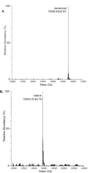
difference between the denaturing and non-denaturing mass of the IBR-RING2, as determined by Paula Pittock (Biological Mass Spectrometry Lab, UWO), was calculated. The native IBR-RING2 had a mass of 15843 Da and the denatured IBR-RING2 was found to have a mass of 15588 Da (Figure 3.5). The difference in mass was 255 Da and division of this value by four resulted in 63.75 Da which is close to the mass of zinc (65 Da), which led to the conclusion that there are four zinc ions coordinated in IBR-RING2.
3.3.2 Backbone Chemical Shift Assignment of IBR-RING2
For structural studies with NMR spectroscopy, each resonance needs to be associated with a specific nucleus in the protein of interest. The backbone chemical shift assignment of IBR-RING2 was accomplished manually using the sequential backbone resonance assignment method. A series of multidimensional NMR experiments (HNCACB, CBCA(CO)NH, HNCO, HN(CA)CO) were used in order to assign the HN, N, Cα and Cβ
Figure 3.5: Deconvoluted ESI-MS of proteins under denaturing and
non-denaturing conditions, which show the mass of both the native and denatured state of IBR-RING2.
A. Denaturing IBR-RING2 B. Non-denaturing IBR-RING2 result.
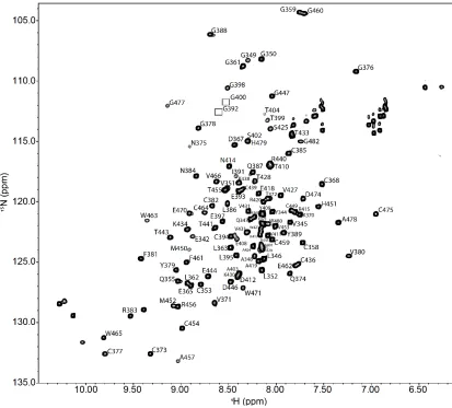
Figure 3.6: Two-dimensional 1H-15N HSQC spectrum of 15N-isotopically labeled IBR-RING2.
3.3.3 Utilization of IBR-RING2 backbone assignment
Assigning chemical shifts of the amide backbone is the first step in solving the structure of proteins. Additionally, the backbone assignment can serve several other purposes, including: conducting interaction studies by titration, the determination of residues involved in zinc coordination, and domain secondary structure. In this section, chemical shifts were used in the prediction of IBR-RING2 secondary structure, as well as zinc-coordinating cysteines.
3.3.4 Prediction of Zinc Coordinating Cysteines
With the backbone assignment completed, identification of cysteines exhibiting zinc coordination became possible. Using the sensitivity of chemical shifts to changes in electronic environment, prediction of zinc ligating cysteines and non-ligating cysteine residues can be made. Kornhaber and colleagues (2006) compiled the differences in values of Cα and Cβ of cysteines when oxidized (Cα: 55.57 ± 2.46 ppm, Cβ:41.17 ± 3.93 ppm), reduced (Cα:59.25 ± 3.06 ppm, Cβ:28.92 ± 2.11 ppm), and or zinc-coordinating (Cα:59.27 ± 2.12 ppm, Cβ: 30.89 ± 1,01 ppm) from the protein data bank (PDB) and BioMagResBank (BMRB). With this statistical data, the probability of the given cysteine’s zinc coordination could be calculated. Through the assignment of Cα and Cβ
Table 2. Cysteines involved in Zn2+ coordination in IBR-RING2 domain, as determined by chemical shifts of Cα and Cβ.
* determined using 3D CBCA(CO)NH and HNCACB experiments
# based on the structure calculation: (I) - involved in 1st zinc coordination site, (II) - in 2nd zinc coordination site, (cat) – suspected catalytic cysteine, (-) - non zinc coordinating cysteine
Cys Chemical Shifts (ppm)* IBR
C353 C358 C368 C373 C377 C382 C385 C394 C407
Actual# I I - I I II II II - Cα 57.731 60.268 58.581 58.504 59.988 57.691 58.112 59.835 58.713 Cβ 31.865 29.375 28.45 28.311 32.365 32.475 32.359 30.761 28.102
Probabilities
Oxidized 0.0% 0.0% 0.0% 0.0% 0.0% 0.0% 0.0% 0.0% 0.0%
Reduced 7.3% 39.8% 98.3% 99.1% 1.2% 7.0% 5.0% 4.1% 99.5%
Zn2+ 92.7% 60.2% 1.7% 0.9% 98.8% 93.0% 95.0% 95.9% 0.5%
Cys Chemical Shifts (ppm)* RING2
C436 C439 C449 C454 C459 C464 C467 C475
Actual# I I cat I I II II II Cα 57.016 59.348 59.291 61.571 60.499 57.776 56.999 64.533 Cβ 31.698 31.196 28 32.045 30.174 32.93 31.87 29.092
Probabilities
Oxidized 0.0% 0.0% 0.0% 0.0% 0.0% 0.0% 0.0% 0.0%
Reduced 13.2% 3.5% 99.5% 0.3% 6.8% 7.8% 12.3% 4.9%
coordinated to zinc ions. In addition, there were those other residues (C358, C436, C467) that had over 60% chance of zinc coordination. The data also clearly showed that several cysteine residues (C368, C373, C407, C468) exist in their reduced states based on Cα and Cβ chemical shifts (Table 2). In total, this data showed that 13 cysteines in IBR-RING2 are involved in zinc coordination. Zinc coordination in a protein structure typically requires four ligands, and it was predicted that there are more than three zinc ions that can bind to the IBR-RING2 structures. Since histidines can also act as zinc binding ligands, the data is in good agreement with the coordination of four zinc ions by IBR-RING2 shown from the ESI-MS results.
3.3.5 Determination of Secondary Structure for parkin IBR-RING2
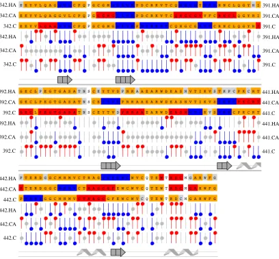
After completing the backbone and side chain chemical shift assignment of IBR-RING2, the secondary structure determination was performed using the chemical shift index (Wishart and Sykes, 1994). This was performed using a macro in the program NMRViewJ that uses chemical shifts of Hα, Cα, and C for each residue of IBR-RING2 to predict the secondary structure of the protein, as described in 2.4.2. In general, large regions of α-helices or β-strands were not present in the structure especially in the linker region between IBR and RING2 (Figure 3.7). The predicted secondary structure shows two β-strands in the IBR domain, between G350-L352 and L362-E365, a β-strands in the linker region Y409-D412, and three α-helices, P437-T441, C454-W471, and T473-A478, and two β-sheets, T433-P435 and F462-W464 in the RING2 region are present.
Figure 3.7: Chemical shift index of IBR-RING2 for secondary structure prediction.
RING2 domains. This is in agreement with the findings for the human parkin IBR structure, that showed little secondary structure (Beasley et al., 2007).
3.3.6 Structure Calculation of IBR-RING2
a)
b)
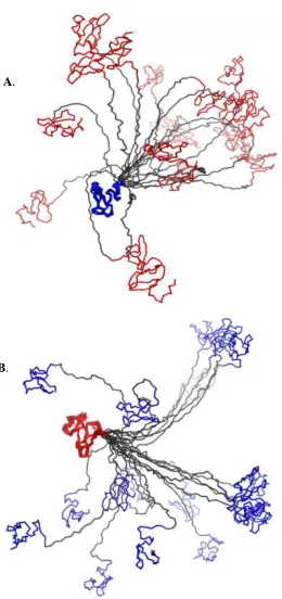
Figure 3.8: Superposition of 20 structures overlay of IBR-RING2, on each of the IBR (blue) and RING2 (red) domains.
Due to the flexible nature of the linker, both domains do not superposition well at the same time, as shown above. A. Superposition of IBR-RING2 to IBR using Cα atoms. B. Superposition of IBR-RING2 to RING2 using Cα atoms.
Figure 3.9: Representative ribbon structure of IBR-RING2 with the flexible linker between the two domains.
Spheres represent zinc ions bound to each of the domains. Magenta represents the β -sheets and cyan represents the α-helix in the RING2 structure.
C
N
Flexible Linker
IBR
Table 3: Structural Statistics for 20 lowest NOE energy structures of IBR-RING2
1 For IBR residues 342-396 / RING2 residues 430-482 / Entire sequence E342-G482 2 Psi/Psi dihedral restraints determined using TALOS+
3 Total of 4 NOE violations > 0.5 Å over all 20 models 4 As reported by Xplor-NIH
5
Using residues V351-I392 for IBR / residues K430-W480 for RING2 (all 20 models)
6 As reported by Procheck using residues V351-I392 for IBR / residues K430-W480 for RING2 /
the entire sequence E342-G482.
Protein Completeness of Resonance Assignments
Backbone (N, CA) (103/112) – 92.0%
Sidechain (C, H) (1073/1219) – 82.8%
HN (122/133) – 91.7%
HA (151/158) – 95.4%
HB (202/202) – 100%
NMR distance and dihedral constraints
Distance constraints1
Total NOE 611 / 447 / 1156
Intra-residue 156 / 143 / 360
Inter-residue
Sequential (|i – j| = 1) 201 / 117 / 353
Medium-range (|i – j| < 4) 65 / 48 / 115
Long-range (|i – j| > 5) 189 / 139 / 328
Intermolecular - / - / 0
Hydrogen bonds 0 / 0 / 0
Zinc restraints 24 / 24 / 48
Total dihedral angle restraints2
φ 22 / 25 / 49
ψ 22 / 25 / 49
Structure statistics
Violations (mean and s.d.)
Distance constraints (Å) 0.040 ± 0.002
Dihedral angle constraints (º) 0.467 ± 0.080
Max. dihedral angle violation (º) 7.5
Max. distance constraint violation (Å)3 0.7 Deviations from idealized geometry4
Bond lengths (Å) 0.006 ± 0.000
Bond angles (º) 0.550 ± 0.022
Impropers (º) 0.758 ± 0.056
Average pairwise r.m.s. deviation (Å)5
Heavy 1.288 ± 0.313 / 1.519 ± 0.399
Backbone 0.800 ± 0.220 / 0.914 ± 0.216
Ramachandran statistics6
Residues in most favored regions Residues in additional allowed regions Residues in generously allowed regions Residues in disallowed regions
Individual IBR and RING2 domains within the IBR-RING2 structure are shown in Figures 3.10 and 3.11. As shown previously in the human parkin IBR structure (Beasley et al., 2007), the IBR did not display any notable secondary structure. The N-terminus at the beginning of the IBR is not as structured, and can be attributed to it being the end part of the linker region between RING1 and IBR. The IBR domain shows a bilobal fold with two zinc ions bound to the protein. It contains two zinc sites in “scissor-like” and “Gag-knuckle-“scissor-like” folds, as described by Beasley et al. (2007). The long ends of the loop for the first zinc site (utilizing C353, C358, C373, and C377) are the origin of the ‘scissor-like’ fold also referred to as zinc ribbon (Krishna et al., 2003). The IBR second zinc binding site (using C382, C385, H390, and C394) is referred to as the zinc knuckle-like fold, based on the compact zinc binding site which occurs due to the sudden turn (zinc knuckle) required to wrap around the zinc ion.
a b) c
Figure 3.10: Ribbon structure of parkin IBR domain with template of zinc binding sites.
A. IBR domain with zinc coordinating cysteines are highlighted in yellow, showing
limited secondary structure. B. Zinc ribbon structure representative figure. C. Representation of gag-knuckle-like fold.
Figure 3.11: Ribbon structure of parkin RING2 domain.
RING2 domain is illustrated with α helix and β sheets, with catalytic cysteine and zinc coordinating cysteines highlighted in yellow.
A. B. C.
C
N
Zn Site I Zn Site II
Zn Site I Zn Site II