ABSTRACT
MACIAS, EVERARDO. The Role of CDKs in Normal and Neoplastic Proliferation. (Under the direction of Dr. Marcelo Rodriguez-Puebla.)
Cyclin-dependant kinases (CDKs) are serine/threonine kinases which play a central role in cell cycle progression. The more characterized function of CDKs is the phosphorylation and inhibition of pRb allowing the transcription of genes needed for cell cycle progression from G1 to S phase. Loss or mutation of pRb
results in deregulated cell growth however; this is also achieved by functional inactivation due to elevated CDK activity. In particular, molecular analysis of some human tumors shows deregulated CDK2 and CDK4 activity. CDK4 is implicated in tumor development since it is frequently found overexpressed, genetically amplified and mutated in human tumors. In contrast, CDK2 is not mutated and is rarely found genetically amplified or overexpressed. Therefore, it remains unclear whether elevated CDK2 kinase activity in human tumors is a causative effect or consequence of the disease. The general purpose of our studies has been to determine the role CDK2 and CDK4 in normal and neoplastic proliferation.
results suggest that the efficacy of using CDK2 inhibitors will depend on the oncogenic pathway involved.
The Role of CDKs in Normal and Neoplastic Proliferation
by
Everardo Macias
A dissertation submitted to the Graduate Faculty of North Carolina State University
In partial fulfillment of the Requirements for the degree of
Doctor of Philosophy
Comparative Biomedical Sciences
Raleigh, NC 2007
APPROVED BY:
_________________________________ ________________________________ Dr. Marcelo Rodriguez-Puebla Dr. Jonathan M. Horowitz
Co-Chair of Advisory Committee Chair of Advisory Committee
ii
DEDICATION
I would like to dedicate the works presented here to my mother, Maria Oralia
Valdez. Your perseverance as a single mother taught me how to face adversity
and inspired me to forge ahead.
To my father, Everardo Macias Becerra, for your advice and support.
To my brothers and sisters, Maria Isabel Sullivan, John Anthony Macias,
Dalileah Caballero, and Alejandro Macias, your love and support made this
work possible.
iii
ACKNOWLEDGMENTS
I would like to acknowledge my advisor, Marcelo Rodriguez-Puebla, for his
guidance and support throughout this process. Thank you for taking a chance on me and
providing me the skills necessary to pursue a career in research. I will work hard to ensure
that your investments in me have not been in vain.
I would like to extend as special thanks to Paula Miliani de Marval for everything
you have taught me in and out of the lab. Thank you for believing in me and being one of
my strongest supporters.
I would also like to express thanks to the College Assistant Migrant Program
(CAMP) at St. Edward’s University for giving me and many others in my position the
opportunity to seek higher education. I don’t know where many of us would be without
programs such as yours. In particular, I would like to extend my gratitude to Esther
Yocaño and LindaValdez.
In addition, I would like to thank the members of my graduate committee, Dr.
Robert Smart, Dr. Jon Horowitz and Dr. Cullen. Your input and participation throughout
this process is deeply appreciated.
iv
BIOGRAPHY EVERARDO MACIAS
EDUCATION
2003-2007 North Carolina State University, Raleigh, NC Department of Molecular Biomedical Sciences Comparative Biomedical Sciences Program Laboratory of Dr. Marcelo Rodriguez-Puebla 1997-2001 St. Edward’s University, Austin, TX
Bachelors of Science in Chemistry
PUBLICATIONS
Everardo Macias, Paula Miliani de Marval, Adrian Senderowicz, Marcelo L. Rodriguez-Puebla. Elevated CDK2 kinase activity increases epidermal proliferation, but does not enhance tumor development or malignant progression. Manuscript in preparation
Everardo Macias, Yongbaek Kim, Paula Miliani de Marval, Andres Klein-Szanto, and Marcelo L. Rodriguez-Puebla. Cdk2 deficiency decrease ras/cdk4-dependent malignant progression, but not myc-induced tumorigenesis. Cancer Research, In Press
Everardo Macias, Paula L. Miliani de Marval, Andrzej Dlugosz, John M. Cullen, and Marcelo L. Rodriguez-Puebla. Transgenic expression of CDK4 and CDK2 in mouse embryonic oral cavity is specifically retained in the adult adenohypophysis and collaborates in pituitary tumorigenesis. Cancer Research, In Press.
Wei-Ming Chien, Everardo Macias, Paula L. Miliani de Marval, Stuart Rabin, Kendra Garrison, Jason Orthell, Marcelo Rodriguez-Puebla, Matthew L. Fero. Genetic mosaics reveal both cell autonomous and non-autonomous functions of murine p27Kip1. Proc Natl Acad Sci U S A. 2006 Mar 14;103(11):4122-7.
Paula L. Miliani de Marval, Everardo Macias, Robert Rounbehler, Piotr Sicinski, Hiroaki Kiyokawa, David G. Johnson, Claudio J. Conti and Marcelo L. Rodriguez-Puebla. Lack of Cyclin-Dependent Kinase 4 Inhibits c-myc Tumorigenic Activities in Epithelial Tissues. Molecular and Cellular Biology, Sept. 2004, p. 7538–7547
v
TABLE OF CONTENTS
List of Figures……….vii
List of Tables ………...x
Chapter I Introduction………1
1.1 Cell Cycle Regulation………2
1.2 Regulation of Cyclin-dependant kinases………7
1.3 CDKs in Human and Experimental Mouse Tumors………12
1.4 c-Myc and the Cell Cycle………14
1.5 Ras Signaling ………..17
1.6 TGF-β Signaling ………..19
1.7 Mouse Skin Model ………..21
1.8 Pituitary Gland ……….23
1.9 Research Focus ………..25
Chapter II Elevated CDK2 kinase activity in the epidermis does not enhance tumorigenesis ………...27
2.1 Introduction ………..28
2.2 Matrials and Methods ………..30
2.3 Results ………..33
2.4 Discussion ……….53
Chapter III CDK2 Ablation reduces CDK4/h-Ras mediated malignant progression, but does not alter c-Myc mediated tumorigenesis……….58
vi
3.2 Materials and Methods ………61
3.3 Results ………..64
3.4 Discussion ………87
Chapter IV Transgenic expression of CDKs in mouse embryonic oral cavity is specifically retained in the adult adenohypophysis and collaborates in pituitary tumorigenesis ………94
4.1 Introduction ………..95
4.2 Materials and Methods………...97
4.3 Results ………101
4.4 Discussion ……….133
Chapter V General Discussion ………141
Bibliography ………152
Appendix I Analysis of CDK4 distribution in high and low molecular weight complexes in normal and neoplastic epithelial cells ………...177
Appendix II Genetic mosaics reveal both autonomous and cell-nonautonomous function of murine p27Kip1 ………...190
Appendix III Enhanced malignant tumorigenesis in CDK4 transgenic mice ……….197
vii
LIST OF FIGURES
Figure 1. The Cell Cycle and CDKs ………...6 Figure 2. Redistribution of Cip/Kip Family Members ………..9 Figure 3. Founder K5CDK2 transgenic mice………35 Figure 4. K5CDK2 skin morphology and immuno-localization of transgene
expression ………36 Figure 5. Histochemical analysis of K5CDK2 skin ………..37 Figure 6. Biochemical Analysis of K5CDK2 Epidermis ………39 Figure 7. Kinetics of papilloma formation and progression to SCCs in K5CDK2 transgenic mice………42 Figure 8. Biochemical analysis of K5CDK2 skin tumors……….46 Figure 9. Western blot and immunoflourescence analysis of TGF-β effector and target proteins ……….51 Figure 10. Epidermal proliferation in K5CDK4/CDK2-/- compound mice ……....67
Figure 11. Biochemical analysis of K5CDK4/CDK2-/- epidermal lysates ……....69 Figure 12. Effect of CDK2 deletion on Ras-CDK4 mediated skin tumorigenesis ………71 Figure 13. Keratin expression in K5CDK4/CDK2-/- mouse skin tumors ………76 Figure 14. Biochemical analysis of K5CDK4/CDK2-/- papillomas ……….78
viii
Figure 18. Biochemical analysis of K5Myc/CDK2-/- mouseepidermis ………...86
Figure 19. Visualization of K5-transgenes in embryonic pituitary………….…..105 Figure 20. CDK transgene expression in adult mouse pituitary glands ……….106 Figure 21. Biochemical analysis of K5-transgenic mouse pituitary glands …108 Figure 22. Keratin 5 expression developing pituitary gland ……….109 Figure 23. CDK4 transgene expression in CDK4 null background ………….110 Figure 24. Hormone and hCDK4 expression in developing pituitary ………..112 Figure 25. Analysis of pituitary stem cells with Hoechst 33342 vital dye ……114 Figure 26. CDK4 transgene and hormone expression in proliferating cells of developing pituitary gland as analyzed by immunofluorescence ……….116 Figure 27. Phenotypic analysis of K5CDK4/p27-/- compound mice ………….119
Figure 28. Immunohistochemical analysis of anterior and intermediate lobe hormones in normal pituitary glands and K5CDK4/p27-/- pituitary tumors ……..121 Figure 29. Dorsal view of K5CDK2/p27-/- pituitary glands ……….123
Figure 30. Hormone and transgene expression in proliferating cells of
K5CDK4/p27-/- pituitary tumors ………..125 Figure 31. Biochemical analysis of K5CDK4/p27-/- pituitary tumors …………129 Figure 32. Collaboration of CDK4 and p27Kip1 loss in thymus ……….131 Figure 33. Expression of T-pit in K5CDK4 pituitary glands………..132 Figure 34. Distribution of CDKs in HMW and LMW fractions of wild type
ix
x
LIST OF TABLES
1
CHAPTER I
2 1.1 Cell Cycle Regulation
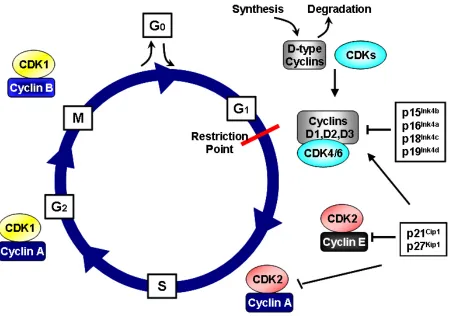
Cell division consists of an organized series of events in which a cell duplicates its contents and then divides into two daughter cells, a process known as the cell cycle. The cell cycle is divided into four phases, first gap phase (G1),
synthesis phase (S phase), second gap phase (G2) and mitosis (M phase)
respectively. The vast amount of DNA is duplicated in S phase, which requires 10-12 hours, approximately half the time of a typical mammalian cell cycle. The segregation of duplicated chromosomes and cell division occurs in M phase, a very fast process consisting of nuclear division (mitosis) and cell division (cytokinesis) (Alberts 2002). Cell growth and duplication of cellular organelles and macromolecules is carried out in gap phases, G1 and G2 phases before S phase and
3
multiple cyclins and cyclin-dependent kinases. Recent analysis of the human genome identified at least 29 genes encoding related proteins that share a conserved stretch of 150 amino acid residues termed the cyclin box (Malumbres and Barbacid 2005). The more well characterized cyclins are those associated with G1 (D-type cyclins, D1, D2, D3), S phase (E1/E2 and A1/A2) and mitosis (A and B1/B2) (Murray 2004). Catalytic subunits Cdk1-11 have been cloned and characterized in addition to multiple highly related molecules have been identified, but have not been formally designated CDKs because their activating subunits have not been identified (Malumbres and Barbacid 2005).
The commitment of cells to undergo another round of cell division is made during G1, at a stage called the restriction point (R-point) (Pardee 1974). Once cells
4
permitting the transcription of cyclin E. This allows activation and complex formation of CDK2/cyclin E, which further phosphorylate pRb. Hyper-phosphorylated pRb releases E2F family transcription factors allowing the transcription of S phase critical genes, including those encoding the A-type and B-type cyclins (Malumbres and Barbacid 2001).
In S phase, CDK2-Cyclin E participate in the initiation of DNA synthesis while CDK2-Cyclin A complexes phosphorylate proteins required for the proper completion and exit from S phase. A few putative substrates of CDK2/Cyclin E involved in DNA synthesis include Cdt1, minichromosome maintenenace protein 2 (MCM2) and Cdc6. CDC6 and Cdt1 are involved in loading MCM proteins onto chromatin, which are also referred to as “licensing factors” for DNA replication. The recruitment of minichromosome maintenance proteins (MCM), which are helicases, is essential for initiation andelongation of the replication fork (Labib, Tercero et al. 2000). MCM2 is phosphorylated by CDK2/Cyclin E, targeting it for further phosphorylation, which activates its helicase activity. CDK2/Cyclin A phosphorylates CDC6, which leads to its export from the nucleus, and targeted degradation by the anaphase-promoting complex/cyclosome (APC/C) (Otzen Petersen, Lukas et al. 1999). Exportation of CDC6 by CDK2/Cyclin A is thought to prevent reloading of MCM proteins and assure that DNA is replicated only once per cell cycle (Aguda 2001).
5
6
Figure 1. The Cell Cycle and CDKs. The cell cycle is divided into four phases, first gap phase (G1), synthesis phase (S phase), second gap phase (G2) and mitosis (M
7
1.2 Regulation of Cyclin-dependant kinases
1.2.1 Cyclin synthesis and regulation
8
Zindy et al. 1997). Interestingly, GSK-3β-dependent turnover of cyclin D1 is inhibited by Ras via PI-3 kinase/Akt signaling (Diehl, Cheng et al. 1998). CDK2 is activated by complex formation with cyclins E1/E2 and A1/A2. Although CDK2 has also been shown to complex with D-type cyclins the preferential binding partners are cyclins E and A. In contrast to growth factor inducible D-type cyclins, synthesis of E and A type cyclins is strictly dependent on E2F transcriptional activation (Ohtani, DeGregori et al. 1995; Schulze, Zerfass et al. 1995). Once the cells enter S phase, cyclin E is targeted for rapid degradation by the SCF-Fbw7 ubiquitin ligase (Koepp, Schaefer et al. 2001). The rapid turnover of Cyclin E is facilitated by auto-phosphorylation and permits the formation of CDK2-Cyclin A complexes.
1.2.2 Cyclin-CDK complex formation and regulation
CDK complexes are also regulated through association with Cyclin-dependent kinase inhibitors, CKIs, which consist of two families the INK4 (Inhibitors for CDK4) and Cip/Kip family. The Cip/Kip family consists of three family members:
p21WAF1/CIP1, p27KIP1 and p57KIP2. The latter are considered broad-spectrum
inhibitors because they bind to both cyclin D-CDK4/6 kinases and cyclin E/A-CDK2 kinases (Ekholm and Reed 2000). In contrast, INK4 family members (p15Ink4b, p16Ink4a, p18Ink4c and p19Ink4d) are considered narrow-spectrum inhibitors because
9
10
members. Thus, INK4 inhibitors not only cause the inhibition of CDK4,6, but also causes a redistribution of Cip/Kip family members to cyclin-E-CDK2 resulting in the inhibition of these complexes (Figure 2)(Sherr and Roberts 1999).
While newly synthesized D-type cyclins quickly form complexes with CDKs and are translocated into the nucleus of proliferating cells, evidence suggests that complex formation requires other proteins. Purified recombinant cyclin D1 and CDK4 proteins bind with very low affinity in vitro and ectopically expressed D-type cyclins and CDKs do not form holoenzymes in serum deprived cells (Kato, Matsuoka et al. 1994; Matsushine, Quelle et al. 1994). Several interactors of cyclins D-CDK4/6 have been proposed to play a role in complex assembly. Chaperone protein, HSP90 via p50cdc37 is thought to aid in the proper folding of CDK4 and compete with p16INK4a
11
Martin et al. 2002; Bagui, Mohapatra et al. 2003). It is also suggested that p21Cip1
and p27Kip1 do stabilize cyclin D3/CDK4 and cyclin D1/CDK4 complexes, but they are not required for their assembly (Bagui, Mohapatra et al. 2003).
1.2.4 Post-translational Modifications
12
1.3 CDKs in Human and Experimental Mouse Tumors
pRb is considered the master switch of the cell cycle and a great deal of literature shows the importance of this protein in tumor development. While direct mutational inactivation of pRb is found in human tumors, such as retinoblastoma and osteosarcomas, alterations of this pathway may also be caused by functional inactivation of pRb by hyperphosphorylation (Gallie, Dunn et al. 1991). This is normally the result of elevated CDK activity caused by the overexpression of cyclins and CDKs, or loss of CKIs.
13
in normal cells (Koff, Giordano et al. 1992; Tsai, Lees et al. 1993). Thus, CDK2 is an attractive target for therapeutic intervention since it is thought to be essential for normal and deregulated cell cycle progression. Surprisingly, ablation of cyclin E and CDK2 in mouse models have shown that both CDK2 and Cyclin E are dispensable for normal mouse development (Berthet, Aleem et al. 2003; Geng, Yu et al. 2003; Ortega, Prieto et al. 2003). Coincidently, Tetsu and McCormick published work suggesting that CDK2/Cyclin E activity is dispensable for cell cycle progression in some cancer cells lines (Tetsu and McCormick 2003). Recent evidence suggests that CDK2 may be dispensable for p21Cip1 and p27Kip1 mediated tumor suppression (Aleem, Kiyokawa et al. 2005; Martin, Odajima et al. 2005). Moreover, CDK2 -/-primary cells remain susceptible to oncogenic transformation (Berthet, Aleem et al. 2003; Ortega, Prieto et al. 2003).
14
2004; Reddy, Mettus et al. 2005; Yu, Sicinska et al. 2006). Also, CDK4R24C knock-in
mice develop a wide spectrum of tumors (Sotillo, Dubus et al. 2001). More specifically, we have shown that papillomas derived from mice overexpressing CDK4 under the K5 promoter progress to squamous cell carcinomas at a very high frequency (Miliani de Marval, Macias et al. 2004). In conjunction, we have shown that epidermal hyperplasia and development of spontaneous oral tumors in K5Myc mice is dependent on CDK4 expression (Miliani de Marval, Macias et al. 2004). Molecular analysis of K5CDK4 skin tumors show that this phenotype is in part mediated by the sequestration of p27Kip1 and p21Cip1 by CDK4, indirectly activating CDK2 kinase activity. We have also shown that epidermal hyperplasia and spontaneous oral tumor formation in K5Myc transgenic mice is dependent on the sequestration mechanism by elevated levels of endogenous CDK4 (Miliani de Marval, Macias et al. 2004).
1.4 c-Myc and the Cell Cycle
15
16
17 1.5 Ras Signaling
18
carcinogenesis protocol (Robles, Rodriguez-Puebla et al. 1998). In addition, Cyclin D1 is targeted for proteosomal degradation by phosphorylation by glycogen-synthase kinase 3β (GSK-3β) (Diehl, Zindy et al. 1997; Diehl, Cheng et al. 1998). GSK-3β dependant turnover of cyclin D1 is also regulated by Ras since the activity of protein kinase B (Akt), which inactivates GSK-3β, is activated by Ras via PI-3 kinase signaling (Diehl, Cheng et al. 1998). Ras further promotes G1phase progression by altering the expression levels of p27Kip1 and p21Cip1 at the transcriptional and proteolytic level (Liu, Martindale et al. 1996; Medema, Kops et al. 2000; Delmas, Manenti et al. 2001).
19 1.6 TGF-β Signaling
Transforming growth factor β (TGF-β) signaling is activated via three independently encoded TGF-β ligands, β1, β2, and β3 (Roberts and Sporn 1990). All TGF-β isoforms initiate signaling of downstream targets via transmembrane serine/threonine kinases, type I and II TGF-β receptors (TβRI and TβRII). TβRI and TβRII are primarily found as homodimers in the endoplasmic reticulum (ER) and on the plasma membrane, although there is some evidence suggesting that they can form heterodimers in the absence of ligands (Gilboa, Wells et al. 1998; Gilboa, Nohe et al. 2000). TGF-β ligands preferentially bind TβRII homodimers, ligand bound TβRII has a high affinity for binding to and transactivating TβRI receptors (Moustakas, Lin et al. 1993). Activation of TβRI leads to auto-phosphorylation and phosphorylation of effecter Smad proteins. Once phosphorylated and released by the active receptor complex, two receptor activated Smads (R-Smads 2/3) and a common Smad4 form a heterotrimeric complex, translocate to the nucleus and regulates genes by DNA binding and recruiting transcriptional co-activators or repressors (Derynck and Zhang 2003).
1.6.1 TGF-β and Cell Cycle
Relevant to understanding the role of TGF-β in tumor development is the
ability of TGF-β to induce growth inhibition. A key step to cell cycle arrest by TGF-β is Smad mediated transcriptional induction of CKIs, p15Ink4b and p21Cip1.
20
p15Ink4b binds to and inhibits CDK4 and CDK6. As detailed above p15Ink4b
displaces Cip/Kip family members from cyclin D-CDK complexes, increasing the pool of unbound p21Cip1 and p27Kip1 allowing them to bind to and further inactivate CDK2 complexes. Furthermore, TGF-β induced cell cycle arrest is also carried out by decreasing the synthesis of CDK4 (Ewen, Sluss et al. 1993; Ewen, Oliver et al. 1995; Sandhu, Garbe et al. 1997). TGF-β also slows cell cycle progression by decreasing the expression of c-Myc and Cdc25a (Frederick, Liberati et al. 2004; Ray, Terao et al. 2005).
1.6.2 TGF-β signaling in Neoplasias
21 1.7 Mouse Skin Model
1.7.1 Mouse skin
The skin is comprised of three primary components, a superficial epithelial lining consisting of several layers called the epidermis, a richly vascularized and innervated connective tissue frequently called the dermis and epithelial structures connected to the epidermis called appendages (hair follicles, sebaceous glands) (Conti 1989). The epidermis consists of four layers of epithelial cells or keratinocytes. The basal cell layer is made up of resting keratinocytes which have proliferative capacity, mitotic or proliferating keratinocytes, early differentiating keratinocytes and unrelated cells such as melanocytes, Langerhan's and Merkel cells. As basal layer keratinocytes divide they detach from the underlying basal lamina and migrate vertically, making up the prickle cell layer (stratum spinosum), the granular cell layer (stratum granulosum) and finally the outermost, horny cell layer (stratum corneum) (Maronpot 1999). As they migrate, keratinocytes become post mitotic, irreversibly differentiated, enucleated, undergo keratinization and are ultimately shed into the environment. This process is dependent on the basal cells of the epidermis to maintain a steady proliferative rate, which compensates for the continual shedding of the outermost squamous cells.
1.7.2 Two-Stage Carcinogenesis Model
22
23
the genetic background benign exophytic epithelial tumors of cauliflower like structure, papillomas, appear 6-8 weeks after the initial application of a tumor promoter. These benign tumors may progress to squamous cell carcinomas after 25-30 weeks depending on the genetic background of the mouse strain.
1.8 Pituitary Gland
24
spatial pattern (Sheng and Westphal 1999; Zhu and Rosenfeld 2004). Proliferation in the pituitary gland is believed to arise from self-mitosis of differentiated cells, the proliferation of undifferentiated and possibly pituitary stem cells (Taniguchi, Yasutaka et al. 2002).
1.8.1 Alteration of Cell Cycle regulator in pituitary tumors
25
organogenesis, as mice nullizygous for CDK4 develop a hypoplastic pituitary gland and display lactotroph dysfunction and infertility (Moons, Jirawatnotai et al. 2002; Moons, Jirawatnotai et al. 2002).
1.9 Research Focus
CDKs are serine/threonine kinases, which play a central role in cell cycle progression. The most characterized function of CDKs is the phosphorylation and inhibition of pRb. Inhibition of pRb releases E2F transcription factors allowing cells to progress from G1 to S phase. Disruption of the pRb pathway renders cells insensitive to antigrowth signals that act through this pathway to block G1 progression. Insensitivity to growth signals leads to deregulated cell proliferation and is considered a hallmark of cancer (Hanahan and Weinberg 2000). It is clear that direct loss/mutation of pRb or pRb functional inactivation due to elevated CDK activity results in deregulated cell growth.
26
activated in K5CDK4 and K5Myc transgenic mice. Molecular analysis suggests that CDK2 is activated due to the sequestration of CKIs by elevated levels of CDK4. This highlights the sequestration or non-catalytic function of CDK4 and suggests that CDK2 may in fact contribute to the development of human tumors. However, since the time our studies were undertaken there have been multiple controversial findings regarding the role of CDK2 in normal and neoplastic cell proliferation. First, CDK2 mice are viable with little phenotype and CDK2 null cells remain susceptible to oncogenic transformation (Berthet, Aleem et al. 2003; Ortega, Prieto et al. 2003). Lastly, certain cancer cell lines were shown to continue to proliferate despite inhibition of CDK2 (Tetsu and McCormick 2003). Altogether, these works have brought about three central questions:
(1) Does elevated CDK2 kinase activity contribute to tumor development? (2) Is CDK2 necessary for tumor development and/or progression?
(3) Which is the more relevant function of CDK4 in tumorigenesis? It’s kinase or sequestration activity?
27
CHAPTER II
Elevated CDK2 kinase activity in the epidermis does not
28 2.1 Introduction
The cyclin/CDK/pRb pathway is frequently deregulated in human neoplasias. In particular, molecular analysis of human and experimental tumors has shown deregulation of CDK4 and CDK2 kinase activity. CDK4 has been clearly implicated in tumor development as found overexpressed in various human tumors and found mutated in the p16Ink4 binding domain, CDK4R24C, in hereditary melanoma (Zuo
1996). While CDK2 kinase activity is found elevated in human tumors, CDK2 is not directly affected by mutations and only a extremely small subset of human tumors have been shown to have CDK2 gene amplification or elevated protein expression (Kitahara, Yasui et al. 1995; Marone, Scambia et al. 1998; Shintani, Mihara et al. 2002). In contrast, CDK2 regulatory binding partner cyclin E is frequently found amplified and overepxressed in human tumors (Akama, Yasui et al. 1995; Keyomarsi and Herliczek 1997; Eguchi, Fujii et al. 1999). Mouse models have further demonstrated that Cyclin E contributes to the development of tumors (Bortner and Rosenberg 1997; Smith, Henze et al. 2006; Ma, Fiering et al. 2007). Likewise, loss of CDK2 inhibitor p27Kip1 is a common event in human tumors and mouse models demonstrate its role as a tumor suppressor. While mouse models of CDK2 regulatory proteins Cyclin E and p27Kip1 are susceptible to tumor development, it remains unclear whether elevated CDK2 kinase activity per se is a cause or a consequence of tumorigenesis.
29
Gimenez-Conti et al. 2001; Miliani de Marval, Macias et al. 2004). Biochemical analysis of K5-CDK4 epidermis and skin tumors show that this phenotype is in part mediated by the sequestration of p27Kip1 and p21Cip1 by CDK4, indirectly activating CDK2 kinase activity. In support, primary human keratinocytes infected with CDK4 and Ras retrovirus produce invasive neoplasias when grafted to nude mice (Lazarov, Kubo et al. 2002). Biochemical analysis of these grafted tumors shows a reduction of p21Cip1 complexed with CDK2 due to CDK4 sequestration and for that reason, increased CDK2 kinase activity. We have also shown that epidermal hyperplasia and spontaneous oral tumor formation in mice K5-Myc transgenic mice is dependent on the sequestration of p27Kip1 and p21Cip1 by elevated levels of endogenous CDK4, indirectly activating CDK2 kinase activity (Miliani de Marval, Macias et al. 2004). Altogether, these data suggest that increased CDK2/Cyclin E activity observed in human and experimental tumors may in fact play a role in tumor progression. Therefore, we hypothesize that deregulated CDK2 kinase activity contributes to the development of experimental mouse and human tumors.
30
demonstrate that contrary to our initial hypothesis elevated CDK2 kinase activity does not increase papilloma formation or enhance the rate of malignant progression to squamous cell carcinomas under a two-stage carcinogenesis model. To conclude, we provide evidence that inhibition of Smad3, a novel CDK4 target, accelerates malignant progression of K5CDK4 skin tumors, a mechanism that is absent in K5CDK2 and K5CDK4D158N experimental skin tumors.
2.2 Materials and Methods
2.2.1 Development of transgenic mice
K5CDK2 and K5-CDK4D158N transgenic mice were develop by cloning human-CDK4 cDNA carrying the D158N mutation or human-CDK2 into the vector pBK5
which contained the 5.2 Kb bovine keratin 5 regulatory sequences, β-globin intron 2
31 2.2.2 Two-stage chemical carcinogenesis
For two-stage carcinogenesis, twenty mice from each group were initiated with a single topical application of 50 μg of DMBA in 50 μl of acetone on the dorsal mouse skin at 21 days of age. Two weeks after initiation, mice received 2.5 μg of TPA in 200 μl of acetone twice a week for 25 weeks. Skin tumors were counted once a week until the end of the experiment at 40 weeks. Malignant progression to SCC was determined by macroscopic observation and further confirmed by histopathological analysis of paraffin-embedded H&E stained cross sections.
2.2.3 Western blots and kinase assays
For Immunoblots protein lysates were collected from epidermal skin scrapes with RIPA lysis buffer, 150 mM NaCl, 1.0% IGEPAL, 0.5% DOC, 0.1% SDS, 50mM Tris (pH 8.0). Papillomas were snap frozen in liquid N2 and crushed with a pestle
and mortar. Homogenates from epidermal scrapes or papillomas were sonicated and centrifuged at 14,000 r.p.m at 4˚C. Supernatants were boiled in 2X lameli sample buffer for western blot analysis or stored at -80°C.
32
Santa Cruz, CA, USA) for 2 hr on an end over end rotor at 4°C, then incubated with 35 µl of protein-A-agarose bead for 1 hr. Beads were pelleted and washed twice each with IP buffer and once with kinase buffer (50 mM HEPES [pH 7], 10 mM MgCl2, 5 mM MnCl2). Then, 30 μl of kinase buffer, 1 µg of pRb (CDK4 assay) or histone H1 (CDK2 assay) substrate (Upstate Biotechnology Inc., Charlottesville, VA.), 5 μCi of [γ-32P]ATP (6,000 Ci/mmol), 1 mM DTT, and 5 μM ATP were added to the bead pellet and incubated for 30 min at 30°C. Then 30 ul of SDS sample buffer was added to each sample and boiled for 3 min to stop reaction and electrophoresed through polyacrylamide gels.
Western blot and kinase assay bands were digitized and quantified using UN-SCAN-IT gel ™ version 6.1 software for windows.
2.2.4 Immunostaining
For immunostaining tissue cross sections of formalin fixed skin or tumors were permeablized using citrate antigen retrieval buffer, blocked with 10% normal serum and stained with antibodies for CDK2 (Santa Cruz Biotechnology, Santa Cruz, CA, USA) or p15Ink4b (Lab Visions Corp, Fremont, CA). Epithelial cell proliferation was measured by intraperitoneal injection of BrdU 30 min before the mice were sacrificed by CO2 asphyxiation. BrdU incorporation was detected by
33
and an avidin–biotin–peroxidase kit (Vectastain Elite, Vector Laboratories, Inc.) with diaminobenzidine as chromogen.
2.2.5 Statistical analysis
Statistical analysis was performed using GraphPad Prism 4 Software (GraphPad Software, San Diego, CA, USA)
2.3 Results
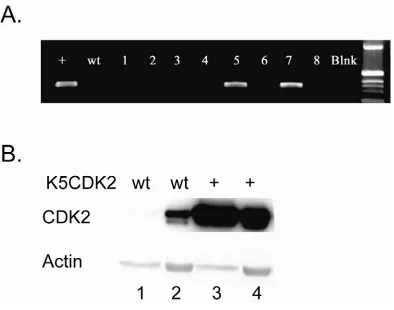
2.3.1 Generation of CDK2 transgenic mice
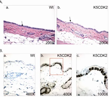
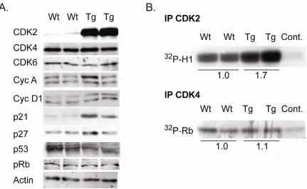
Expression of human CDK2 was targeted to the stratified epithelium using the 5’ regulatory sequence of the Keratin 5 gene. The bovine K5 promoter was previously shown to target transgene expression to the basal cell layer of the stratified squamous epithelia (Ramirez, Bravo et al. 1994). Based on PCR results of integration positive mice, two founders were used to establish lines K202 and K203 of K5CDK2 transgenic mice in an FVB/N background (Figure 3). Immunohistochemistry using antibodies against CDK2 of paraffin-embedded skin verified that exogenous CDK2 was overexpressed and restricted to the keratin 5 expressing basal cell layer of the epidermis (Figure 4B). Line K202 was amplified and used for all the work presented. Western blot analysis verified that K5CDK2 mice exhibit increased CDK2 protein expression in skin epidermis and thymus (Figure 3).
34
35
36
37
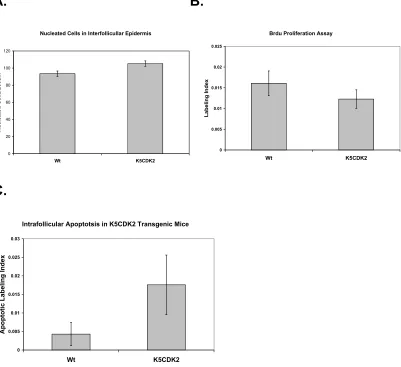
A. B.
Nucleated Cells in Interfollicullar Epidermis
0 20 40 60 80 100 120 Wt K5CDK2 N u c lea ted Ce lls/2 00 um
Brdu Proliferation Assay
0 0.005 0.01 0.015 0.02 0.025 Wt K5CDK2 La be ling In dex
C.
Intrafollicular Apoptotsis in K5CDK2 Transgenic Mice
0 0.005 0.01 0.015 0.02 0.025 0.03 Wt K5CDK2 A p o p totic L abe ling Ind ex
38
2.3.2 Biochemical analysis of K5CDK2 mouse skin
Western blot analysis was used to verify that K5CDK2 mice exhibited increased CDK2 protein expression in the epidermis. Epidermal lysates from K5CDK2 mice show a 11.3-fold increase of CDK2 protein expression in mouse epidermis in comparison to wild type littermates (Figure 6A). In order to study whether CDK2 affects protein levels of related cell-cycle regulators we also determined the levels of CDKs, CKIs and cyclins. CDK4, CDK6, and CDK2 cyclin binding partners cyclin A and E protein levels remain at similar levels in comparison to wild type mice. CDK2 inhibitor p27Kip1 appears unaltered in comparison to wild type mice, however p21Cip1 expression increased 5-fold increase in K5CDK2 epidermis.
39
40
We conclude that forced expression of CDK2 results in increased protein levels and CDK2 kinase activity, but does not affect CDK4 protein levels or kinase activity.
2.3.3 Elevated CDK2 kinase activity does not enhance mouse skin
carcinogenesis
41
mice and 95% for wild type (Figure 7A). Macroscopic observation and histopathological analysis of skin tumors showed that unlike K5CDK4 transgenic mice K5CDK2 papillomas do not show increased susceptibility to squamous cell carcinoma formation. In fact, K5CDK2 and wild type mice developed SCC at a similar frequency, 25% and 28% at 40 weeks, respectively (Figure 7C). Histophathological analysis of paraffin-embedded papillomas shows no difference in the degree of differentiation between K5CDK2 and wild type papillomas (data not shown).
42
43 A.
44
C.
Percentage of Mice with SCC
0 10 20 30 40 50
1 3 5 7 9 11 13 15 17 19 21 23 25 27 29 31 33 35 37 39
45
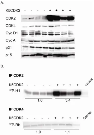
2.3.5 Biochemical analysis of K5CDK2 papillomas
Biochemical analysis of K5CDK2 papillomas shows that CDK2 protein levels are elevated, on average 8.4-fold, compared to wild type papillomas (Figure 8A). Due to the heterogeneous nature of papillomas (dermal and epidermal compartments) expression varies within samples of each group, but CDK2 is consistently overexpressed in papillomas from transgenic mice. Unlike our observations in normal epidermis, immunoblot analysis of papilloma extracts did not reveal any persistent increase in p21 protein levels (Figure 8A). CDK2 in vitro kinase assays show on average a 3.4-fold increase in kinase activity in papillomas from transgenic mice in comparison to wild type papillomas (Figure 8B, IP CDK2). CDK4 kinase activity is similar in wild type and K5CDK2 papillomas (Figure 8B, IP CDK4). Our initial hypothesis was that overexpression of CDK2 would enhance papilloma formation and accelerate malignant progression to SCCs. However, despite increased CDK2 protein levels and kinase activity, K5CDK2 transgenic mice do not exhibit increased susceptibility to the formation Ras-induced skin tumors.
2.3.6 Elevated CDK2 kinase activity in K5CDK4D158N transgenic mice
46
47
Histopathological analysis of paraffin embedded skin cross sections shows that K5CDK4D158N epidermis is hyperplastic in comparison to wild type mice. In fact, quantification of the number of nucleated cells per 200 μm of interfollicular epidermis showed elevated number of nucleated cells in K5CDK4D158N in comparison to wild type littermates, 87.5 and 74.6 cells/200 μm, respectively. Consistent with the observed hyperplasitic phenotype, in vivo proliferation assays demonstrate that K5CDK4D158N have a four-fold increase in the number of proliferative keratinocytes (BrdU positive cells) in comparison to wild type littermates. Biochemical analysis of K5CDK4D158N epidermal lysates clearly shows an increase in CDK2 kinase activity as demonstrated by in vitro kinase assays. While, CDK4 kinase activity was similar in K5CDK4D158N and wild type epidermis, showing that CDK4 D158N does not act as
a dominant negative isoform. As expected, overexpression of kinase inactive CDK4 results in the activation of CDK2 kinase activity via the sequestration of CKIs. This was confirmed by co-immunoprecipitation experiments showing increased levels of p27Kip1 and p21Cip1 complexed with CDK4 in K5CDK4D158N epidermal lysates.
48
papillomas from K5CDK4D158N progress to SCCs at a similar rate to those derived
from wild type littermates.
2.3.7 Inhibition of Smad2/3: an alternative mechanism for CDK4 mediated
malignant progression
We had previously hypothesized that the indirect activation of CDK2 in K5CDK4 transgenic animals contributes to enhanced malignant progression of papillomas to SCCs. However, to this point we have shown in two transgenic mouse models that elevated CDK2 kinase activity does not enhance tumor formation or progression. These data suggest that the malignant phenotype observed in K5CDK4 transgenic mice is due to CDK4 kinase activity as opposed to the described sequestration of CKIs from CDK2-Cyclin E. The main target of CDK4, 6 and CDK2 is the pRb protein family, thus, we hypothesized that CDK4 plays a unique role in tumorigenesis targeting additional substrates. Therefore, we decided to investigate additional putative CDK4 targets that may contribute to the observed malignant phenotype in K5CDK4 skin tumors. It has recently been shown that CDK4 and CDK2 can phosphorylate and inhibit the antiproliferative effects of
49
phosphorylation in vivo in comparison to wild type mice (Figure 9A). In an independent study, Ras transduced Smad3 null keratincoytes developed tumors which underwent rapid conversion to malignant carcinomas (Vijayachandra, Lee et al. 2003). This rapid conversion to malignant carcinomas is consistent with the increased malignant progression observed in K5CDK4 mice (Miliani de Marval, Macias et al. 2004). Thus, our results suggest that overexpression of CDK4 also inhibits the transduction of TGF-β1 anti-growth signaling in K5CDK4 papillomas via the inhibition of Smad3; mimicking Smad3 ablation in Ras transduced keratinocytes. To determine whether this mechanism is active in K5CDK4 skin tumorigenesis, we performed biochemical studies using protein extracts from 30-week-old papillomas from wild type and K5CDK4 mice. Immunoblot analysis shows elevated phospho-Smad3 at Thr 179, CDK specific site, in K5CDK4 papillomas in comparison to wild type tumors (Figure 9B). Correspondingly, immunoblot and immunofluorescence analysis for TGF-β1 downstream target p15Ink4b shows a clear reduction in K5CDK4
50
51
Figure 9. Western blot and immunoflourescence analysis of TGF-β effector and target proteins. (A) Immunoblot analysis of K5CDK4 and wild type epidermal lysates for phospho-Smad2/3 T179 and CDK4. (B) Immunoblot analysis of 30 week wild type and K5CDK4 papillomas for phosphor-Smad2/3 Thr 179, total Smad2/3, CDK4, p15. (C) Immunoblot analysis of 30-week K5CDK4D158N and wild type papillomas for p15Ink4b, phospho-Smad2/3 Thr 179 and CDK4. (D)
53 2.4 Discussion
CDK2 is not generally found overexpressed, amplified or affected by mutations in human tumors. However, amplification and altered expression of Cyclin E is frequently found in human tumors and its expression serves a good prognostic indicator (Keyomarsi, Conte et al. 1995). Proteolytic processing of Cyclin E into low molecular weight isoforms is a frequent event in breast tumors (Harwell, Porter et al. 2000). These low molecular weight isoforms of cyclin E have been found to activate CDK2 kinase activity and are refractory to Cip/Kip inhibitors (Harwell, Mull et al. 2004). Decreased protein levels of p27Kip1 (CDK2 inhibitor) is also a frequent event in human tumors. Furthermore, elevated levels of CDK4 have been shown to sequester CKIs indirectly activating CDK2-Cyclin E. Altogether, these alterations of cell cycle regulatory proteins provide mechanisms in which CDK2 is indirectly deregulated. For these reasons, it is widely believed that elevated CDK2 kinase activity is a contributing factor in human tumors. However, it has recently been shown that CDK2/Cyclin E activity is dispensable for cell cycle progression in cancer cells (Tetsu and McCormick 2003).
54
that elevated levels of CDK2 protein induce a compensatory mechanism, which may keep CDK2 activity in check. To this effect, we observed elevated p21Cip1 protein expression in K5CDK2, but not K5CDK4D158N epidermal lysates. It has been suggested that p21 and p53 act as an inducible barrier for deregulated cyclin E (Minella, Swanger et al. 2002). Likewise, it has been shown that co-transfection of cyclin E and CDK2 in cell culture results in the transactivation of p53 signaling (Segawa, Hokuto et al. 1993). Interestingly it has been shown that overexpression of CDK4 and CDK6, but not CDK2, can abrogate p53 mediated growth arrest in culture (Latham, Eastman et al. 1996). Therefore, is is possible that overexpression of CDK2 may induce a p53 response which up regulates p21 levels. This response appears to be specific for CDK2 overexpression and was not detected in the case of hyperactivation of CDK2 through and indirect mechanism (K5CDK4D158N). Western blot analysis did not detect any notable differences in total p53 expression or differences in protein mobility associated with phoshorylation (Figure 6A). It is important that future studies determine the status of p53 signaling using phospho-specific p53 antibodies keratinocytes overexpressing CDK2.
55
56
The fact that pRb family members are substrates of both CDK4 and CDK2, leads us to hypothesize that other CDK4 substrates may be participating in CDK4-induced malignant progression. There is growing evidence suggesting that overexpression of CDK4 in epithelial cells causes resistance to TGF-β treatment. Ectopic expression of CDK4 in mink lung epithelial cells results in TGF-β1 resistance and primary keratinocytes from mice overexpressing CDK4 binding partner, Cyclin D1, are also resistant to TGF-β1 treatment (Ewen, Sluss et al. 1993; Martinez, Chen et al. 2000). Notably, K5CDK4 transgenic mice exhibit dermal fibrosis, although there is no transgene expression in dermis (Miliani de Marval, Gimenez-Conti et al. 2001). It is well established that while TGF-β1 inhibits epithelial cells, it stimulates proliferation of dermal fibroblasts (Roberts and Sporn 1990). Thus it is probable that overexpression of CDK4 in mouse keratinocytes may lead to a TGF-β1 resistance and stimulate TGF-β1 expression and secretion resulting in fibroblast proliferation associated with dermal fibrosis. In agreement, primary keratinocytes derived from K5CDK4 newborn mice demonstrated partial resistance to TGF-β1 treatment in comparison to wild type keratinocytes. These preliminary results need to be repeated therefore data was not shown here. However, we showed that K5CDK4 epidermis has an elevated level of Smad3 phosphorylation at Thr 179, a CDK specific site. This result suggested that CDK4 might block the TGF-β pathway in mouse keratinocytes. Therefore, we decided to focus on the role of CDK4 in TGF-β
57
Immunoblot and immunofluorescence experiments demonstrated that Smad3 downstream target p15Ink4b is at very low levels in K5CDK4 papillomas. The latter suggest that phosphorylation Smad2/3 phosphorylation at Thr 179 influences Smad2/3 transcriptional activity. In this sense, tumors derived from CDK4-Ras transfected primary keratinocytes also show altered levels of Smad3 transcriptional target telomerase reverse transcriptase (TERT) (Lazarov, Kubo et al. 2002). In our mouse models, we have shown the induction of TGF-β downstream target, p15Ink4a in both K5CDK2 and K5CDK4D158N papillomas despite having elevated CDK2 kinase activity. Altogether, these data suggest that inhibition of Smad2/3 and the inability to induce p15Ink4b is a direct function of CDK4 kinase activity. The latter provides a new epigenetic mechanism, which leads to loss of TGF-β anti-growth signaling in normal and neoplastic growth.
We conclude that CDK2 does not behave as an independent oncogene nor synergizes with Ha-Ras to induce tumor development or malignant progression to SCC. In addition, our results suggest that CDK4 mediates malignant progression not only through inibithion of pRb family of proteins, but also via inhibition of TGF-β
58
CHAPTER III
CDK2 ablation reduces CDK4/Ha-Ras mediated malignant
59 3.1 Introduction
The pRb family of proteins, pRb, p107 and p130, are key substrates for G1 cyclin/CDK complexes, and negatively regulate the passage of cells from G1 to S phase (Weinberg 1995). The Cip/Kip family of CDK-inhibitors, p21Cip1, p27Kip1 and p57Kip2, form inactive complexes with CDK2-Cyclin E and CDK2-Cyclin A. Cip/Kip family members p21Cip1 and p27Kip1 also bind CDK4,6/Cyclin D complexes but do not
interfere with their kinase activities (Blain, Montalvo et al. 1997; Labaer, Garret et al. 1997). Thus, cyclin D-CDK4 may sequester p27Kip1, controlling the amount of p27Kip1 available for inhibition of CDK2 activity. We and others have demonstrated that indirect activation of CDK2 occurs by sequestration of p27Kip1 upon forced expression of CDK4 (Bouchard, Thieke et al. 1999; Perez-Roger, Kim et al. 1999; Miliani de Marval, Gimenez-Conti et al. 2001).
60
CDK2 through expression of dominant-negative (DN) CDK2 or antisense oligonucleotides did not prevent cell proliferation of cancer cells in vitro (Tetsu and McCormick 2003). On the other hand, genetic ablation of CDK4 has a more dramatic effect on normal cell proliferation and inhibition of tumorigenesis. CDK4-/- mice are viable, but showed reduced body size suggesting growth retardation in several tissues. Severe defects leading to degeneration of pancreatic islet cells and proliferative defects in the pituitary gland were observed (Rane, Dubus et al. 1999; Tsutsui, Hesabi et al. 1999; Moons, Jirawatnotai et al. 2002; Moons, Jirawatnotai et al. 2002). In addition, we and others have demonstrated the inhibition of tumor development in CDK4-/- mice (Rodriguez-Puebla, Miliani de Marval et al. 2002; Zou, Ray et al. 2002; Miliani de Marval, Macias et al. 2004). Altogether, these data lead to the general concept that CDK4, but not CDK2 is a more favorable target for therapeutic intervention.
61
may be necessary for K5CDK4 and K5Myc tumorigenesis. To investigate this
hypothesis, we have generated K5CDK4/CDK2-/- and K5Myc/CDK2-/- compound mice. In this chapter, we demonstrate that CDK2 plays an important role in CDK4-induced keratinocyte proliferation. In addition, we show that CDK2 contributes to the progression of chemically induced mouse skin tumors carrying Ha-ras activating mutations. A decreased number of skin tumors and a strong reduction of malignant progression to squamous cell carcinomas (SCCs) is clearly observed in K5CDK4/CDK2-/- mice. In contrast, we found that CDK2 is dispensable c-Myc-induced tumorigenesis and keratinocyte hyperproliferation. Overall, we have established that ras-induced tumors are more susceptible to CDK2 ablation than c-Myc-induced tumors, suggesting that the efficacy of targeting CDK2 in tumor development and malignant progression is dependent on the oncogenic pathway involved.
3.2 Materials and Methods
3.2.1 Mouse Experiments and Pathological Analysis
K5CDK4, K5-Myc and CDK2-null mice were generated as previously
described (Miliani de Marval, Gimenez-Conti et al. 2001; Rounbehler, Schneider-Broussard et al. 2001; Ortega, Prieto et al. 2003). K5CDK4/CDK2-/- and K5Myc/CDK2-/- compound mice were obtained by interbreeding the respective
62
application of 50 μg of DMBA in 50 μl of acetone on dorsal mouse skin. At day 21, mice received 2.5 μg of TPA in 200 μl of acetone twice a week for 25 weeks. Skin tumors were counted once a week until the end of the experiment at 40 weeks. Malignant progression to SCC was determined by macroscopic observation and further confirmed by histopathological analysis of paraffin-embedded H&E stained cross sections.
3.2.2 Western Blots and Kinase Assays.
For Immunoblots protein lysates were collected from epidermal skin scrapes with RIPA lysis buffer, 150 mM NaCl, 1.0% IGEPAL, 0.5% DOC, 0.1% SDS, 50mM Tris (pH 8.0). For immunoblot analysis of skin tumors, papillomas were snap frozen in liquid N2 and crushed with a pestle and mortar. Homogenates from
63
washed twice each with IP buffer and kinase buffer (50 mM HEPES [pH 7], 10 mM MgCl2, 5 mM MnCl2). Then, 30 μl of kinase buffer, 1 µg of pRb or histone H1 (Upstate Biotechnology Inc., Charlottesville, VA.) substrate, 5 μCi of [γ-32P]ATP (6,000 Ci/mmol), 1 mM DTT, and 5 μM ATP were added to the bead pellet and incubated for 30 min at 30°C. SDS sample buffer was added, and each sample was boiled for 3 min to stop reaction and electrophoresed through polyacrylamide gels. Western blot and kinase assay bands were quantified using UN-SCAN-IT gel ™ version 6.1 software for windows.
3.2.3 Immunostaining
For immunofluorescence tissue cross sections of formalin fixed skin tumors were permeablized using citrate antigen retrieval buffer, blocked with 10% normal serum and stained with antibodies for keratin 5 (Covance Research Products, Berkeley, CA, USA) and keratin 13 (Novus Biological, Littleton, CO, USA) followed by incubation with Alexafluor secondary antibodies (Molecular Probes). Epithelial cell proliferation was measured by intraperitoneal injection of BrdU 30 min before the mice were sacrificed by CO2 asphyxiation. BrdU incorporation was detected by
64 3.2.4 Statistical analysis
Statistical analysis was performed using GraphPad Prism 4 Software (GraphPad Software, San Diego, CA, USA)
3.3 Results
3.3.1 Biochemical and histological evaluation of K5CDK4 / CDK2-/- mice
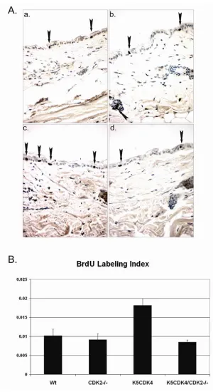
CDK2 expression has been largely implicated in induction of cell proliferation (Tsai, Lees et al. 1993; van den Heuvel and Harlow 1993; Ohtsubo, Theodoras et al. 1995), however, ablation of CDK2 does not affect cell cycle progression in mice, only the meiotic process (Berthet, Aleem et al. 2003; Ortega, Prieto et al. 2003). We previously demonstrated that overexpression of CDK4 in mouse skin results in increase epidermal proliferation associated with elevated CDK4 and CDK2 kinase activity, presumably due to sequestration of p27Kip1 and p21Cip1 (Miliani de Marval, Gimenez-Conti et al. 2001; Miliani de Marval, Macias et al. 2004). In order to determine if CDK2 is necessary for CDK4-induced epidermal proliferation and malignant transformation, we have generated K5CDK4/CDK2-/- compound mice. Consistent with previous results, analysis of epidermal proliferation demonstrates a two-fold increase in the number of S-phase cells (BrdU-positive) in K5CDK4 epidermis compared with wild type mice (Figure 10). On the other hand, CDK2-null mice do not show a difference in the level of keratinocyte proliferation compared with wild type mice, however, ablation of CDK2 in K5CDK4 background (K5CDK4/CDK2
-/- mice) diminishes proliferation triggered by CDK4 overexpression (K5CDK4 mice)
65
does not affect transgenic expression of CDK4 in K5CDK4/CDK2-/- compound mice,
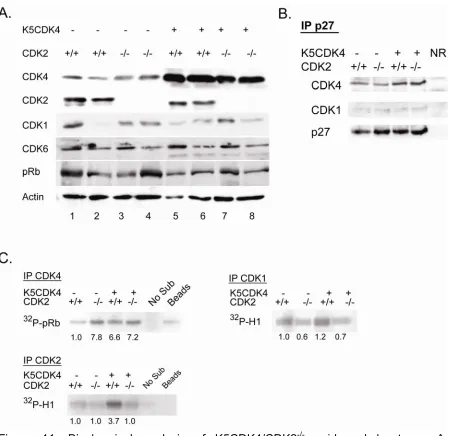
which remains higher compared to wild type littermates (Figure 11). Moreover, loss of CDK2 expression is not compensated by increased levels of endogenous CDK4 or CDK6 which although heterogeneous among siblings of same genotype, the levels of expression were similar between CDK2-/- and wild type mice (Figure 11). Consistent with our previous description of K5CDK4 model, we did not observe differences in Rb protein levels (Miliani de Marval, Gimenez-Conti et al. 2001). In addition, we did not detect changes in protein mobility consistent with pRb-phosphorylation. In vitro kinase assay shows 3.7-fold increase in CDK2 activity in epidermis from K5CDK4 mice (Figure 11C, IP CDK2) (Miliani de Marval, Gimenez-Conti et al. 2001). K5CDK4 mice shows increase CDK4 kinase activity (6.6-fold) compared with wild type mice and importantly, CDK2 ablation does not reduce this kinase activity (K5CDK4/CDK2-/- mice, 7.2-fold increase) (Figure 11C, IP CDK4). Interestingly, the kinase activity of endogenous CDK4 is also augmented, 7.8-fold, in CDK2-/- epidermis compared with wild type epidermis. While p27Kip1 and p21Cip1 inhibit CDK2-Cyclin E, they act as assembly factors for CDK4-Cyclins D. Therefore, to determine if elevated CDK4 kinase activity in CDK2-/- is due to a redistribution of Cip/Kip proteins we conducted co-immunoprecipitation experiments. Immunoprecipitation of p27Kip1 does not show increased complex formation in CDK2
-/- compared to wild type (Figure 11B). In addition, CDK4/p27Kip1 complex formation
in K5CDK4/CDK2-/- epidermis is similar to that found in K5CDK4 epidermis. At
66
CDK2-/- cells warrant further investigations. Recently, it has been demonstrated that
67
Figure 10. Epidermal proliferation in K5CDK4/CDK2-/- compound mice. A. BrdU incorporation of representative paraffin skin sections from wild type (a), CDK2-/- (b),
68
In fact, a mild reduction in CDK1 kinase activity was observed in keratinocytes lacking CDK2 expression, although we did not detect increased p27Kip1 complexed with CDK1 (Figure 11B). We conclude that ablation of CDK2 does not affect normal keratinocyte proliferation, but reduces CDK4-induced keratinocyte hyperproliferation.
3.3.2 CDK2 deficiency reduces skin tumor development and malignant
progression
69
Figure 11. Biochemical analysis of K5CDK4/CDK2-/- epidermal lysates. A.
70
Reduced tumor incidence was observed in CDK2-/- and K5CDK4/CDK2-/- littermates
that reached a plateau of 82% and 70% of mice with tumors (Figure 12A). Ablation of CDK2 also results in decreased number of tumors per mouse (multiplicity). At 20 weeks of promotion the four groups of mice reached a plateau where reduced papilloma multiplicity was observed in K5CDK4 mice compared with wild type littermates (8.5 and 6 papillomas per mouse respectively) (p<0.05; Mann-Whitney U test) (Figure 12B). CDK2 deletion results in the development of fewer papillomas in both CDK2-/- and K5CDK4/CDK2-/- mice (3.5 tumors/mouse) compared with wild type and K5CDK4 mice (p<0.05; Mann-Whitney U test) (Figure 11B). Consistent with our previous studies, SCCs develop rapidly and in greater number in K5CDK4 transgenic mice (Figure11C) (Miliani de Marval, Macias et al. 2004). The first SCC appeared at 22 weeks of promotion and 70% of K5CDK4 mice had developed SCC by 40 weeks of promotion. In sharp contrast, CDK2 ablation leads to a strong reduction of malignant progression since only 28% of K5CDK4/CDK2-/- mice
71
74
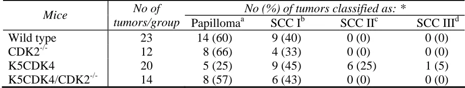
Histopathological analysis was performed with 30-40 week old skin tumors graded as papillomas or SCCs according to a modified Broder's classification (Klein-Szanto 1997; Klein-(Klein-Szanto and Conti 2002). This classification system divides SCCs into four grades: 1) SCC I: well differentiated SCC, most of the tumor mass composed of differentiated cells; 2) SCC II: moderately differentiated with approximately 50% of differentiating cells; 3) SCC III: poor differentiated tumor with little keratinization and 4) SCC IV: poorly differentiated tumors with marginal or no keratinization, including spindle cell carcinomas (Klein-Szanto, Larcher et al. 1989; Klein-Szanto 1997). Most of the wild type tumors analyzed were benign papillomas (60%) and 40% were classified as SCC I. On the other hand, analyzed tumors from K5CDK4 mice were classified as SCC I (45%), SCC II (25%) and SCC III (5%) and papillomas (25%). These data are consistent with our previous observation that forced expression of CDK4 increases the rate of malignant conversion (Miliani de Marval, Macias et al. 2004). In sharp contrast, only 43% of the K5CDK4/CDK2
75
Table 1. Histopathologic analysis of K5CDK4/CDK2-/- skin tumors
No (%) of tumors classified as: *
Mice No of
tumors/group Papillomaa SCC Ib SCC IIc SCC IIId Wild type 23 14 (60) 9 (40) 0 (0) 0 (0) CDK2-/- 12 8 (66) 4 (33) 0 (0) 0 (0) K5CDK4 20 5 (25) 9 (45) 6 (25) 1 (5) K5CDK4/CDK2-/- 14 8 (57) 6 (43) 0 (0) 0 (0) χ2, p< 0.05. a No atypia in basal layers. b Well differentiated SCC, most of the tumor mass composed of differentiated
cells. c Moderately differentiated with approximately 50% of differentiating cells. d Poorly differentiated tumor with little
76
77
78
Figure 14. Biochemical analysis of K5CDK4/CDK2-/- papillomas. Protein lysates
79
3.3.3 Lack of CDK2 expression does not inhibit oral tumor development
Overexpression of the murine c-myc gene in the basal cell layer of epithelium (K5Myc mice) results in epidermal hyperplasia and hypertrophy (Rounbehler, Schneider-Broussard et al. 2001; Miliani de Marval, Macias et al. 2004). In addition, a high incidence of spontaneous tumors was observed in the oral mucosa of K5Myc transgenic mice (Rounbehler, Schneider-Broussard et al. 2001; Miliani de Marval, Macias et al. 2004). These results showed that c-myc acts as an oncogene in the stratified epithelium, but the mechanisms leading to the malignant phenotype are not fully understood. Interestingly, K5Myc mice exhibit elevated CDK4 protein levels and ablation of CDK4 inhibits c-myc tumorigenic activities in epithelial tissues and reduces CDK2 activity, presumably through redistribution of p27Kip1 that bind and
inhibit CDK2 (Miliani de Marval, Macias et al. 2004). In order to investigate whether CDK2 mediates the oncogenic activities of c-myc, we developed K5Myc transgenic mice that lack the expression of CDK2 (K5Myc/CDK2-/- mice). These mice, along
80
develop tumors with the same frequency as K5Myc mice (Figure 15B). Cell proliferation analysis of oral cavity tumors did not reveal any obvious differences between the two genotypes (Figure 16). Wild type and CDK2-/- littermates did not show any signs of spontaneous tumor development up to 18 months of age. Thus, mice with genetic ablation of CDK2 remain fully susceptible to Myc-driven tumorigenesis of the oral mucosa. Collectively, these data indicate that CDK2 does not play a relevant role in the development of the spontaneous oral tumors observed in K5Myc mice.
3.3.4 CDK2 is dispensable for c-myc mediated keratinocyte proliferation
We also asked whether CDK2 mediates the epidermal hyperproliferation phenotype triggered by Myc overexpression. Thus, we analyzed the epidermis of K5Myc and K5Myc/CDK2-/- mice. The skin of K5Myc mice shows hyperplasia (increase cell number) (Figure 17). Abrogation of CDK2 expression (K5Myc/CDK2-/-)
81
CDK2 inhibitors p27Kip1 and p21Cip1 (Miliani de Marval, Macias et al. 2004). In order
82
83
84
85
86
87 3.4 Discussion
The concept that CDK2 is essential for controlled entry into S phase and the onset of DNA replication was challenged a few years ago when two independent groups reported the generation of CDK2-/- mice (Berthet, Aleem et al. 2003; Ortega, Prieto et al. 2003). These mice are viable, develop normally and only show meiotic defects. These results were surprising, as CDK2 was thought to be required for promoting G1/S transition. In addition, it was also demonstrated that cancer cells can proliferate in absence of CDK2 (Tetsu and McCormick 2003). Thus, data has accumulated in the last few years introducing the concept that CDK2 is not an adequate target for therapeutic intervention. In this chapter, we have investigated the role of CDK2 in two oncogenic pathways. We have clearly shown that CDK2 contributes Ras-CDK4 malignant progression but is dispensable for c-Myc driven oral squamous cell carcinomas.
88
Kiyokawa et al. 2005). Here, we have examined the skin of CDK2-/- mice which is
structurally and functionally similar to that of wild type siblings. It has been reported that CDK1 activation compensates for the absence of CDK2 in thymocytes, although this mechanism may be dependent on the absence of p27Kip1(Aleem, Kiyokawa et al. 2005). Our biochemical analysis of CDK2-/- epidermis shows a reduction rather than activation of CDK1. However, we observed increased activity of CDK4 in the epidermis of CDK2-/- mice (Figure 11C, IP CDK4 ). Since p27Kip1 stabilize CDK4/D-type cyclin complexes (Cheng, Olivier et al. 1999), it was possible that in absence of CDK2, redistribution of p27Kip1 increases CDK4/D-type cyclin/p27Kip1 complex formation. Nevertheless, biochemical analysis did not show increased formation of those complexes. Whether increased CDK4 kinase activity compensates for CDK2 in the epidermis merits further investigation. The importance of the CDK4-CDK2 link in keratinocyte proliferation was demonstrated by the activation of CDK2, through titration of p27/p21, by forced expression of CDK4 (Miliani de Marval, Gimenez-Conti et al. 2001; Miliani de Marval, Macias et al. 2004) and the reduction of the total number of proliferative cells in K5CDK4/CDK2-/- epidermis compared to K5CDK4 mice. Therefore, activation of CDK4 and CDK2 kinases are necessary for epidermal hyperproliferation observed in K5CDK4 mice.
89
multiplicity in both, K5CDK4/CDK2-/- and CDK2-/- mice. More importantly, lack of
CDK2 expression in a K5CDK4 background (K5CDK4/CDK2-/- mice) results in clear reduction of CDK4-induced malignant progression to SCCs. Therefore, lack of CDK2 affects papilloma development and more importantly, CDK2 expression is required for CDK4-induced malignant progression. Histopathological analysis of tumors from the four genotypes confirms the role of CDK2 in SCC progression. Tumors from K5CDK4 mice were classified as regular papillomas and SCC I, II, and III; whereas most of the K5CDK4/CDK2-/- tumors were regular papillomas and no SCCs II or III were observed. In addition, K5CDK4 carcinomas showed areas of strong positive immunofluorescence staining for keratin 13 (K13), a malignancy marker in squamous epithelia (Nischt, Roop et al. 1988), whereas SCCs from K5CDK4/CDK2
-/-mice were K13-negative.
90
K5CDK4 transgenic lines exhibiting different levels of CDK4 protein expression develop SCCs at a similar rate (Miliani de Marval, Macias et al. 2004). Similar to CDK1 expression in epidermis, CDK1 protein levels were variable among tumors from the same genotype, but there are no relevant differences in CDK1 levels among the four genotypes.
In chapter 2 we suggested that K5CDK4 mediated malignant progression is due to the inhibition of Smad3 antiproliferative effects. We demonstrated that K5CDK4 papillomas but not K5CDK2 or K5CDK4D158N papillomas had reduced protein levels of p15Ink4b. Transcription of p15Ink4b can be down-regulated via phosphorylation and inactivation of Smad3 by CDK4 (Liu and Matsuura 2005; Liu 2006). However, decreased p15Ink4b was observed in both K5CDK4 and
K5CDK4/CDK2-/-, suggesting that its downregulation is not responsible for CDK4-induced malignant progression.
91
have recently shown that CDK2 influences survival of cells under genotoxic stress by inhibiting the forkhead transcription factor, FOXO1. Importantly, CDK2 phosphorylates and inhibits the pro-apoptotic function of FOXO1 (Huang, Regan et al. 2006; Huang and Tindall 2007). Thus, CDK2 ablation could restore or increase FOXO1 activity leading to reduced tumorigenesis.