Copyright 0 1994 by the Genetics Society of America
A
Yeast
Artificial
Chromosome Clone Map
of
the Drosophila Genome
Haini
Cai,'Paula
Kiefel, Joyce Yee
and
Ian Duncan
Department of Biology, Washington University, Saint Louis, Missouri 63130Manuscript received September 8, 1993 Accepted for publication January 1, 1994
ABSTRACT
We describe the mapping of 979 randomly selected large yeast artificial chromosome (YAC) clones of Drosophila DNA by in situ hybridization to polytene chromosomes. Eight hundred and fifty-five of the clones are euchromatic and have primary hybridization sites in the banded portions of the polytene chromosomes, whereas 124 are heterochromatic and label the chromocenter. The average euchromatic clone contains about 21 1 kb and, at its primary site, labels eight or nine contiguous polytene bands. Thus, the extent as well as chromosomal position of each clone has been determined. By direct band counts, we estimate our clones provide about 76% coverage of the euchromatin of the major autosomes, and 63% coverage of the X . When previously reported YAC mapping data are combined with ours, euchromatic coverage is extended to about 90% for the autosomes and 82% for the X . The distribution of gap sizes in our map and the coverage achieved are in good agreement with expectations based on the assumption of random coverage, indicating that euchromatic clones are essentially randomly distributed. However, certain gaps in coverage, including the entire fourth chromosome euchromatin, may be significant. Het- erochromatic sequences are underrepresented among the YAC clones by two to three fold. This may result, at least in part, from underrepresentation of heterochromatic sequences in adult DNA (the source of most of the clones analyzed), or from clone instability.
T
HE yeast artificial chromosome (YAC) cloning tech- nique (BURKE et al. 1987) allows the cloning and faithful propagation in yeast cells of fragments of exog- enous DNA hundreds of kilobases in length. In this re- port, we apply this cloning technology to the mapping of the Drosophila genome. Our approach has been a very simple one: random large YAC clones of Drosophila DNA are mapped by in situ hybridization to polytene chromosomes. Since the average YAC clone mapped contains about 211 kb and the average polytene chro- mosome band contains about22
kb per chromatid, most YAC clones label eight or nine contiguous bands, or ap- proximately a lettered unit on BRIDGES (1935) map [see also LEFEVRE (1976) and SORSA (1988)l. Thus, the cyto- logical extent as well as chromosomal position of each clone can be determined by in situ hybridization. With the use of biotinylated probes, the resolution of this method is remarkably good. In finely banded regions, resolution likely can be within 5-10 kb (SPIERER et al.1983).
A major advantage of our cytological approach is that continuity of the clone map is provided at all stages by the polytene chromosomes themselves. Thus, at least for a crude map, clones need only be mapped by their site of in situ hybridization, and do not need to be mapped relative to one another and placed into contigs. In ad- dition, the cytological mapping of clones is little affected by the presence of repeats; with few exceptions, the pri- mary site of hybridization is unambiguous even when
I Present address: Department of Biology, University of California at San Diego, 9500 Gilman Drive, La Jolla, California 92093.
Genetics 1 3 6 1385-1401 (April, 1994)
repetitive sequences are present. Indeed, the method can provide substantial information about the repeats themselves. A major drawback is that the method is use- ful only for mapping euchromatic clones and provides little information about the approximately 25% of the genome composed of heterochromatin. This material either does not replicate in polytene cells (a-hetero- chromatin) or replicates but has a diffuse poorly banded morphology (pheterochromatin) (GALL et al. 1971) [for reviews see SPRADLING and RUBIN (1981) and ASHBURNER (1989)
1.
Although many heterochromatic clones are identified in our study by hybridization to the chromocenter, their relative chromosomal locations have not been determined.In this report, we present the in situ hybridization pattern of 979 Drosophila YAC clones. For 855 of these, the primary site of hybridization is euchromatic, whereas for 124, the primary site is the chromocenter. The eu- chromatic clones appear to be essentially randomly dis- tributed, and, as estimated by direct band counts, pro- vide about 76% coverage of the autosomes and 63% coverage of the X . When combined with the YAC clones reported by AJIOKA et al. (1991), these should extend coverage of the autosomal euchromatin to about 90% and the X chromosome euchromatin to about 82%.
MATERTALS AND METHODS
1386 H. Cai et al.
liquid nitrogen and ground to a fine powder. The frozen pow- der was then homogenized in cold nuclear isolation buffer (BINGHAM et al. 1981) using a Dounce B pestle. Bulk fly parts were removed by brief centrifugation and discarded. The sus- pension of nuclei and other cellular debris was washed 1-2 times in cold nuclear isolation buffer and then lysed in 2%
Sarkosyl. CsCl was gently dissolved to 1 g/ml in the lysate. The mixture was then centrifuged at 45,000 rpm in a Vti65 vertical rotor for 16 hr at 25". DNA was collected from the CsCl gra- dient, dialyzed extensively against 1 X TE (10 mM Tris, 1 mM EDTA, pH 8.0), and fractions were checked for nuclease con- tamination by incubating with 1 X EcoRI buffer at 37" for 20 min followed by field inversion gel electrophoresis (see be- low). Fractions showing significant degradation of high mo- lecular weight DNA after incubation in EcoRI buffer were dis- carded. Fractions devoid of nuclease activity were then pooled and aliquots containing 2-5 pg DNA were used in test EcoRI partial digestions. Test digestions were done for 10 min at 37" with EcoRI concentrations ranging from 0.5 to 20 units/ml. Digestion conditions were identified that lowered the average size of DNA by 200 kb or more depending on the quality of the input DNA. Partial digestions were scaled up to 200-500 pg DNA and were stopped by adding EDTA to 50 mM and placing the reaction on ice. Digestion products were then size frac- tionated over 5-12.5% sucrose velocity gradients and fractions containing DNA greater than 200 kb were pooled, concen- trated and dialyzed in UHlOO collodion bags (Schleicher &
Schuell). The vector pYAC4 (BURKE et al. 1987) was digested to completion with EcoRI and BamHI and treated with calf intestinal alkaline phosphatase (Boehringer). After treatment at 75" for 10 min to inactivate phosphatase, followed by phenol extraction, vector DNA was added to partially digested high molecular weight Drosophila DNA at a 3:l weight ratio and ligated for 1-2 hr at 25". Ligation reactions were used directly to transform yeast strain AB1380 (BURKE et al. 1987) essentially as described by BURGERS and PERCIVAL (1987). Transformants were selected on -Ura plates containing 1 M sorbitol (Fisher Biotech.). Both top and bottom agars were 2.5% agar.
Transformant selection: All YAGcontaining cells were grown on YCD medium, which lacks uracil and tryptophan. The recipe is modified from the AHC' medium of BROWNSTEIN
et al. (1989) and is as follows: for 1 liter, 20 g glucose, 6.7 g Bacto yeast nitrogen base without amino acids (Difco), 10 g casein hydrolysate (Sigma), 20 mg adenine, 50 mg lysine, 20 mg histidine, 40 mg arginine, 60 mg isoleucine, 60 mg leucine, 20 mg methionine, 50 mg phenylalanine, 200 mg threonine, and 50 mg tyrosine. Primary transformants were picked onto YCD/2% agar plates and grown for 2-3 days at 30". Patches showing strong red growth were harvested into 96-well micro- titer plates containing 15% glycerol in water and stored at -80". A Replaclone 96-prong transfer device (L.A.O. Enter- prises) was used to replicate cultures from these master plates into 1-ml YCD cultures in micro-test tubes in racks of 96 (Bio- Rad 223-9395). Cultures were grown at 30" with vibration
(Bellco mini-orbital shaker, setting 4). These 1-ml cultures were used for initial size screening.
Initial sizing: When the 1-ml cultures had reached 1-3 X
10' cells/ml, agarose plugs of the YAC clones were prepared as described by CARLE and OLSON (1984). Cells were pelleted gently and incubated 1 hr at 37" in 0.05 ml of 1 mg/ml zy-
molyase (lOOT, ICN) in SCE (1 M sorbitol, 0.1 M citric acid, 10 mM EDTA, pH 5.8) plus 25 mM /3-mercaptoethanol to form spheroplasts. The suspension was then mixed with an equal volume of 1.2% molten low melting temperature agarose and poured into plug molds (Pharmacia LKl3 Biotechnology). Plugs were digested in 1.5 ml 1 mg/ml proteinase K (Boeh- ringer) in 1 % Sarkosyl, 0.45 M EDTA, 10 mM Tris-HC1, pH 9.0,
for 24-48 hr at 55". Digested plugs were equilibrated in 0.5 X
TBE (45 mM Tris, 45 mM boric acid, 1 mM EDTA, pH 8.0) by multiple buffer changes for 6-8 hr followed by field inversion gel electrophoresis (FIGE) (CARLE et al. 1986) using a Bio-Rad
Pulsewave 760 switching module. One percent agarose gels were run at 10-15" at a constant voltage of 250 V. Throughout electrophoresis, the 0.5 X TBE buffer was continuously recir- culated and cooled through a closed system similar to that described by CARLE and OLSON (1984). To insure temperature uniformity, electrophoresis tanks were kept in Styrofoam chests during electrophoresis runs. For the first 12 hr, a pulse ramp of 3-30 sec forward time and a constant pulse ratio of 3: 1 was used. To increase separation in the 50-200-kb range, a second ramp of 3-60 sec forward time with changing pulse ratio of 3-12:1 followed for 6 hr. DNA was visualized by ethidium bromide staining/deionized water destaining after electrophoresis.
Verification sizing and DNA purification: Master plate cul- tures showing clones of 180 kb or larger were streaked to single colonies on YCD plates. A single colony was then grown in 5 ml YCD on a roller drum for 3 days at 30". One milliter of this culture was embedded in agarose, processed as above and subjected to FIGE as for the initial sizing (ex- cept the two ramps were each for 8 hr). Agarose gel bands containing YAC clones were excised, and the DNA was puri- fied using the sodium iodide-glass powder method (Gene- clean, Bio 101) (VOGELSTEIN and GILLESPIE 1979). For long term maintenance, the settled pellet from the remaining 4 mls of culture was resuspended in 1.0 ml 15% glycerol in wa- ter and stored at -80" in cryovials (Nalge 5000-0020).
Polytene chromosome squashes: Polytene chromosome squashes were prepared essentially as described in a protocol generously provided byJoHNG LIM. Slides were cleaned in No- chromix (Godax Laboratory) /sulfuric acid according to manufacturer's instructions, rinsed well in deionized water, and air dried. Slides were then subbed by dipping in a solution of 0.1% gelatin/O.Ol% chrome alum in water, followed by air drying. Most slides were further treated by incubating in 3 X SSC/l X Denhardt's solution (MANIATIS et al. 1982) for 2-3 hr at 65". Slides were then rinsed in deionized water, dipped in 3:l ethanol/acetic acid and dried. Coverslips were siliconized under vacuum in a desiccator using dimethyldichlorosilane as described by MANIATIS et al. (1982). Slides and coverslips were kept as dust-free as possible during processing. Climbing third instar larvae from uncrowded cultures were collected, washed in water and dissected in a drop of 45% acetic acid on a sili- conized slide. Two or three glands were transferred to a drop of 1:2:3 solution (lactic acid:water:acetic acid) on a subbed slide, covered with a siliconized coverslip and squashed by tap- ping the coverslip 10-20 times with blunt forceps. Excess 1:2:3 solution was then blotted off the slide, the edge of the coverslip held with a folded Kimwipe and the tip of blunt forceps dragged over the coverslip in a serpentine pattern. Most slides were then flattened in a custom slide press fabricated from a small arbor press by the Washington University Biology ma- chine shop. Slides were then left at 4" for 3-4 hr to promote flattening, followed by freezing on dry ice, flipping off the coverslip with a razor blade and dehydration in 95% ethanol. Finally, slides were air dried and examined by phase contrast microscopy. Only slides with well flattened, nonrefractile, chromosomes were chosen for in s i t u hybridization.
Biotinylation and in situ hybridization: DNA was labeled with biotin-dCTP (ENZO Biochem Inc.) by the random hex- amer labeling method (FEINBERG and VOGELSTEIN 1983) for 24-48 hr at 25". In s i t u hybridization to polytene chromosome squashes was carried out as described in LANCER-SAFER et al.
Drosophila YAC Clone Map 1387
ENZO Biochem Inc.) and diaminobenzidine (DAB) (Sigma) to visualize the hybridization. For most slides, hybridization signal was intensified using 0.08% NiCI,. All hybridizations were to polytene chromosomes from a C a n t o n s strain iso- genic for chromosomes 2 and 3.
Error minimization: During pilot experiments, it became apparent that errors could be a significant problem. Since most errors in these experiments occurred during transfers of cultures or solutions to new tubes, our procedures were revised
so as to minimize transfers and consequent tube relabelings. Use of a centrifuge that accommodates 96tube boxes elimi- nated one particularly error-prone tube transfer. An addi- tional important change was to keep all tubes involved in pro- cessing each clone until completion of the in situ slides, so that the slides and tubes could be checked against one another. These changes appear to have helped, since no errors were detected among the approximately 5% of clones for which in situs were repeated.
Other DrosophilaYAC clones used in mapping: Clones des- ignated with the prefix R or Rx were produced as described above using DNA from our strain of Cantons. Clones desig- nated Rt were derived by the same method from a
T(Y;2) CB25, cn/y;cn bw strain (provided by TERRY LYITLE). Clones denoted DY are from CAW! et al. (1989) and were made from randomsheared Oregon RC adult DNA using the pYACP-1 vector. Clones designated with the prefix N were con- structed by ANDREW LINK from Not1 fragments of Oregon RC embryo DNA as described by DANILEVSKAYA et al. (1991).
RESULTS
Construction
of
YAC libraries: Production of large YAC clones of Drosophila DNA has proven to be rather difficult, apparently because of high nuclease levels. After a number of attempts, we succeeded in producingtwo sets of clones, designated DYR and DYRx. The DYR library is the major source of clones used in our m a p ping, and consists of nearly 1,000 clones of average insert size about 200 kb. The DYRx library, although large (6,000 clones), has an average insert size of only about 140 kb and contributed relatively few clones to the m a p ping.
Also
mapped were a few clones (designated DYRt) constructed from DNA from males of a T(Y;2)CB25,cn./y;cn bw stock. In an attempt to circumvent nuclease contamination, we also tried (without success) to pre- pare YAC clones from the double nuclease mutant
DNase-2"' DNase-1'" (GRELL 1976).
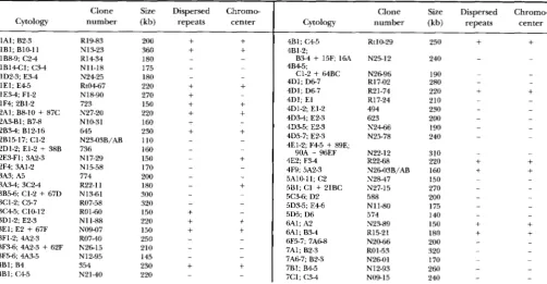
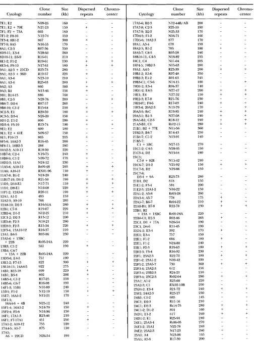
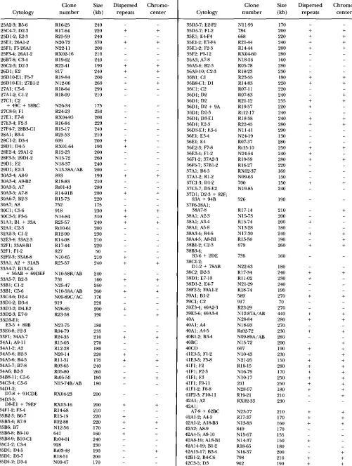
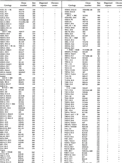
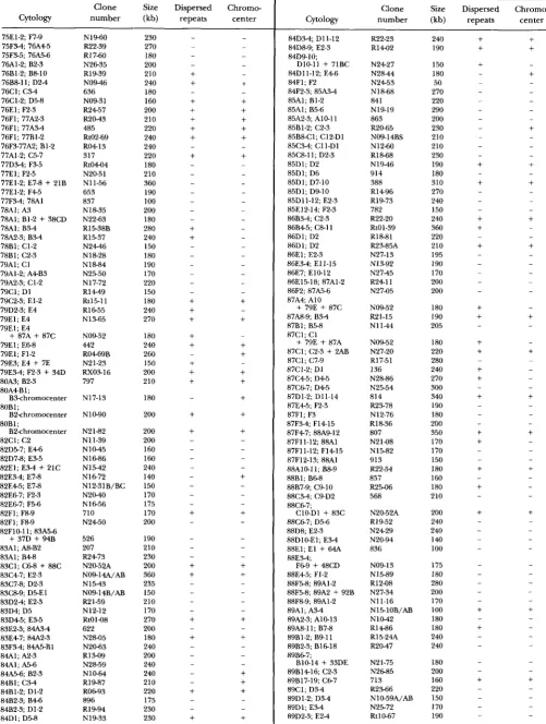
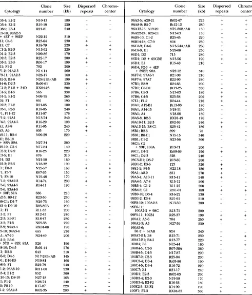
Euchromatic clones: The primary sites of hybridiza- tion of 855 euchromatic clones are listed in the APPENDIX
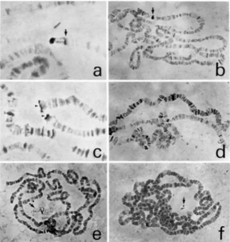
(see Table 2). The average size of these clones is 21 1 kb. The appearance of a typical in situ hybridization is shown in Figure la. Although many of the clones con- tain repetitive sequences, and label numerous sites, the primary site of hybridization is almost always clear by its
extent and strength of staining (see Figure l b ) . It is dif- ficult to convey using black and white photographs just how easily the primary site can usually be recognized. In our preparations, the primary site is almost always jet black, whereas secondary sites have a grayish or purplish cast. Of the 855 euchromatic clones, at least 39 are chi- meric and show two (34 clones) or three (five clones) primary sites of hybridization. Additional chimeric
FIGURE 1.-In situ hybridizations to polytene chromosomes. The reproducibility of the following hybridization patterns has not been tested. (a) Typical appearance of an in situ hybrid- ization signal. DY609 (260 kb) is shown hybridized to its locus at 28C1-2; D M . Note that the large heavy band indicated by the arrow (actually a cluster of five o r six bands including 28B and 28C1, 2) is only partially labeled. The importance of ex- amining well stretched chromosomes is illustrated here, since in unstretched chromosomes DY609 often appears to label this entire complex of bands. (b) In situ hybridization of a clone (Dk902) containing dispersed repeats. Note that the primary site of hybridization (42C3-5; D3) (see arrow) is easily distin- guished from dispersed repeats by the strength of hybridiza- tion. Confirmation is provided by well stretched chromo- somes, which show a continuous block of labeling at the primary site. (c) In situ hybridization of DY628, one of five clones that labels all telomeres. Shown is hybridization signal at the tips of 3 R (top) and 2R (bottom) (see arrowheads). All such clones also label the chromocenter and dispersed eu- chromatic repeats. DY628 is likely derived from heterochro- matin, since it shows no major euchromatic site of hybridiza- tion. (d) A clone (DYN17-71) containing a repeat or repeats enriched on the X chromosome. The X (at top) shows s u b stantially more label than the autosomal arms, including 2L (in the middle) and 3R (at the bottom). Although the primary site of this clone is on the X (at 11D1; DlO-El), similar labeling was also seen with certain autosomal clones. (e) Labeling of the nucleolus (arrow) by DYN15-24. Eleven other clones show similar speckled labeling of the nucleolus.
(0
Labeling of an island within the nucleolus by DYN27-08. This labeling pattern is unique among our clones, and is not seen in all nuclei. DYN27-08 also labels the base of the X at 20AB.1388 H. Cai et al.
TABLE 1
Clone coverage by chromosome arm
Chromosome arm
X 2L 2R 3L 3R 4
~
Total bands:" 1,120 1,152 1,073 1,233 50
Bands not covered: 414 252 262 269 258 50
Percent coverage by band: 63 72.8 77.3 74.9 79.1 0
Sites mapped/arm: 147 174 163 191 224 0
Sum of clone sizes (kb):b 28,960 36,180 32,090 37,930 45,340 0
"
Taken from SORSA (1988). These numbers assume all BRIDGE'S doublets are real.~~~ _ _ _ _ _ ~
927
______ ______ _ _ _ _ _ _ _ ~ _ _ _ _ _ _ ~ _ _ _ _ _ _ ______ ~
Calculated assuming chimeric clones split equally among different sites.
Using the SOWA map, we estimated by direct counts the number of bands in each chromosome arm covered by our clones. These counts and the percent of bands included in clones for each arm are summarized in Table 1. We find that coverage of the euchromatin of the autosomal arms is, on average, about 76%, whereas cov- erage of the Xchromosome is about 63%. Reduced cov- erage of the X results from a lower number of X chro- mosomal clones: there are only 147 primary sites mapped on the X, as compared to an average of 188 for the autosomal arms. A deficit of X chromosomal clones is to be expected because the DNA used for cloning was from a mixed population of males and females, in which X chromosomal DNA is present at only 75% the level of autosomal DNA (AJIOKA et al. 1991). Quite unexpected, however, is the complete absence of clones from chro- mosome 4, which constitutes the largest gap in coverage (see below). Also unexpected is an apparent deficit in sites mapped in the right arm of chromosome 2. 2R is second only to 3R in band count and estimated DNA content (SORSA 1988), and yet has the fewest sites mapped of any major autosomal arm. We have no ready explanation for this deficit, which is not seen in the YAC mapping data of AJIOKA et al. (1991).
Based on our observed band coverage of 63% for the X , and 76% for the major autosomes, the Poisson dis- tribution can be used to calculate that our clones should contain 0.99 equivalents of Xchromosome euchromatic DNA and 1.44 equivalents of autosomal euchromatic DNA, assuming random coverage. These estimates of DNA content agree reasonably well with physical mea- surements. The generally accepted figure for the size of the Drosophila genome is 1.65 X
lo5
kb (RASCH et al.1971). From 25 to 30% of this is heterochromatic DNA and is not present in the banded regions of polytene chromosomes [see SPRADLINC and RUBIN (1981) for re- view]. Of the remaining DNA, about a fifth (SOMA 1988), or 0.23-0.25 X
lo5
kb, is located in the X eu- chromatin, and four fifths, or 0.92-0.99 Xlo5
kb, in autosomal euchromatin. Our clones contain about 28,960 kb of X euchromatic DNA, and 151,530 kb of autosomal euchromatic DNA (assuming chimeric clones are equally subdivided), corresponding to 1.16- 1.26 equivalents of X euchromatic DNA, and 1.5-1.65equivalents of autosomal euchromatic DNA. The rea- sonably good agreement between X and autosomal equivalents calculated by the Poisson distribution and by physical measurement indicates that the mapped YAC clones are essentially randomly distributed. The slightly higher DNA equivalent values estimated by physical measurements than by the Poisson distribution may in- dicate that the Drosophila genome is slightly larger, or
the fraction of heterochromatic sequences slightly lower, than is commonly thought. Alternatively, cover- age may not be entirely random.
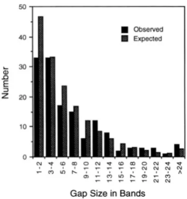
The size distribution (in band numbers) of gaps in our map of the major autosomal arms is summarized in Figure
2.
These gaps were estimated by assuming clone limits lay at the midpoint of the band uncertainties listed in the APPENDIX. Most of the gaps are relatively small, and could be spanned by cosmids, P1 clones (STERNBERG 1992), or small YACs. Also shown in Figure 2 are the numbers of gaps expected based on the assumption of random clone placement. These expectations were cal- culated using the exponential distribution, as described in the legend to Figure2.
There is reasonably good agreement between observed and expected values(x'
=12.38, degrees of freedom = 8,
p
0.14), with the ex- ception that we see fewer very small (1 or 2 band) gaps than expected. This disparity is not surprising, however, because many small gaps lying within the uncertainties of adjacent YACs would be missed. The good agreement between gaps observed and gaps expected according to the exponential distribution provides additional evi- dence that the YACs are essentially randomly distrib- uted, at least on the major autosomes.Drosophila YAC Clone Map 1989
1 the chromocenter. Finally, it is possible that the large
40
ti
E30
I)
3 20
10
0
" " . - N C U
Gap Size in Bands
FIGURE 2.-Distribution of gaps in coverage of the major autosomes. Black columns represent the observed numbers of gaps of the indicated sizes. Hatched columns represent the expected numbers calculated according to the exponential distribution. Expected values were calculated using the fol- lowing parameters: total number of autosomal bands = 4385; number sites mapped on the autosomes = 752; average cov- erage at each site = 201.5 kb; average size of band = 22.5 kb. Note that, because chimeric clones have been taken into ac- count, the number of sites mapped exceeds the number of clones mapped, and the average coverage at each site is less than the average clone size. To calculate the expected distri- bution, each mapped site was initially treated as a point and the distribution of gaps calculated by evaluating the exponen- tial distribution integral -e-".lilx (where 0.171 is the number of
sites mapped per band). Since the average coverage at each mapped site was about nine bands (201.5/22.5 = 8.96), gaps in the distribution so derived would be expected to be short- ened by this same distance. Accordingly, the distribution was truncated at nine bands to produce the expected distribution shown. The goodness of fit of the observed gap distribution to the expected distribution was tested by
2.
Classes in the tail of the distribution were pooled in this test so that the expected number in each group always exceeded five. The Pvalue o b tained (=0.14) indicates the observed distribution is not sig- nificantly different from expectation. Note we have not at- tempted to incorporate variability in clone size or band size into our calculations. However, doing so could only improve the fit of the observed results to expectation.natively, the presence of many dispersed repetitive se- quences in these regions could obscure the primary site of hybridization. If repeats abundant in the chromo- center were present at these sites, clones from them could have been misclassified as heterochromatic. The possibility of such misclassification is very real for the euchromatin of chromosome 4, which is strongly en- riched in repeats (MIKLOS et al. 1988). Misclassification is most likely to occur with smaller (<150 kb) clones, since the primary sites of large clones are normally quite unambiguous, even when many dispersed repeats are labeled. Indeed, a few smaller clones initially classified as heterochromatic were found, upon reexamination, to be euchromatic clones containing repeats abundant in
size in terms of band counts of the four gaps above is misleading; these regions may be no larger physically than many other gaps in the map. This would appear to be the case at least for the section 31 gap. AJIOW et al. (1991) report that a single clone of 220 kb covers most of four lettered divisions in this finely banded region.
About 38% of the mapped euchromatic YACs clearly label dispersed middle repetitive sequences in addition to the primary site. If such sequences were randomly distributed, then based on their abundance in the ge- nome [see SPIL~DIJNG and RUBIN (1981) and RUBIN
(1983) for reviews), one would expect a far higher per- centage of clones to contain repeats. Indeed, 38% is almost certainly an underestimate, since the labeling of repeats in our in situs is highly dependent on hybrid- ization conditions. The same clone can show no trace of mobile elements in one slide, and strong labeling of such elements in another. A number of our slides show weak X-specific labeling in addition to the primary site. Hybridization sites are clustered in numbered sections 1-14, and resemble the distribution of a repetitive se- quence family described by WARINC and POLLACK (1987) (see Figure Id). About 28% of all euchromatic YACs, and 66% of those showing dispersed repeats, label the chromocenter. Presumably such clones hybridize to mo- bile elements present in heterochromatin. About 3% of the clones in Table 1 labeled the chromocenter in ad- dition to a euchromatic primary site, but did not label dispersed repeats. Very likely, dispersed repeats are present in most or all of these clones, but went unde- tected in euchromatin because the in situ signal was too weak. A ~ I O K A et al. (1991) report a very low percentage
of euchromatic YACs labeling dispersed repeats (6%)
and the chromocenter (7%). This difference with our data is not understood, but may result from weak label- ing of their slides, or incomplete data collection.
Heterochromatic clones: Of the 979 YAC clones ex- amined by in situ hybridization, 124, or about 13%, were classified as heterochromatic since they labeled the chromocenter and had no obvious euchromatic primary site.
As
described above, these assignments are probably not totally accurate, since euchromatic clones very rich in repeats could be misclassified as heterochromatic. The chromocentric staining patterns for heterochro- matic clones are remarkably diverse, and include strong, almost uniform labeling; granular staining; weak, fi-1390 H. Cai et al.
the heterochromatic clones labeled dispersed repetitive sequences in euchromatin in addition to the chromo- center. This is probably an underestimate, since in situ
labeling was so weak for ten of the remaining clones that euchromatic repeats would not have been seen if present. Ten of the heterochromatic clones show stippled labeling of the nucleolus (see Figure l e ) . Two clones with euchromatic primary sites (N11-66, N27-12) also label the nucleolus, perhaps because they are chi- meric, or contain repeats also present in the nucleolus organizer. Clone N27-08, whose primary site includes the most proximal portion of the X euchromatin (20&5;B1-3) labels an island within the nucleolus (see Figure
If).
The fraction of YAC clones that is heterochromatic is almost certainly less than the 13% estimated above, since the DY and DYN libraries are badly contaminated with multiple isolations of identical clones. Duplicate clones of euchromatic DNA are easily identified by their iden- tical in situ localizations and extents, and have been removed from the data in the APPENDIX. Synonymous clones of heterochromatic DNA are not so easily iden- tified, and many duplicates or triplicates of DY and DYN clones are likely present among our heterochromatic clones. Based on the frequency of reisolations of euchromatic clones (duplicates comprised about 25% of the DY and 10% of the DYN clones mapped), it is probable that only about 11
%
of unique YAC clones are heterochromatic.Four of the clones classified as heterochromatic label all telomeres (see Figure IC) as well as the chromocenter and dispersed euchromatic repeats. These clones pre- sumably contain He-T sequences (RUBIN 1977; YOUNG et al. 1983; RENKAWITZ-POHL and BIALOJAN 1984; TRAVERSE and PARDUE 1989; BEISSMANN et al. 1990), which are lo- cated at all telomeres and in the pericentric heterochro- matin, as well as one or more mobile elements present in euchromatin. Clone R19-83, whose primary site (1Al; B2-3) is at the tip of the X , also labels all telomeres, the chromocenter, and euchromatic repeats.
Finally, in addition to the 979 clones described above, we have identified a number that, after repeated at- tempts, do not label polytene nuclei. These clones could contain sequences that are severely underreplicated, or perhaps deleted (&EN and SPRADLING 1990; GLASER
et al. 1992), during polytenization. Alternatively, these clones may contain DNA from some source other than Drosophila.
Clone stability. The large majority of Drosophila YAC clones appear completely stable. However, a few in- stances of instability have been seen. Many of our initial cultures, picked directly from the transformation plates, were mixed, and showed more than one clone on sizing gels. By streaking to single colonies, almost all of these cultures could be resolved into separate clones, which were given lettered designations. In most cases, sepa- rated clones labeled completely different sites and ap-
pear unrelated. In nine cases, however, subclones of dif- ferent size labeled the same site by in situ. Presumably, these have undergone some type of rearrangement in yeast. In two of these cases, smaller derivatives labeled a visibly shorter region, and appear to have arisen by ter- minal deletion. In our entire analysis, we found only one clone (Rt05-24A; primary site 13A5-6
+
13B4; C47) that showed a gap in a primary hybridization site, and that may, therefore, have undergone a large internal dele- tion. Initial instability of heterochromatic clones would have been rather difficult to detect in our work, since rearranged heterochromatic clones would probably show the same chromocentral labeling as their progeni- tors. Nonetheless, we find no evidence of size instability upon subculturing of heterochromatic clones. The fre- quency of apparent rearrangement of YAC clones seen here (about 1%) is comparable to that seen by others working on Drosophila and other systems (VILAGELIU and TILER-SMITH 1992).DISCUSSION
We report the mapping of 979 randomly selected large YAC clones by in situ hybridization to polytene chromosomes. Of these, 855 clones of average size 211 kb map to euchromatic sites. By direct band counts, we estimate that these clones provide about 76% coverage of the euchromatin of the major autosomes, and 63% coverage of the X euchromatin. Two lines of evidence indicate that the euchromatic clones are essentially ran- domly distributed. First, coverage (as estimated by band counts) is in good agreement with the Poisson expec- tation, calculated using the generally accepted figure of 165,000 kb as the genome size. Second, the distribution of gaps in the coverage matches well the distribution expected if coverage were random (i. e . , the distribution predicted by the exponential distribution). Although euchromatic coverage appears essentially random, three of the largest gaps (6B;6F, 16A, 16F, and all of chromo- some # ) may be significant, as similar gaps are present in the YAC mapping data of AJIOKA et al. (1991).
In the study of AJIOKA et al. (1991), 500 euchromatic YAC clones were mapped. Thirty-eight of these clones were also mapped in our study, and are described again here because, in most cases, our mapping is somewhat different. When the clones from the two projects are pooled, they contain about 42,700 kb of DNA from X euchromatin and 228,000 kb of DNA from autosomal euchromatin. Assuming a genome size of 165,000 kb and a heterochromatin fraction of 25%, these amounts correspond to about
1.7
equivalents of X euchromatic DNA and about 2.3 equivalents of autosomal euchro- matic DNA. Assuming random distribution of clones, the combined sets of YACs should, according to the Pois son distribution, provide about 82% coverage of the X ,and 90% coverage of the major autosomes.
Drosophila YAC Clone Map 1391
of average size 198 kb comprise one euchromatic ge- nome equivalent. This claim is highly suspect, however, since their central argument to support it is incorrect. Assuming random clone distribution, the proportion of sequences not covered by clones should be equal to the zero term of the Poisson distribution, e-‘, where G is the number of genome equivalents of DNA present in the clones analyzed. AJIOKA et al. consider the fraction of lettered divisions not labeled by any of their YACs (212,’ 600 for the major chromosome arms) to be equal to the proportion of all sequences not covered, and calculate G as 1.04. However, it is clear from their own data that many of the lettered divisions that are labeled are only partially so. Thus, their calculation substantially overes timates the coverage achieved. We calculate that their clones constitute about 0.8 euchromatic DNA equiva- lents, rather than the 1.04 claimed, and their euchro- matic coverage at about 55%. Unfortunately, it is not possible to estimate their coverage by direct band counts, since many of their localizations are only to let- tered division, not to band.
One hundred and twenty-four of the 979 YAC clones (13%) mapped in this report are heterochromatic and label the chromocenter as the primary site. As described above, because of contamination of the DY and DYN clone sets with duplicate clones, probably only about 11% of unique YACs are heterochromatic. Since 25- 30% of the genome is composed of heterochromatin, there would appear to be an underrepresentation of het- erochromatic sequences in YAC clones by a factor of two or three. Taken together, the heterochromatic YACs contain only about 0.6 equivalents of heterochromatic sequences in the genome. A number of factors could contribute to this apparent underrepresentation. First, it could be that the distribution of EcoRT and Not1 sites precludes the efficient cloning of some heterochromatic sequences using these enzymes. Second, it is possible that many heterochromatic clones went unrecognized because they contain sequences not replicated in poly- tene cells, and fail to label any site by in situ hybridiza- tion. Third, it is quite possible that many heterochro- matic clones are highlyunstable, and break down before they can be analyzed. We note, however, that the het- erochromatic YACs in our collection appear stable and are of even larger average size (216 kb us. 21 1 kb) than the euchromatic clones.
The deficit of heterochromatic clones may be more apparent than real. Because most of our YACs were constructed using DNA from adults, the expected fraction of heterochromatic clones is not entirely clear. BLUMENFELD and FORREST (1972) found one sat- ellite of Drosophila melanogaster to be underrepre- sented in total adult DNA by about 50%. Similarly, H A ” O N D and LAIRD (1985) found the 1.705 g/cm3 sat- ellite to be underrepresented in nurse and follicle cells from adults. These instances presumably result from underreplication in polytene (e.g., Malpighian tu-
bules) or polyploid (e.g., midgut, hindgut, salivary gland, ovary) tissues of the adult. In Drosophila uirilis,
it would appear that different satellites are under in- dependent replication control, since specific satellites are underrepresented to different levels in different adult tissues (BLUMENFELD and FORREST 1972; ENDOW and GALL 1975). Thus, although underrepresentation of het- erochromatic YACs is to be expected among clones made from adult DNA, the extent of this underrepre- sentation is difficult to predict, and is probably sequence-specific.
The fraction of YAC clones labeling the nucleolus (about 1.3%) is close to the fraction of adult DNA com- posed of ribosomal DNA repeats (about 2%) (SPRADLING and RUBIN 1981; TARTOF 1973). Some nucleolar clones were probably incorrectly scored as chromocentral, since nucleolar labeling would likely have been missed
in weakly labeled slides.
On first impression, it would seem reasonable to use the data the APPENDIX to estimate clone overlaps and de- fine contigs. Indeed, this is exactly what was done to generate the gap distribution shown in Figure 1. How- ever, we believe it would be a mistake to take the details of such an analysis too seriously, since several factors limit the resolution of our cytological data. Because of duplicate clones in the DY and DYN libraries, we unin- tentionally did a fairly large test of the reproducibility of our cytological localizations. Although independent mappings of the same clone were always approximately the same, they usually differed in detail. Similarly, al- though rough agreement is seen for the 38 clones mapped by both AJIOKA et al. (1991) and ourselves, when considered in detail, direct contradictions exist for 24 of the 33 of these clones mapped to band by AJIOKA et al.
In practice, the greatest limitation to the accuracy of mapping was the variable quality of in situ slides. Every effort was made to examine highly stretched chromo- somes, since localizations precise to a fraction of a band can often be made in such preparations. However, for some clones, stretched chromosomes could not be found, rendering the mapping much less accurate. Al- most always in such cases clones appear to cover a greater area than is real. The magnitude of this effect was a surprise, and caused us to reexamine most of our
in situs.
As
a result, we significantly downsized the cov- erage of many, perhaps most, of our clones described in preliminary reports (HARTL et al. 1992; ASHBURNER et al. 1991). Another important variable is the strength of thein situ signal. If slides are overdeveloped, stain obscures the underlying banding pattern and can spread beyond the primary site. Conversely, if staining is weak only ma- jor bands are labeled, and coverage can be underesti-
1392 H. Cai et al.
be mistakenly included within the primary site. For all of these reasons, we believe determination of clone over- laps and generation of contigs cannot be done convinc- ingly from cytological data [although see HARTL (1992) for such an attempt using our preliminary data], and must await molecular studies.
A major motivation for our mapping has been to pro- vide materials to aid other Drosophila researchers in their cloning efforts. As shown by VAN DER BLIEK and MEY- EROWITZ (1991) in their cloning of the shibire locus from one of our YACs, gel-purified YAC DNA can be rapidly subcloned into cosmids. These subclones can then be ordered to generate a complete “walk” and, if appro- priate mutations exist, the gene of interest identified. A detailed protocol for subcloning from YACs is presented by WHITTAKER et al. (1993).
An
alternate approach, use- ful for YACs not containing repeats, has been to use YACs as probes to screen existing phage or cosmid li- braries [as, for example, in the cloning of buttonhead(E. A. WIMMER, personal communication)]. YACs should be particularly useful in cloning genes for which break- point alleles exist, since it should be relatively easy, by i n s i t u hybridization to polytene chromosomes, to identify subclones that span such breakpoints. Although the techniques required for subcloning from YACs are rela- tively straightforward, YACs have not received wide use in the Drosophila community, probably because most labs are not set up to run the pulse field gels required to purify the clones. To encourage the use of YACs, de- tailed protocols describing the culturing of YAC clones and the purification of YAC DNA by field inversion gel electrophoresis are included with all YAC shipments.
Obviously, of greater utility for gene cloning would be the development of a complete genome map using more convenient clones, such as cosmids or P1 clones. The YACs reported here are potentially of great use in the development of such finer scale maps, and have already played a significant role in the isolation and ordering of X chromosomal cosmids in the mapping project of KAFATOS et al. (1991) and SIDEN-KIAMOS et al. (1990). As in the nema- tode genome project (COULSON et al. 1988) ), YACs may also help by bridging gaps in PI or cosmid maps caused, for example, by sequences not clonable in bacteria. The large size and stability of heterochromatic Y4Cs suggest that Y4Cs will be of central importance in the analysis of het- erochromatin. A particularly exciting prospect in this re- gard is the cloning of a Drosophila centromere, as this could facilitate the development of yeast-Drosophila shuttle vectors, and allow application of the powerful tech- niques of yeast genetics to Drosophila.
This work was initiated as a collaboration with DANIEL L. HARTL, and
we would like to thank him for generously providing clones from the
DY and DYN libraries. We are also grateful to ANDREW LINK for pro-
viding the DYN library to our groups for mapping. DAN GARZA gave us
much advice on YAC cloning in the early phases of the project, and
MAWARD OLSON and GEORGES CARLE were very helpful in setting up the
pulse field gel apparati. We thankJoHNc LIM for generously providing
an important unpublished protocol for in situ hybridization, and
TERRY L aand ED GRELL for providing fly stocks. Finally, we thank
ALAN TEMPLETON and JOY BERGELSON for advice on statistics, DIANNE
MATTSON for discussions, and DIANNE MA’ITSON, FLAWANO GIORGINI and the anonymous reviewers for comments on the manuscript. Our work was supported by a grant from the National Institutes of Health.
LITERATURE CITED
AJIOKA, J. W., D. A. SMOLLER, R. W. JONES, J. P. CARULU, A. E. C. VELLEK
et al., 1991 Drosophila genome project: One-hit coverage in
yeast artificial chromosomes. Chromosoma 100: 495409.
ASHBURNER, M., 1989 Drosophila: A Laboratory Handbook. Cold
Spring Harbor Laboratory, Cold Spring Harbor, N.Y.
ASHBURNER, M., D. M. GLOVER, R. D. C. SAUNDERS, I. DUNCAN, D. HARTL et al., 1991 Genome Maps 1991: Drosophila melanogastercom-
ponent. Science 254: 246-262.
BIESSMANN, H., J. M. MASON, K. FERRY, M. D’HULST, K. VALGEIRSDOT~R
et al., 1990 Addition of telomere-associated HeT DNA se-
quences “hea1s”broken chromosome ends in Drosophila. Cell 64:
663-673.
BINGHAM, P. M., R. LEVIS and G . M. RUBIN, 1981 Cloning of DNA
sequences from the white locus of D. melanogaster by a novel and
general method. Cell 25: 693-704.
BLUMENFELD, M., and H. S. FORREST, 1972 Differential under-
replication of satellite DNAs during Drosophila development.
Nature New Biol. 2 3 9 170-172.
BRIDGES, C. B., 1935 Salivary chromosome maps with a key to the
banding of the chromosomes of Drosophila melanogaster.
J. Hered. 26: 60-64.
BROWNSIXIN, B. H., G . A. SILVERMAN, R. D. L I ~ L E , D. T. BURKE, S. J.
KORSMEYER et al., 1989 Isolation of singlecopy human genes
from a library of yeast artificial chromosome clones. Science 244:
BURGERS, P. M., and K. J. PERCIVAL, 1987 Transformation of yeast
spheroplasts without cell fusion. Anal. Biochem. 163: 391-397.
BURKE, D. T., G . F. CARLE and M. V. OLSON, 1987 Cloning of large
segments of exogenous DNAinto yeast by means of artificial chro-
mosome vectors. Science 2 3 6 806-812.
CARLE, G . F., and M. V. OLSON, 1984 Separation of chromosomal
DNA molecules from yeast by orthogonal-field-alternation gel
electrophoresis. Nucleic Acids Res. 12: 5647-5664.
CARLE, G . F., M. FRANK and M. V. OLSON, 1986 Electrophoretic sepa- rations of large DNA molecules by periodic inversion of the elec-
tric field. Science 232 65-68.
COULSON, A,, R. WATERSTON, J. Km, J. SULSTON and Y. K o m ,
1988 Genome linking with yeast artificial chromosomes. Nature
DANILEVSKAYA, 0. N., E. V. KURENOVA, M. N. PAVLOVA, D. BEBEHOV, A. J.
LINK et al., 1991 He-T DNA sequences in the Y chromosome of
Drosophila melanogaster share homology with the X-linked Stel-
late genes. Chromosoma 100: 118-124.
ENDOW, S. A,, and J. G . GALL, 1975 Differential replication of satellite
DNA in polyploid tissues of Drosophila virilis. Chromosoma 50:
175-192.
FEINBERG, A. P., and B. VOGELSTEIN, 1983 A technique for radiolabel-
ing DNA restriction endonuclease fragments to high specific ac-
tivity. Anal. Biochem. 1 3 2 6-13.
GALL, J. G . , E. H. COHEN and M. L. POW, 1971 Repetitive DNA se-
GARZA, D., J.
w.
AJIOKA, D. T. BURKE and D. L. W m , 1989 Mappingquences in Drosophila. Chromosoma 33: 319-344.
the Drosophilagenomewith yeast artificial chromosomes. Science
GLGER, R. L., G . H. -EN and A. C. SPRADLING, 1992 Replication
forks are not found in a Drosophila minichromosome demon-
strating a gradient of polytenization. Chromosoma 102: 15-19.
GRELL, E. H., 1976 Genetics of some deoxyribonucleases of Dro-
sophila melanogaster. Genetics 83 (Suppl.): s28-~29.
HAMMOND, M. P., and C. D. URD, 1985 Chromosome structure and
DNA replication in nurse and follicle cells of Drosophila mela-
nogaster. Chromosoma 91: 267-278.
HARTL, D. L., 1992 Genome map of Drosophila melanogaster based
on yeast artificial chromosomes, pp. 39-69 in Genome Analysis,
1348-1351.
335: 184-186.
Drosophila YAC Clone Map 1393
Vol. 4: Strategies f o r Physical Mapping, edited by K. E. DAVIES and
S. M. TILGHMAN. Cold Spring Harbor Laboratory, Cold Spring
Harbor, N.Y.
HARTL, D. L., J. W. AJIOKA, H. CAI, A. R. LOHE, E. R LOZOVSKAW et aL,
1992 Towards a Drosophila genome map. Trends Genet. 8: 70-75.
KAFATOS, F. C., C. LOUIS, C. SAVAKIS, D. M. GLOVER, M. ASHBURNER et al.,
1991 Integrated maps of the Drosophila genome: progress and
prospects. Trends Genet. 7: 155-161.
KARPEN, G. H., and A. C. SPRADLING, 1990 Reduced DNA polyteniza-
tion of a minichromosome region undergoing positioneffect var-
iegation in Drosophila. Cell 63: 97-107.
L A N C E R ~ A F E R , P. R., M. LEVINE and D. C. WARD, 1982 Immunological
method for mapping genes on Drosophila polytene chromo-
somes. Proc. Natl. Acad. Sci. USA 79: 4381-4385.
LEFEVRE, G., JR., 1976 A photographic representation and interpre-
tation of the polytene chromosomes of Drosophila melanogaster
salivary glands, pp. 31-37 in The Genetics and Biology of Dro-
sophila, Vol. la, edited by M. ASHBURNER and E. NOVITSKI. Aca-
demic Press, New York.
MANIATIS, T., E. F. FRITSCH and J. SAMBROOK, 1982 Molecular Cloning:
A Laboratory Manual. Cold Spring Harbor Laboratory, Cold Spring Harbor, N.Y.
MIKI.OS, G. L. G., M.-T. YAMAMOTO, J. DAVIES and V. PIRROTTA, 1988 Mi-
crocloning reveals a high frequency of repetitive sequences char-
acteristic of chromosome 4 and the Pheterochromatin of Dro-
sophila melanogaster. Proc. Natl. Acad. Sci. USA 85: 2051-2055.
~ C HE. M., , H. J. BARR and R. W. RASCH, 1971 The DNA content of
sperm of Drosophila melanogaster. Chromosoma 33: 1-18.
RENKAWITZ-POHI., R., and S. BIALOJAN, 1984 A DNA sequence of Dro-
sophila melanogaster with a differential telomeric distribution.
Chromosoma 89: 206-21 1.
RUBIN, G. M., 1977 Isolation of a telomeric DNA sequence from Dro-
sophila melanogaster. Cold Spring Harbor Symp. Quant. Biol. 42:
RLJBIN, G. M., 1983 Dispersed repetitive DNAs in Drosophila, pp.
329-361 in Mobile Genetic Elements, edited by J. A. SHAPIRO. Aca-
demic Press, New York.
rt al., 1990 Towards a physical map of the Drosophila melano- gaster genome: mapping of cosmid clones within defined
genomic divisions. Nucleic Acids Res. 18: 6261-6270.
1041-1046.
%DEN-~AMOS, I., R. D. C. SAUNDERS, L. SPANOS, T. kfA]ERUS,J. TREANEAR
SOMA, V., 1988 Chromosome Maps of Drosophila, Vol.11. CRC Press,
Boca Raton, Fla.
SPIERER, P., A. SPIERER, W. BENDER and D. S. HOGNESS, 1983 Molecular
mapping of genetic and chromomeric units in Drosophila mela-
nogaster. J. Mol. Biol. 168: 35-50.
SPRADLING, A. C., and G. M. RUBIN, 1981 Drosophila genome
organization: conserved and dynamic aspecrs. Annu. Rev. Genet.
STERNBERG, N. L., 1992 Cloning high molecular weight DNA frag-
ments by the bacteriophage P1 system. Trends Genet. 8: 11-16.
TARTOF, K. D., 1973 Regulation of ribosomal RNA gene multiplicity
in Drosophila melanogaster. Genetics 73: 57-71.
TRAVERSE, K. L., and M. L. PARDUE, 1989 Studies of He-T DNA se-
quences in the pericentric regions of Drosophila chromosomes.
Chromosoma 97: 261-271.
VAN DER BLIEK, A. M., and E. M. MEYEROWITZ, 1991 Dynamin-like pro-
tein encoded by the Drosophila shibiw gene associated with ve-
sicular traffk Nature 351: 411-414.
VILAGEIJU, L., and C. TYLER-SMITH, 1992 Structural instability of YAC
clones and the use of recombinationdeficient yeast host strains,
pp. 93-112 in Techniques f o r the Analysis of Complex Genomes,
edited by R. ANAND. Academic Press, San Diego.
VOGEISTEIN, B., and D. GILLESPIE, 1979 Preparative and analytical pu-
rification of DNA from agarose. Proc. Natl. Acad. Sci. USA. 76:
WARING, G. L., and J. C. POLLACK, 1987 Cloning and characterization
of a dispersed, multicopy, X chromosome sequence in Drosophila
melanogaster. Proc. Natl. Acad. Sci. USA 84: 2843-2847.
WHITTAKER, P. A,, M. MATHRUBUTHAM and L. WOOD, 1993 Construc-
tion of phage sublibraries from nanogram quantities of YAC DNA
purified by preparative PFGE. Trends Genet. 9 195-196.
YOUNG, B. S., A. PESSION, K. L. TRAVERSE, C. FRENCH and M. L. PARDUE,
1983 Telomere regions in Drosophila share complex DNA se-
quences with pericentric heterochromatin. Cell 34: 85-94.
1 5 219-264.
615-619.
Communicating editor: M. T. FULLER
APPENDIX
The primary sites of hybridization of 855 euchromatic clones are listed in Table 2.
TABLE 2
Primary sites of hybridization of 855 euchromatic clones
Clone Size Dispersed Chromw
Cytology number (kb) repeats center
1Al; B2-3 1B1; Bl0-ll 1B8-9; C 2 4 1B14C1; C3-4 1D2-3; E 3 4 1 E l ; E 4 5 1E34; F1-2 2A1; B8-10
+
87C 1F4; 2B1-2 2A3-Bl; 87-8 283.1; BIZ-16 2D1-2; El-2+
38B 2B15-17; C1-2 2E3-Fl; 3A2-3 2F4; 3A1-2 3A3; A5 3A3.1; 3C2-4 3B56; Cl-2 + 67D 3C1-2; C5-7 3C45; C10-123D1-2; E2-3 3E1; E2
+
67F 3F1-2; 4A2-3 3F5-6; 4A3-5 3F3-6 4A2-3+
62F 4B1; B44B1; C 4 5
R I M 3 R1434 N13-23 Nll-18 N2425 Rt0467 N18-90 723 N27-20 N10-31 645 N2303B/AB 736 N 1 7-29 N15-58 774 R22-11 N13-61 R07-58 RO 1-60
N11-88
N09-07 R0740 N2615 N12-95 N2140 354
200 360 180 175 220 180 270 150 220 230 160 110 160 150 170 200 300 180 320 220 150
250 150 210 145 230 220
Clone Size Dispersed Chromo-
Cytology number (kb) repeats center
4B1; C 4 5 4B1-2; 4B45; 4D1; D 6 7 4D1; D 6 7 4D1; E l 4D1-2; El-2 4D3-4; E2-3 4D3-5: E?-3 4E1-2; F 4 5
+
89E; 4D5-7; E2-390A
+
96EF 4E2; F 3 4 5A10-11; C2 4F9; 5A2-35C3-6; D2 581; C1 + 21BC 5D3-5; E 4 6 5D5; D6 6A1; B3-4 6A1; A2 6F5-7; ?A68 ?AI; B2-3 7A67; B2-3 7B1: B 4 5 7C1; C.W
B3-4 + 1 5 F 16A C1-2
+
64BCRt10-29 N25-I2 N2696 R17-02
R2 1-74 494 R17-24 623 N2466 N25-78 N22-12 R22-68 N26-03B/AB N28-47 N27-15 588 N11-80 574 N23-89 N20-66 R15-21 R0 1-53 N26-01 N12-93 N09-15
1394 H. Cai et al.
TABLE 2-Continued
Clone Size Dispersed Chromo- Cytology number (kb) repeats center
7E1; E2 7E1; E2
+
79E 7F1; F2+
73A 7F1-2; F8-10 7 F 3 4 8B1-2 7F7-9; 8A5 8A1; C2-3 8D9-11; E 3 4 8D10-11; E 6 9 8E1-2; F1-2 8E3-4; F9-10 9Al; A2-3+
21CD 9Al; A2-3+
36D SA 1 ; A3-4 9A1; A 3 4 9A2; A3 9A3; B1 9B1; B1415 9B1; C2-3 9B67; D2-4 9B8-10; C1-2 9C2-3; E l 9C3-5; D 3 4 9D1-2; El-2 9D34; F5-10 9E1; E2 SEI; E2+
61E 9E1; F10-I3 9F5-6; 10.42-3 9F8-11; 10B2-310.42-3; A10-11 10B7-8; C 2 4 10B8-9; C1-2 10D2-3; l l A l llA5-6; A10-12 l l A 6 A10-ll 11A7-8; B1-2 llBl7-19; Dl-2 11D1; D10-El 11D1; D8-El 11F1-2; 12A5-6 12A1; A1-2 12A2-3; AS-10 12A8-10; D2-3 12B1; C7-8 12B2-4; Dl-2 12C1-2; D2-3 12E5-8; F2-3 12E8-9; F2-3 13A1; 8 4 5 13.45-6
+
13BC+ 22B
13B3; C1-2 13B4; C 4 7 13D3-6; E 4 5 13E1-2; F7-15 13El0-11; 14.445 14B1; B15-18 14B1; BY-4 14B5-6; C6-7 14B3-4; C1-2 14F1-3; 15B1 15D1; F1-2 15E7; 16A1-2 15F1-3; 15F14; 16A1-2 15F24; F5-6 16F1; 17A1-3 16F1; F7-17A1 17AI-2; A10-12 17A45; A5-7 17A5; 12F.34; 13A10-12
+
13A + 22816A46 + 4B
A6
+
22CDN09-26 N21-23 665 N12-74 867 N 10-55 RO7-36 N28-60 R l 6 O l R19-61 N17-61 R2373 R19-57 N23-10 R25-03 860 N1346 886 N28-76 R07-57 R15-64 N26-20 R20-59 806 R13-74 600 N09-57 805 R I M 5 288 R18-90 N19-75 N09-72 N2412 Rt0948 N2420 R X O 1-96 R21-50 N17-71 N10-08 R20-11 483 766 Rl651A R19-07 N12-25 R13-12 N19-21 R15-34 R1637 RO3-06 Rt05-24A 581 Rt05-24A 75 1 822 922 699 802 R17-25 R1608 N10-89 N12-19 N12-21 N25-12 N 1 6 9 6 45 1 R2340 755 875
N2654 ~ 1 8 - 7 0
160 150 160 150 300 170 350 260 210 230 180 280 220 210 200 180 150 160 200 260 210 180 150 280 190 150 180 205 200 280 220 240 170 230 205 150 240 190 150 220 190 150 280 280 220 210 200 280 220 210 230 220 190 220 190 300 275 220 200 210 190 240 150 170 240 150 150 210 250 220 150 190
Clone Size Dispersed Chromo- Cytology number (kb) repeats center
17A5-6; B2-3 17A7-8; C2-3
17A7-9; B2-3 17D45; F1-2 17D5-6; 18A2-3 18A1; A 3 4 18A2-3; B1-2 18A5-7; C 4 5 18B10-11; C 4 5 18C1; C4 18345; 19B2-3 19A1; A 4 5 19B1-2; E5-6 19B2-3; El-2 19B3-Cl; C3-6 19D1-2; E3-4 19D1-2; E 4 5 19E1; E4 19E2-3; E 7 4 19E9-Fl; F 4 6 1 9 F 3 4 2OA2-3 2OA3-5; B C 20A3-5; B1-3 20A5-Bl; C2-3 21A3-Bl; C1 21B1; B2
+
77E 21B2-3; B 4 7 21B47; C1-2 21B5-7;C1 + 5BC 21C1-2; C 4 5 21C3-4; D2 21C5; 21C67; Dl-2 21C7-8; D2 21C7-8; 21D1; D2 21E1-2; F3-4 21E2-3; 22A1-2 22Al-2; A5-8 22A3-4; A5-7 22A5-7; B 6 7 22A8-81; B7-8
C7-8 + 82E
D 3 4
+
9A22B1; B2 22B841; E2-3
+
13A + 13BC 22C1; D l + 17A 22C1; D4-6 22D1-2; E 3 4 22E1; E3-4 22E1; F1-2 22E1; F1-2 22E1; F2-3 22E2-3 F34 22F1; 23A2-3 22F1-2; 23A1-2 22F1-2; 23A5-7 22F3-4; 23A2-3 22F3-4; 23B2-3 22F3-4; 23C2-3 23A1; A I 2 23A2-3; C1 23D1-2; E3-4 24B1; C1-2 24C1; D2-3 24C1; D2-3 24D1; El-2 24D1-2; E l 24E1; 25A3-4 24E1-2; 25A1 25A1; A4 25A1; A5-8 23F2; 24A2-324C1-2; Dl-2
24E2; 25A2-3
Drosophila YAC Clone Map
TABLE 2-Continued
1395
Clone Size Dispersed Chromc-
Cytology number (kb) repeats center
25A2-3; B5-6 25C47; D2-3 25D1-2; E 2 5 25E1; 26A1-2 25F1; F5-26A1 25F3-4; 26A1-2 26B7-8; C3-4 26C2-3; D2-3 26D1; E2 26D10-El; F5-7 26D10-El; 27B1-2 27A1; C5-6 27A1-2; C1-2 27C1; C2
+ 49C
+
58BC 2 7 c a 9 FI 27E1; E7-8 27E54; F2-3 27F47; 28B3C1 28A1; B 3 4 28C1-2; D3-4 28D1; D 4 5 28E2-4; 29A1-2 28F3-5; 29D1-2 29D1; E2 29D1; E2-3 3 0 A 3 4 A8-9 30A3-4; A9-B2 30A3-5; A7 30A3-5; A 7 4 30A67; B2-3 30A7; A8 30C1; C5-6 3OC3-5; F3-6 31A1; B1+
33A 3Z.41; C2-332A2-3; C1-2 32E3-4; 33A2-3 32F1; 33A8B1 32F2-3; 33A6-8 32F1; F1-2
33A47; B13C6 33A1; A2 + 31AB 33A5-7; B2-3 33B1; C 5 6 33B1; C1-2 3 3 C 4 6 D 2 4 33D1-2; D3-4 33D1-2; D 4 E 2 33132-3; E7-9 33D3-El; 33E6-8; F2-3 33F1; 3 M - 7 34A1; A9-11 34A1-2; A2 34A5S; B 4 5 34A56; B2-3 34A5-7; B7-8 34A6 B2-3 34B8-11; C5-6 3 4 C 3 4 C5-6 34D1-2; 34D3-5; 34F1-2; F3-4 35B2-3; B 6 7 35B3.1; B7-9 35B6 B7 35B68; B9-10 35B8-9 BlOCl 35D1; D 4 5 35C1-2; C3-4 35D1; D5-7 35D1-2; D3-1
+ 56AB
+
69DEFE 3 6 + 89B
D7-8
+
9lCDE D8-El+
79EFR1625 R17-64 E 5 5 9 N20-72 N22-11 RXO2-16 R19-62 R22-41 817 R19-84 N12-06 R18-64 R18-09 N2634 R2423 RX0495 R16-84 R15-17 R25-53 609 RX01-64 N13-72 R12-23 N18-37 N13-38A/AB 893 R18-83 RtOl43 R1491B R15-73 752 918 N1484 R25-57 Rt10-61 R12-90 R14-08 827 R1744 N1C-65 R2557 N10-58B/AB 73 1 N2547 N10-58A/AB N09-89C/AC 919 N26-65 R23-58 N21-75 R0479 R2435 R15-05 R12-28 N20-14 R03-65 Rll-31 R05-80 Rt05-50 N15-74B/AB RX0423 R14-68 RXO3-16 R22-88 R1519 N12-56 641 928 Rt0401 Rt0348 R18-51 NO947 240 220 240 370 200 210 240 190 240 200 260 290 210 175 250 200 220 240 210 260 190 200 260 240 200 190 210 280 200 220 175 230 310 240 200 230 210 220 210 50 240 240 160 260 260 170 220 200 190 180 235 210 270 180 220 170 240 260 180 180 200 200 210 220 220 170 160 240 230 190 200 170
Clone Size Dispersed Chromc-
Cytology number (kb) repeats center
35D5-7; EZ-FZ 35D5-7; F1-2 35E1; E4F4 35E1-2; E7-F4 35E1-2; F2-3 35F2; Fs-12 36A3; A7-8 36A5-6; B2-3 36.49-10; (22-3 36B1; C1 3 6 B W 1 ; D l 36C1; C2 36D1; D2 36D1; D2 36D1; D2
+
9A 36D1; D 3 E 1 36D1; D2-3 36D1; E2-3 36D3-El; E 3 4 36E1; E M 36E1; E4 36E2-3; F7-8 36E3-4; F1-2 36F67; 37B1-2 36F1-2; 37A2-3 37A1; B 4 5 37A1-2: B1-2 37C5-7; D5-E2 37C1-3; Dl-2 37D1; D2-3+
82F;83A
+
94B 37F638A1;38A7-8 38A1; A2-3
38A1; A3-4 38A3-4; B 4 6 38.41; A 5 8 38.445; A8B1 38B1-2; (22-3 38B3-4; 38C1-2; 38C2; D2-3 38D1; E7-10 38D1-2; E 4 7 38F2-5; 39A1-2 39C1; C2 39A1; B1-2 39E34; 40.42-3 39E5-6; 40A3-4 40A 40A1; A4 40A1; A 4 5 40B1-2; B 3 4 40BC 40CD 41E3-5; F5-8 41E3-5; F1-2 41F1; F2 41F1; F2-3 41F1; F3 41F1; F9-11 41F1-2; F6-8 41F2-3; F10-11 42A1; 42.41; A2
A7-9 + 62BC 42A1-2; A 4 5 42A2; A8-9 42.41-2; AlBB3 4Z.445; A810 42A8-10; Al&Bl 42A1419; B1-2 42A15-17; B3-4 42B1-2; B4C6 42C3-5; D3
B5-6 + 2DE Dl-2
+
78ABN11-95 784 668 R2144 R1444 RXo460 R05-78 R1483 R23-55 RO7-11 R07-63 R21-12 R19-57 Rt12-17 ~ 1 8 - 5 6 ~ 1 a 2 3
R18-38 N1141 R2245 N2419 R07-37 R t l 5 1 0 N2454 R1627 R19-59 RX02-37 N09-63 700 N19-85 526 R17-14 N15-73 R15-74 N13-28 N17-30 R15-50 679 736 N2263 R17-34 R11-02 N21-29 589 917 N12-87A/AB N28-84 N1893 ~ 1 8 7 4
R23-29
H. Cai et al.
TABLE 2-Continued
Clone Size Dispersed Chromo- Cytology number (kb) repeats center
42C69; D5-6 42C7-10; D5-6 42C8-9; E l 42E3-5; 43A2-3 42F1-2; 43A2-3 43E1-3; E7-8 43E8-14; F1-2 43E1417; 44A2-3 44C1; C2-3 44C1-2; Dl-2 44D1; D M 44El-2; F 3 4 44E1-3; F1-2 45A2; C1-2 44F2-3; 45A2-3 45C1; El-2 45D1-2; F1-2 45D7-9; 46A5-4 46A1; AS4 46A1; A4 46B3-4; D2-3 46Cll-Dl; F67 46E7-9; 47A2 46F10-47AI; A5-8 4793-4: D 3 4 4794; 87-8 47B4; D l 4795-6; C 6 7 47Dl-2; D5-6 47E6F1; 48A1 47F3-5; 48A2-3 47F10-14; 48A4 48A1; B 4 6 48A3-4; B 6 7 481134; C1 48k5-Bl; C5-6 4881-2; C'L-3
48C1; C3 48C1; C 4 7 48C1; C5-8 48C1; D l + 88EF 48D 1; E2-3 48DL-3; E l 48D67; F2-3 48E2-3; F 6 7 49A34; 910-12 49A9-11; B1-2 49Cl; C2-3 49C1-2; El-2 49D1; E7-F1 49D1-2; E 6 7 49E5-7; F M 49F.34; 50A3-4 50A5-7; B 6 9 50A10-11; C 3 4 50A12-14; C 4 5 50B1; C1-2 50B1-2; C3-4 50C1; C10-12 50C1; C 4 5 50C3; C4 + 59D 5Oc8-9; C23-DI 50C9-1 I ; D2-3 50C1618; D67 50D1; E 6 7 50FI4; 51A5-6 50F8; 51A45 51A67; C1
+ 92F; 93A 5191; C2-3 51CI; D2-3 51D9-11; E 2 9 5lD9-11; E 6 7 51E1; E5-6 51E45; ElO-Fl
+ 27C + 58B
N28-13 R12-56 N23-37 RIG60 N10-80 714 Rt0141 619 N10-10 898 N2241 N12-63 N2690 R19-72 N20-67 R25-46 N14-48 N24-84 N17-62 626 N1640 R15-68 R14-41 R25-07 K03-20 728 912 R17-69 R19-34 R2476B R17-63 R12-87 N1462 829B R1749 R19-41 N09-89B/AB R0343 N 10-34 N09-13 N15-20 N12-62 R23-77 R19-66 R l 6 4 1 878 N2634 R0667 N12-92A/AB N25-60 R05-85 R12-14 N10-95 R16-07 N09-10 N 17-88 N2748 N21-05 N18-25 N 13-78 N1427 N 1 7-22 Rt01-57 N17-51 ROf-34 686 NOS-90 N28-28 R1472 N20-27 N 17-08 R15-51 N20-75 N22-62 I90 150 180 200 200 210 190 190 190 280 185 180 I90 170 250 240 240 280 200 270 220 240 210 190 220 140 200 200 270 200 180 160 160 I70 270 180 200 220 190 170 175 180 180 200 180 220 150 175 190 260 170 230 195 220 220 170 160 180 210 170 240 140 150 240 270 180 210 160 170 240 200 230 140 150 Cytology 51F1-2; 52A5-7 51F7-10; 5'2B3-4 52A4; B2-3 52B1-2; C 6 9 52Cl; Dl-2 52C1-2; Dl-2 5 2 C 1 4 CED2 52C8-Dl; El-2 52D7-8; E9-11 52Dl-2; El-2 52E1; 53A2-3 53B3-CI; C7-11 53C1; C E l l 53C1-2; D2-3 53D10-11; E2-3 53D10-13; Fl-2 53Dl2-14 E9-11 53E1;
Fll-13
+
69AB 54A1; B2-3 54A2-Bl; B1618 5481-2; Dl-2 54810-16; D2-3 54B1618; D2-3 55A1; A4B3 5 5 8 3 4 ; C 6 7 5585-10; C5-8 55C1; C5 5 5 C M ; E2-3 55F1-2; 56B1-2 56A1-2: B1-2 56A1-2; B1-2+ 33AB + 69DEF 56A2-3; Bl-2 56Dll-E2; E5-6 56D 1 ; E3-5 56F2-3; F7-8 56F4-6; Fl3-I5 56F8-9; F1617 57A1: A4 57Bl-'L; 95-6
57A5-6; B 3 4 57845; 810-13 57845; Cl-2 57B8-11; D2-3 57C2-4; E 2 4 57D10-11; E 4 5 57Dll-El; F2-3 57E1; E2 57E1; F 4 7 57E5-7; F 6 8 57F8-9; 58A3-4
58A2-3; B 6 7 5892-3; B4C4 5 8 B 3 4 D2-3 58B67; D2-3 58C1-2; D2-4 58D1; D7-8 58D.W; D7-8 58D6E2; F47 59A1; A3-4 liYC4-5; D 3 4 59B46; C3-5 59D1-2; D 3 4 + 50C 59D7-10; F2-3 60A8-11; B10-12 60A10-13; B12-13 60D9-10; D14-15 60A15-16; 811-12 6 0 D 1 4 1 6 E7-9 60F1-2; F3 6lAI; A5-6 60F2; F3 6lC4-7; Dl-2 61D1; D2
+
27C + 49CClone Size Dispersed Chromo- number (kb) repeats center
R07-3 1 N22-16 NlM5A/AB N28-02 551 R07-60 N27-23 510 R17-20 664 R15-76 R22-59 R24-56 R21-11 R18-80 K14-17 N 1 3-66 853 N 12-75 N28-01 R0645 R24-60 Rt09-29 843 KO65 1 N2240 R17-59 R03-14 N19-69 139 N10-58B/AB 848 N10-14 N25-59 R18-15 N26-70 R21-33 mol-81 R17-43B 586 R1877 N17-58 R24-34 R23-92 K21-93 N28-96 R15-93 R17-75 N1434 N2426 RX02-27 N2634 74 1