Copyright 0 1990 by the Genetics Society of America
The
rZZA Gene of Bacteriophage T4.
I. Its DNA Sequence and Discovery of a New Open Reading Frame Between
Genes
60
and rIIA
Patrick Daegelen' and Edward Brody
Centre de Ginitique Moliculalre d u C.N.R.S., Laboratoire propre associi a l'universiti Pierre et Marie Curie, 91 190 Gif-sur-Yvette, France
Manuscript received October 30, 1989 Accepted for publication February 16, 1990
ABSTRACT
We have determined the DNA sequence of the rZZA gene and have discovered a small open reading frame, rZZA. I , between genes 6 0 and rZZA. T h e predicted molecular weights of these proteins are 82,840 for rIIA and 8,124 for rIIA. 1. The rIIA protein has a repeated motif which suggests that the gene has evolved by duplication. It also has a motif which suggests that it belongs to a group of ompR- like proteins that control regulation of gene expression in response to changes in the external environment. We have sequenced three different missense mutants whose mutations lie in the Ala segment of the rZZA genetic map. All three changes are found within the first 35 bp of the rZZA coding sequence. T h e region of control of protein synthesis is identical in the rZZA gene and in gene 44 of T 4 . We relate this finding to the high sensitivity of both RNAs to translational repression by the T 4 regA gene product.
B
ENZER's (1959, 1961) analyses of the rZZ loci of bacteriophage T 4 are one of the cornerstones of modern molecular genetics. T h e rZZA and rZZB genes have served as tools for numerous studies since that time, including the nature of the genetic code (CRICK et al. 1961), mechanisms of gene expression [see SINGER, SHINEDLING and GOLD 1983 for a review] and the mechanism of frameshift mutagenesis induced by acridine dyes (RIPLEY et al. 1988).
T h e rZZ muta- tions serve to define the rex genes of bacteriophage X(MATZ, SCHMANDT and GUSSIN 1982), although the nature of this interaction remains unknown. Both the rIIA and rIIB proteins copurify with membranes from T4-infected Escherichia coli, partitioning with the true inner membrane rather than into the cell wall (ENNIS
and KIEVITT 1973; TAKACS and ROSENBUSCH 1975;
WEINTRAUB and FRANKEL 1972). In addition, the rII proteins have been reported to be DNA binding pro- teins (HUANG and BUCHANAN 1974) and to be part of a membrane-free DNA-protein complex containing newly replicated DNA (MANOIL, SINHA and ALBERTS
1977). The rII proteins are bound to such a DNA- protein complex even when parental DNA cannot replicate (UZAN et al. 1985).
T h e rIIB gene has been sequenced (PRIBNOW et al.
198 1 ; HUANG 1986) and has been the subject of recent studies on translation (SHINEDLING et al. 1987a,b) and transcription (SHINEDLING, WALKER and GOLD 1986). T h e wealth of genetic information concerning the
Biotechnologie, I.N.R.A. Donlaine de Vilvert, 78350 Jouy en Josas, France.
' Present address: Groupe SystPmes ParallPles et Biologie, Institut de
Genetics 125: 237-248 (June, 1990)
rZZA gene has not yet been fully exploited. T h e DNA
sequence of a carboxy-terminal region of the rZZA gene has been published (PRIBNOW et al. 198 1 ; SUGINO and DRAKE 1984), leaving two-thirds of the gene unsequenced. We have taken a first step in remedying this situation by sequencing the region between gene 60 and the carboxy-terminal portion of rZZA. This completes the DNA sequence of the rZZA gene. We have found, as well, a small open reading frame (ORF) between genes 60 and rZZA. In a companion study (DAEGELEN and BRODY 1990) we analyze the tran- scriptional control of rZZA gene expression.
MATERIALS AND METHODS
Enzymes and biochemicals: Restriction endonucleases were purchased from New England Biolabs and Bethesda Research Laboratories (BRL). T 4 DNA ligase and E. coli DNA polymerase I Klenow fragment were purchased from Amersham or BRL. Avian myeloblastosis virus (AMV) re- verse transcriptase came from Genofit (Geneva). T 4 poly- nucleotide kinase, deoxy- and dideoxynucleotides, 17-mer M 13 primer, desoxyadenosine 5'-[a-35S]thiotriphosphate
([a3'S]dATP), and adenosine 5'-[y-''P]triphosphate (["P] ATP) were all obtained from Amersham.
Plasmids: DNA of plasmid pTBlOl was a gift from R.
H . EPSTEIN (Geneva)
.
Plasmid pTBlO1 is a derivative of pBR313 (BOLIVAR et al. 1977); it contains a 2-kb EcoRI- Hind111 fragment of T 4 DNA. This fragment, which we shall call 60-A, includes the distal portion of gene 6 0 and the proximal two-thirds of gene rZZA (SELZER et al. 1978, 198 1).
We shall use the name p60A for this plasmid.238 P. Daegelen and E. Brody
TABLE 1
TCspecific oligodeoxynucleotides
Name Length Sequence Location”
r2A 1 ZO-mer 5’-GATAACTTGATGCACGGCTG-3’ 1703-1723
r2A2 19-mer 5’-GAACCATTACCAAGAATTG-3’ 572-590
r2A3 23-mer 5”CATTAAGTGCATGAGCATCAATC-3’ 702-724
r2A6 20-mer 5”GCAGCTTTAGGACGAGGAGC-3’ 2014-2033
T h e location refers to the sequence shown in Figure 2.
lated after 2-aminopurine treatment (AP80, AP129) and the other (F120) is a spontaneous mutant. Phage M13 deriva- tives mpl0 and mpl 1 (MESSING and VIEIRA 1982) and mp18 and mp19 (NORRANDER, KEMPE and MESSING 1983) were obtained from Amersham.
Bacteria: T h e p60A plasmid (SELZER et al. 1978, 1981) was transferred into the E. coli coli strain MC1061 ( F -
araOlIr del (araABOIC-le~)767~ del (lac),74 galK rpsL hsr- hsm+; CASADABAN and COHEN 1980)
.
E. coli JM 101 and J M 105 strains obtained from Amersham were used as therecipient for M13 phage DNA transformations and as the hosts for the propagation of M13 phages. E. coli BE (su-) was the host for the growth of T4B and T4D phages.
T4 phage infection and RNA purification: E. coli BE was grown in M9 medium supplemented with 1-casamino acids (DAEGELEN and BRODY 1976) at 30”. When bacteria reached 5 X lo8 cells per ml, they were infected with T 4 phages at a multiplicity of infection of five. Five minutes later, 10 ml of infected cells were added to 2 ml of lysis buffer containing 5% (wt/vol) NaDodS04 and 1.5 M sodium acetate (pH 5.2), in a boiling water bath (adapted from UZAN, FAVRE and BRODY 1988). T h e samples were held at 100” for 2-3 min, then an equal volume of phenol saturated with 0.25 M sodium acetate (pH 5.2) was added; RNA was then extracted at 65”. Two and in some cases three subse- quent extractions with phenol and chloroform were carried out before ethanol precipitation of the RNA (in the presence of 7% acetic acid to solubilize M9 salts). Finally the RNA pellet was washed once with 70% ethanol, dried, and resus- pended in distilled water.
DNA sequencing: All DNA sequence determinations were done by the chain-termination method (SANGER, NICK-
LEN and COULSON 1977) according to the procedures de- scribed in the DNA sequencing handbook from Amersham. Recombinant M 13 phage DNAs carrying T 4 inserts were used as single-stranded DNA templates, for chain-extension and termination with the Klenow fragment of DNA polym- erase I, and 17-mer M13 primer. In some cases we have used, in place of this primer, T4-specific synthetic oligode- oxynucleotides (see Table 1). T h e annealing and sequencing reactions were done at room temperature. Chain termina- tion products were analyzed on 5 or 6% polyacrylamide, 7
M urea sequencing gels. T h e gels, fixed and washed with a mixture of 7% acetic acid, 10% methanol in water, were dried prior to autoradiography.
Primer-extension reactions: T h e r2A1, r2A2, r2A3 and r2A6 oligodeoxynucleotides were 5’-end labeled by T 4 polynucleotide kinase with [yJ2P]ATP and purified on 20% polyacrylamide, 7 M urea sequencing gels. T h e annealing reaction was performed with 50 pg of RNA in a total volume of 10 111, containing 1 pM of the labeled oligonucleotide and 2 pl of the 5X concentrated annealing buffer (250 mM Tris- HCI (pH 8.3), 300 mM NaC1, 50 mM dithiothreitol) . T h e mixture was heated for 3 min at 85” and then rapidly frozen in a solid COn/ethanol bath. After thawing of the samples on ice, 2 pI of the annealing reaction were distributed in
each of four tubes; then we added 1 pl of a solution of all four deoxynucleotide triphosphates (each at 2 mM in 1X annealing buffer) plus 1 PI of one of the dideoxynucleotide triphosphates (at 250 PM in 1 X annealing buffer). Afterward
1 PI of AMV reverse transcriptase mixture (containing, in 25 pl, 25 units of AMV reverse transcriptase and 5 pl of the 5x reverse transcriptase reaction buffer: 250 mM Tris-HCI (pH 8.3), 300 mM NaCI, 50 mM dithiothreitol and 150 mM magnesium acetate) was added. Incubation was carried out for 30 min at 37 O , after which reaction mixtures were frozen before loading onto sequencing gels (MCPHEETERS et al.
1986)
In vitro synthesis of T4 RNA: T 4 RNA was synthesized in vitro by using T4D+ DNA at 28 pg/ml and E. coli RNA polymerase holoenzyme at 15 &ml under standard reac- tion conditions (BRODY, RABUSSAY and HALL 1983) at 0.2
M NaC1. Incubation was for 2 hr at 37 O . T 4 RNA was then purified by standard phenol extraction methods, precipi- tated with ethanol, and resuspended in water.
Restriction site mapping of 60-rZZA: First, 1 pg of p60A DNA was completely digested with EcoRI nuclease. Each liberated extremity was labeled with 10 pM of [a-’”S]dATP and 5 units of Klenow fragment (for 30 min using 100 pmol of dCTP, dGTP and dTTP in the same buffer used for the digestion by EcoRI nuclease). T h e linearized vector was phenol-extracted, precipitated with ethanol, and resus- pended in the Hind111 restriction nuclease buffer. After total digestion with this enzyme, the two fragments gener- ated were separated by electrophoresis in a 0.7% agarose gel. After sufficient separation, the gel was stained with ethidium bromide; then the bands were visualized under UV-irradiation. A small piece of DEAE-paper (Schleicher and Schuell, NA45, pretreated by soaking 10 min in 10 mM EDTA (pH 8.0), then 5 min in 0.5 M NaOH, acd finally washed extensively with water until pH 7.0), was inserted in front of each DNA band. DNA fragments were then trapped on DEAE paper by further electrophoresis (DRETZEN et al.
1981). This labeled DNA was then redigested partially and completely by a variety of restriction endonucleases. Prod- ucts of these digestions were analyzed on polyacrylamide sequencing gels as described above.
Computer analysis: Most of the analyses were done using the Sequence Analysis Software Package of the University
of Wisconsin Genetics Computer Group (UWGCG; DEV-
DNA Sequence of rZZA Gene
6 0 r I I A ;1 r I I A
2 0 0 4 0 0 6 0 0 8 0 0 1 0 0 0 1 2 0 0 1 4 0 0 1 6 0 0 1 8 0 0 2 0 0 0
r 2 A 2 r 2 A 3
r 2 A 1 r 2 A 6
“ I I
E c o R I A l u I
H p a I I A l u I
C l a I X h o I
H i n d I I I
1 I I I I I I
I I
A c c I H p a I A l u I I
3 6 3 1 5
”
-
-
2 3 4
- -
1-D 4
_ _ _ - _ _ _ _ _ _
3 4
-
3”
-
1
4
+””””_
- - -
2 -- - -
DAYHOFF, BARKER and HUNT (1 983) or RISLER et al. (1 988). In some cases we have used equivalently the computing facilities of the “Base Informatique sur les Sequences d’Ac- ides Nucleiques pour les Chercheurs Europ6ens” at the CITI2 (Centre Inter-Universitaire d’hformatique i Orien- tation Biomedicale; Paris). Secondary-structure predictions of rIIA and rIIA.l proteins were determined by the team of J. GARNIER according their most recent published work (BIOU et al. 1988). Analysis of hydrophobic clusters was done using the program of GABORIAUD et al. (1987).
RESULTS
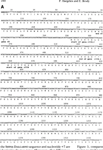
We have sequenced the 2-kb EcoRI-HindIII60-RIZA fragment by subcloning in M 1 3 and using the dideoxy chain termination method. T h e map of this fragment and the cloning strategy are outlined in Figure 1 .The regions which were also sequenced using RNA from T 4 infected cells and AMV reverse transcriptase are indicated by dotted arrows. T h e complete DNA se- quence of this fragment plus the previously published sequence of the distal part of the rZZA gene (starting at the HindIII site at nucleotide 1970; PRIBNOW et al.
1981; SUGINO and DRAKE 1984) are shown in Figure 2. Between the end of gene 60 and the beginning of the rZZA gene is an ORF which we call rZZA. 1 . This ORF codes for a hypothetical protein ( M , = 8124) whose amino acid composition is given in Table 2. The ATG of this ORF is separated by seven nucleo- tides from an AGGA SHINE-DALGARNO sequence,
239
FIGURE 1.-Restriction map of the EcoRI-Hind111 60-rIIA fragment of T 4 DNA and the sequencing strat- egy of the region. T h e distal part of gene 60, the small ORF rIIA.1, and the proximal part of gene rIIA are shown as shaded areas above the nu- cleotide scale. Intercistronic regions between genes 60 and rIIA. 1 , and between genes rIIA.1 and rIIA are shown as black boxes. T h e restriction map of the 1.97 kb shows the sites used for subcloning in M13 during the sequencing project. Recombinant M 13 phage DNA carrying inserts of
T 4 DNA were used as single- stranded templates for the SANGER
dideoxy chain-termination reactions
(SANGER, NICKLEN and COULSON
1977). Sequences obtained from this method are shown with tilled arrows. In some cases, we have used in place of the 17-mer M I 3 primer the oli- godeoxynucleotides r2A1, r2A2,
r2A3 and r2A6 complementary to T4-specific regions of the DNA ge- nome; they are shown as small bars on the map. R2A1, r2A2, r2A3 and r2A6 have been also used as primers for AMV reverse transcriptase using T 4 RNA as templates. In all cases, the digits shown above each arrow represent the number of times each T 4 region was sequenced.
which suggests that this ORF codes for a T 4 protein. HUANG et al. (1988 and personal communication) have independently sequenced part of this gene and have obtained evidence that this protein is synthesized after transcription from a T 7 promoter on a plasmid. T h e early and middle rZZA promoters which are ana- lyzed in the accompanying article are found, respec- tively, just before and in the coding portion of this ORF. T h e rZZA gene codes for a protein ( M , = 82,840) whose amino acid composition is also given in Table 2.
We have analyzed the rIIA protein derived from our DNA sequence with respect to its structure, its possible function (or functions) and the control of its biosynthesis. T h e structure of the rIIA protein has been analyzed using the algorithms of BIOU et al.
(1988) and GABORIAUD et al. (1987). T h e structure predictions for these two methods are shown in Fig- ures 3a and 4a.
Region around the rZZA initiator A T G One of the
240 P. Daegelen and E. Brody
A
10 30 50 70 90
~ G C T T T G T C A A A R C T C C T G T A A T C A T C G C T C A G G T C G G T A A A A A A C A A G A A T G G T T T T A T A C A G T C G C T G A A T A T G A G A G T G C C A
I R F V K T P V I I A Q V G K K Q E W F Y T V A E Y E S A K
AAGATGCTCTACCTAARCATAGCATCCGTTATATTAAGGGACTTGGCTCTTTGGAARAATCTGAA"iTCGTGAGATGATTCAAAACCCAG
D A L P K H S I R Y I K G L G S L E K S E Y R E M I Q N P V
110 130 150 170
190 210 230 250 270
T A T A T G A T G T T G T T A A A C T T C C T G A G A A C T G G A A A G A G C T T G C T G A C C T T C G T A A A G A A T G G A T G A
Y D V V K L P E N W K E L F E M L M G D N A D L R K E W M S
end of
290 310 330 350
GCCAGTAGTTTACTTTACCACAAGGATGTGGTATAATTAATTGGGCAAATGAGGATATTGAAATGAAATCATATAAAGTAAATTTAGAAC
Q * M K S Y K V N L E L
gene 60
370 390 410
start of gene r I I A J
430 4 s n
TTTTTGATAAAGCAGTTCATCGAGAATATAGAATCATTCAACGCTTTTTCGATATGGGAG~GCCGAAGAATTTAAAACCCGCTTTAAAG
F D K A V H R E Y R I I Q R F F D M G E A E E F K T R F K D
."
470
ATATTAGAGATAAAATTCAATCCGACACCGCAACTAARGATGAACTACTAGAAGTTGCTGAAGTTATTAAGCGTAATATGAATTAATGAG
I R D K I Q S D T A T K D E L L E V A E V I K R N M N - *
490 510 530
end of gene rIId.l 550 570 590 610 630
GAAATTATGATTATCACCACTGARAAAGAAACAATTCTTGGTAATGGTTCT~ATCAAAAGCATTTAGCATCACAGCATCTCCTAAAGTA
.tart of gene rIIA
M I I T T E K E T I L G N G S K S K A F S I T A S P K V
650
TTTARAATTCTGTCATCTGATTTGTATACAAACAAGATTCGCGCAGTAGTCCGTGAATTGATTACTAACATGATTGATGCTCATGCACTT
F K I L S S D L Y T N K I R A V V R E L I T N M I D A H A L
670 690 710
730 750 770 790 810
AATGGAAATCCTGAAAAATTTATCATACAAGTTCCTGGACGTTTAGACCCACGATTTGTTTGTCGAGATTTTGGTCCGGGTATGAGTGAT
N G N P E K F I I Q V P G R L D P R F V C R D F G P G M S D
TTTGATATTCRAGGTGATGATAATTCTCCTGGGTTGTATRATTCATACTTCAGTTCATCTAAAGCTGAATCTAATGACTTTATTGGCGGA
F D I Q G D D N S P G L Y N S Y F S S S K A E S N D F I G G
830 850 810 890
910
TTTGGTTTAGGTTCTAAATCTCCGTTTAGTTATACTGATACGTTTAGTATTACTTCGTATCATAAAGGTGAAATTCGTGGTTATGTAGCT 930 950 970 990
F G L G S K S P F S Y T D T F S I T S Y H K G E I R G Y V A
lolo 1030 1050 1070
TACATGGATGGTGATGGTCCACAGATTAAACCTACATTCGTAAAAGAAATGGGTCCAGATGATAAAACTGGTATTGAAATCGTAGTTCCA
Y M D G D G P Q I K P T F V K E M G P D D K T G I E I V V P
GTTGAAGAAAAAGACTTTAGAAACTTTGCTTATGAAGTTTCTTATATCATGCGACCGTTCAAAGATTTGGCTATCATTAATGGTCTTGAC
V E E K D F R N F A Y E V S Y I M R P F K D L A I I N G L D
1090 1110 1130 1150 1170
1190
CGCGAAATTGATTATTTTCCGGATTTTGATGACTATTACGGTGTAAATCCAGAAAGATACTGGCCTGATCGTGGTGGATTATATGCTATC
R E I D Y F P D F D D Y Y G V N P E R Y W P D R G G L Y A I
1210 1230 1250
1270 1290 1310 1330 1350
TACGGTGGTATTGTTTATCCTATCGATGGTGTTATTAGAGACCGTAACTGGCTAAGCATTCGCAATGAAGTGAATTACATTAAGTTTCCA
Y G G I V Y P I D G V I R D R N W L S I R N E V N Y I K F P
ATGGGTTCACTTGATATTGCTCCATCTCGCGAGGCTCTTTCACTGGATGATCGCACTCGTAAAAATATTATTGAACGAGTTAAAGAACTC
M G S L D I A P S R E A L S L D D R T R K N I I E R V K E L
1370 1390 1410 1430
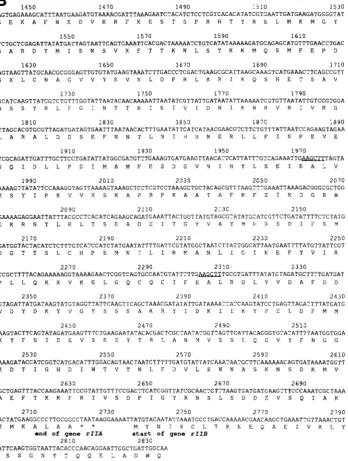
FIGURE 2.-The complete nucleo- tide sequence of gene rIIA and O R F rIlA. I , and their corresponding amino acid sequences. T h e sequence is num- bered from the left end of the EcoRI-
HindIII 60-rIIA fragment which falls into the distal part of gene 6 0 already sequenced (HUANG et al. 1988) . O u r sequence continues up to the first
Hind111 site (located a t nucleotide 1970) contained in the rIIA gene. For conven- ience we also present the previously pub- lished sequence distal to this HindIII site
(PRIBNOW et al. 1981; SUGINO and
DRAKE 1984), as well as the beginning of the rIIB gene sequence (PRIBNOW et al. 1981). Based on the D N A and R N A
sequences, the amino acid sequence of
rIIA protein is shown under the nucleo- tide sequence, as is that of the rIIA.1
protein. T h e BcoRI and HindIII sites at the ends of the 60-A fragment, and the next Hind111 site are underlined.
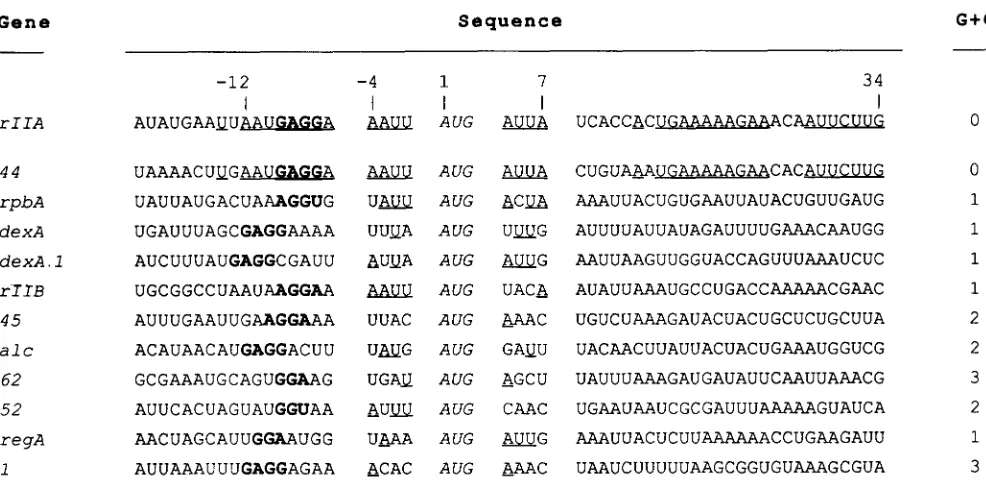
the SHINE-DALGARNO sequence and nucleotide +7 are identical in the two genes. Moreover, after differences between +8 and +12, the coding sequences of the two genes between + 13 and +34 are identical in 18 of 22 nucleotides (20 of 22 if one allows a one-base gap once in each sequence). We find this identity particu- larly remarkable because the 44 protein and the rIIA protein are reported to be the T 4 proteins most sensitive to the translational repression mediated by
the regA gene of T4 (KARAM and BOWLES 1974;
W I B E R G ~ ~ ~ KARAM 1983; WINTER^^^^. 1983). More- over, very recently, WEBSTER, ADARI and SPICER (1989) have shown that the region in gene 44 RNA between -1 1 and +9 is sufficient to specify regA recognition. RNase protection experiments show in- teraction between regA protein and this RNA be- tween positions -10 and +2. Therefore we have, in
Figure 5 , compared the rZZA RNA sequence in this region to similar regions from other proteins sensitive
to regA translational repression. They are arranged
roughly in decreasing order of sensitivity to regA inhibition (MILLER et al. 1987; WINTER et al. 1987; WEBSTER, ADARI and SPICER 1989)
. A
number of correlations of sequence to sensitivity to regA inhibi- tion appear; such correlations could help define what constitutes strong and weak regA binding sites. We note the following:1. AAUU before the AUG and AUUA after the AUG are features only of the two strongest sites (rZZA and 4 4 ) .
DNA Sequence of rZIA Gene
1 4 5 0 1 4 7 0 1 4 9 0 1 5 1 0
24 1
1 5 3 0 AGTGAGAAAGCATTTAATGAAGATGTAAAACGATTTAAAGAATCTACATCTCCTCGTCACACATATCGTGAATTGATGAAGATGGGGTAT
S E K A F N E D V K R F K E S T S P R H T Y R E L M K M G Y
TCTGCTCGAGATTATATGATTAGTAATTCAGTCAAATTCACGACTAAAAATCTGTCATATRRAAAGATGCAGAGCATGTTTGAACCTGAC
S A R D Y M I S N S V K F T T K N L S Y K K M Q S M F E P D
1 5 5 0 1 5 1 0
1 5 9 0 1 6 1 0
1 6 3 0 1 6 7 0 1 6 5 0 1 6 9 0
1 7 1 0
AGTAAGTTATGCAACGCGGGAGTTGTGTATGAAGTAAATCTTGACCCTCGACTGAAGCGCATTAAGCAAAGTCATGAAACTTCAGCCGTT
S K L C N A G V V Y E V N L D P R L K R I K Q S H E T S A V
1 7 3 0 1 7 5 0
1 1 7 0
GCATCAAGTTATCGTCTGTTTGGTATTAATACAACAAAAATTAATATCGTTATTGATAATATTAAAAATCGTGTTAATATTGTCCGTGGA
A S S Y R L F G I N T T K I N I V I D N I K N R V N I V R G
1 7 90
1810
T T A G C A C G T G C G T T A G A T G A T A G T G A A T T T A A T A A C A C T T T G A A T A T T C A T C A T A A C G A A C G T C T T C T G T T T A T T A A T C C A G A A G T A G A A 1 8 3 0
1 8 5 0 1 8 7 0
1 8 9 0
L A R A L D D S E F N N T L N I H H N E R L L F I N P E V E
1 9 1 0 1 9 3 0 1 9 7 0 1 9 5 0
T C G C A G A T T G A T T T G C T T C C T G A T A T T A T G G C G A T G T T T G A A A G T G A T G A A G T T A A C A T T C A T T A T T T G T C A G A A A T T G ~ T A G T A
S Q I D L L P D I M A M F E S D E V N I H Y L S E I E A L V
A A A A G T T A T A T T C C A A A G G T A G T T A A A A G T A A A G C T C C T C G T C C T A A A G C T G C T A C A G C G T T T A A G T T T G A A A T T A A A G A C G G G C G C T G G
K S Y I P K V V K S K A P R P K A A T A F K F E I K D G R W
1 9 9 0 2 0 1 0
2 0 3 0 2 0 5 0
2 0 1 0
2 0 9 0
G A A A A G A G G A A T T A T T T A C G C C T C A C A T C A G A A G C A G A T G A A A T T A C T G G T T A T G T A G C G T A T A T G C A T C G T T C T G A T A T T T T C T C T A T G
E K R N Y L R L T S E A D E I T G Y V A Y M H R S D I F S M FIGURE 2.--Continued.
2 1 1 0 2 1 3 0
2 1 5 0
2 1 7 0 2 1 9 0
2 2 1 0 2 2 3 0
2 2 5 0
G A T G G T A C T A C A T C T C T T T G T C A T C C A T C T A T G A A T A T T T T G A T T C G T A T G G C T A A T C T T A T T G G C A T T A A T G A A T T T T A T G T T A T T C G T
D G T T S L C H P S M N I L I R M A N L I G I N E F Y V I R
2 2 7 0 2 2 9 0
2 3 1 0
C C G C T T T T A C A G A A A A A G G T A A A A G A A C T C G G T C A G T G C C A A T G T A T T T T T G ~ T G C G T G A T T T A T A T G T A G A T G C T T T T G A T G A T
P L L Q K K V K E L G Q C Q C I F E A L R D L Y V D A F D D
2 3 3 0
2 3 5 0
G T A G A T T A T G A T A A G T A T G T A G G T T A T T C A A G T T C A G C T A A A C G A T ~ T A T T G A T A A A A T T A T C A A G T A T C C T G A G T T A G A T T T T A T G A T G 2 3 7 0
2 3 9 0 2 4 1 0
2 4 3 0
V D Y D K Y V G Y S S S A K R Y I D K I I K Y P E L D F M M
2 4 5 0 2 4 7 0
2 4 9 0 2 5 1 0
A A G T A C T T C A G T A T A G A T G A A G T T T C T G A A G A A T A T A C A C G A C T C G C T A A T A T G G T T A G T T C A T T A C A G G G T G T A T A T T T T A A T G G T G G A
K Y F S I D E V S E E Y T R L A N M V S S L Q G V Y F N G G
AAAGATACCATCGGTCATGACATTTGGACAGTAACTAATCTTTTTGATGTATTATCAAATAATGCTTCAAAAAACAGTGATAAAATGGTT 2 5 3 0
2 5 5 0 2 5 7 0
2 5 9 0 2 6 1 0
K D T I G H D I W T V T N L F D V L S N N A S K N S D K M V
2 6 3 0 2 6 5 0
2 6 7 0 2 6 9 0
GCTGAGTTTACCAAGAAATTCCGTATTGTTTCCGACTTCATCGGTTATCGCAACTCTTTAAGTGATGATGAAGTTTCCCAAATCGCTAAA
A E F T K K F R I V S D F I G Y R N S L S D D E V S Q I A K
2 7 1 0 2 1 3 0
2 7 5 0 2 7 7 0
2 7 9 0
ACTATGAAGGCCCTTGCGGCCTAATAAGGAAAATTATGTACAATA~'rAAATGCCTGACCAAAAACGAACAAGCTGAAATTGTTAAACTGT
T M K A L MA YA N* C* K C L T K N E Q A E I V K L Y
end of gene r I I A
2810
start of gene r I I B
2 8 3 0 ATTCAAGTGGTAATTACACCCAACAGGAATTGGCTGATTGGCAA
S S G N Y T Q Q E L A D W Q
3. T h e box between -4 and
+7
has no Gs or Cs for the rIIA and 44 sequences (disregarding the G of the initiator AUG). For the others the G+
C content varies (in this box) from 1 to 3. Again there is a rough inverse correlation between the number of (G + C)s and the regA effect.Structural motifs in the rIIA protein: Mutants in
the rIIA and rIIB genes have similar phenotypes. T h e proteins seem to have similar characteristics. Could the two be structurally related? Analysis of the rIZA and rIIB nucleic acid and protein sequences shows no region of extensive similarity, certainly no more than is found when other T4 genes are compared to rIIA. We do, however, find a hint that the present rIIA gene may have evolved by gene duplication. When tyrosine 10 1 is aligned with tyrosine 48 1 (tyrosine 48 1
is 1 18 nucleotides downstream of the midpoint amino
acid 363 of the rIIA gene), a similarity between the two halves of the molecule becomes evident (Figure 6). Although the overall similarity is only 28% (cal- culated with BESTFIT using the matrix of RISLER),
the clustering is impressive, because there are no gaps in the sequence alignment. T h e predicted structures for these two zones (Figure 3, a and b) do not show extensive similarity.
242 P. Daegelen and E. Brody
TABLE 2
Amino acid composition of rIIA and rIIA.1 proteins
rIIA r ll A . I
Amino acid Mole Mole
residue Number percent Number percent
Ala 38 5.241
C)’S 5 0.690
ASP 57 7.862
Glu 45 6.207
Phe 39 5.379
GlY 41 5.655
His 10 1.379
Ile 63 8.690
Leu 48 6.621
Met 23 3.172
Asn 42 5.793
Pro 29 4.000
Gln 1 1 1.517
LYS 54 7.448
Arg 41 5.655
Ser 61 8.4 14
T h r 31 4.276
Val 44 6.069
Tyr 39 5.379
Isoelectric point 6.58
T r p 4 0.552
4 5.970
0 0.000
6 8.955 8 1 1.940
5 7.463
1 1.493
1 1.493
5 7.463
8 1 1.940
4 5.970
3 4.478
3 4.478
0 0.000
2 2.985
6 8.955
2 2.985
3 4.478 4 5.970 0 0.000
2 2.985
7.91
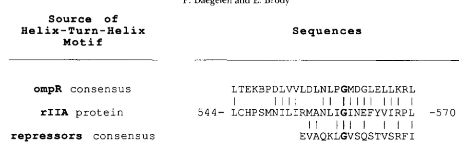
for sigma factors are compared to this rIIA motif. T h e most striking similarity, however, is found when this rIIA motif is compared to a series of proteins, the prototype of which is ompR, which act as regulators of gene expression in response to environmental change (IKENAKA et al. 1988). This extended helix- turn-helix motif is thought to be the DNA-binding part of these proteins which activate gene expression in response to the external environment. Leucine 544 would correspond to the highly conserved leucine
(7
out of 9) in these proteins (see Figure 4 of IKENAKA et al. 1988)
.
Also noteworthy is the conservation of isoleucine 553 which corresponds to a highly con- served hydrophobic amino acid (valine, leucine or isoleucine) and which, when mutated in the ompR protein, leads to the loss of the cell’s ability to regulate porin synthesis in response to changes in the osmolar- ity of the culture medium. It has been known for many years that the phenotype of rZZA mutants is suppressed by the salt composition and concentration in the culture medium (GAREN 196 1; SEKIGUCHI1966; see SINGER, SHINEDLINC and GOLD 1983 for a discussion of the salt effect). The similarity found here raises the possibility that this sequence mediates DNA binding of the rIIA protein in response to changes in the ionic composition of the culture medium.
Since the rIIA protein is strongly associated with the bacterial inner membrane, does it contain the hydrophobic a-helical regions associated with integral membrane proteins? Using GES analysis for identify- ing hydrophobic a-helices, it was found that the most
hydrophobic 17-amino-acid sequence in rIIA had a hydrophobicity of 0.85 kcal/amino acid (ENGELMAN, STEITZ and GOLDMAN 1986). This makes it unlikely that rIIA is an integral membrane protein.
We have carried out extensive computer compari- sons between the rIIA protein sequence and the fol- lowing sequence banks: NBRF, PASTEUR, and our T4 protein sequences bank (168 protein sequences derived from the genes and ORFs contained in the 95-kbp of sequence in our possession). Although short regions of similarity are found with a number of proteins in these banks, no similarity extended over a large proportion of the rIIA protein. One region of short similarity deserves mention. A strong similarity is seen between amino acids 155- 18 1 of the methyl- accepting chemotaxis protein I of E. coli, tsr (BOYD, KENDALL and SIMON 1983), and the motif centered on the duplicated EI-(R or T)-GYVAYM sequence in rIIA (Figure 6)
.
This reinforces the idea that the rIIA protein plays a role in reacting to the external environment of T 4 infected cells. This same motif is also found in the region of amino acids 222 to 236 of the T4 protein 63 (RNA ligase).The rZZA.1 ORF: This ORF is a hydrophilic, some-
what basic peptide containing no proline, cysteine, or tryptophan (Table 2). Secondary structure analysis, by either the method of BIOU et al. (1988) or of GABORIAUD et al. (1987), predicts this peptide to be almost entirely a-helical (Figures 3b and 4b). Exten- sive comparative searches reveal short similarities to a number of E . coli proteins. The greatest similarity (60% with 4 gaps) is to a region between amino acids 279-349 of citrate synthase of E . coli (see Figure 8 ) .
Less impressive, but perhaps more intriguing, is a similarity to the VirG protein of Agrobacterium tume- faciens. T h e VirG locus codes for a protein necessary
for virulence, and its sequence shows it to be homol- ogous to the OmpR group of protein activators which respond to the cells’ environment. In fact, the region of similarity of the VirG protein with rIIA.l lies just downstream (amino acids 102-169; data not shown) of the presumed helix-turn-helix region of this protein (amino acids 67-93) with apparent homology to the helix-turn-helix region of rIIA.
Mutants in the Ala region of the rZZA gene: T h e
unexpected finding of a small ORF between genes 60
DNA Sequence of rIIA Gene
r I I A protein.
50 100
MIITTEKETILGNGSKSKAFSITASPKVFKILSSDLYTNKI~VVRELITNMIDAHALNGNPEKFIIQVPGRLD~RFVCRDFGPGMSDFDIQGDDNSPGL CECEHCCCEEHCCCCCCCEEEEECCCHEEEEHHHHCCCHHHHHHHHHHHHHHHHHHHCCCCCCHEEEHCCCCCCCCEEECCCCCCCCCECCCCCCCCCCE
1 2 1 2 1 2 2 1 3 3 1 3 4 5 5 5 4 2 1 1 1 1 1 3 5 3 1 1 2 3 4 3 1 2 1 1 2 1 1 1 2 3 4 4 5 4 5 4 4 3 2 3 3 4 3 1 1 3 4 5 5 4 1 1 1 2 3 1 2 4 5 5 4 5 4 2 2 3 2 3 4 4 5 5 5 3 4 3 2 2 2 3 5 5 5 5 5 5 5 2
1 5 0 2 0 0
Y N S Y F S S S K A E S N D F I G G F G L G S K S P F S Y T D T F S I T S Y H K G E I R G Y V A Y ~ G D G P Q I K P T F V K E M G P D D K T G I E I W P V E E K D F ~ F A Y E V S Y I M R P F ~ ECCCEECCCCCCCCEEEEEEECCCCCEEEEEEEEEEEECCCHHEEEEEECCCCCCCCCCCCHHCCCCCCCCCEEEEECCCHCCHHHHHHHHHHHHCCCCC
3 2 2 1 3 1 3 4 2 3 3 4 3 3 3 2 1 1 2 1 1 5 4 4 4 5 2 1 3 3 2 3 4 4 3 3 2 3 4 2 4 1 1 1 1 3 3 3 2 2 4 5 5 5 5 5 5 3 3 1 1 1 3 1 3 5 5 5 5 4 4 4 1 3 3 3 2 2 2 1 1 1 1 1 2 1 2 1 3 2 2 3 3 3 2 2 1 1 2 2
2 5 0 300
LAIINGLDREIDYFPDFDDYYGVNPERYWPDRGGLYAIYGGIVYPIDGVIRDRNWLSI~EVNYIKFPMGSLDIAPSREALSLDDRTRKNIIERVKELSE
HHHHCCCCCCCCECCCCCCCCCCCCCCCCCCCCCCEEEECCEEECCCEEEECCCEECECCCCCCECCCCCCCCCCCCCHCHCCHHHHHHHHHHHHHHHHH 2 3 3 3 2 3 2 2 2 2 2 3 2 3 3 4 3 3 3 2 2 3 3 5 4 3 2 2 2 5 5 3 4 3 1 3 3 2 2 3 2 2 3 1 3 2 2 1 2 2 2 3 3 3 1 2 1 1 1 2 2 1 3 2 1 2 2 3 3 5 4 3 3 3 5 4 3 1 4 1 3 1 2 1 3 2 1 1 2 1 3 4 4 4 4 4 3 3 1 3
3 5 0 4 0 0
KAFNEDVKRFKESTSPRHTYRELMKMGYSARDYMISNSVKFTTKNLSYKKMQSMFEPDSKLCNAGWYEVNLDPRLKRIKQSHETSAVASSYRLFGINTT
HHHHHHHHHHHHCCCCCCCCHHHHHHCCCCCCEEECCCCCCCCCCCCHCCHHHHHCCCCCCCCCCEEEEECCCHHHHHHCCCCCCCCCCCEEEEEEECCC 4 2 3 2 3 1 3 3 3 2 1 1 4 4 5 4 4 4 3 1 1 1 1 1 3 1 4 3 3 4 4 2 1 1 3 1 4 3 2 2 1 2 2 2 2 2 2 1 2 3 1 1 1 1 1 3 3 4 3 3 1 2 3 5 3 1 1 2 3 1 3 3 4 1 1 3 3 1 1 1 2 3 2 2 2 1 1 1 1 2 2 3 2 3 2 1 2 3 3 1
4 5 0 5 0 0
KINIVIDNIKNRVNIVRGLARALDDSEFNNTLNIHHNERLLFINPEVESQIDLLPDIMAMFESDEVNIHYLSEIEALVKSYIPKWKSKAPRPKAATAFK CHHHHHHHHHCCHHHHHHHHHCCCHHHHHHHHHHHHHHHHEEECCHHHCCHHHCCHHHHHHCCCHHHHHHHHHHHHHHHHHCCHHHCCCCCCHHHHHHHH
1 1 1 1 1 2 3 2 2 2 3 3 2 3 2 3 2 2 2 2 1 3 3 3 1 1 3 3 2 1 1 2 1 1 3 1 3 1 1 2 2 1 1 5 1 2 2 2 1 1 2 3 3 2 1 2 4 4 4 3 3 1 2 3 1 1 3 3 2 3 4 5 5 5 5 5 5 5 5 4 4 3 2 2 1 1 1 2 4 4 4 4 1 3 3 4 4 4 3 4
5 5 0 6 0 0
FEIKDGRWEKRNYLRLTSEADEITGYVAYMHRSDIFSMDGTTSLCHPSMNILIRMANLIGINEFYVIRPLLQKKVKELGQCQCIFEALRDLYVDAFDDVD HHHHCCHHHHCCECEEECCCHHHHHHHEEECCECEECCCCCCCCCCCCHHHHHHHHHHCCCCHHHHHHHHHHHHHHHHCCHHHHHHHHHHHHHHHHCCCC
3 4 3 1 2 3 1 3 3 1 2 2 1 1 2 1 1 3 2 2 2 1 2 1 2 3 3 1 2 1 3 3 2 3 2 2 3 2 4 4 4 2 3 3 4 4 5 5 1 3 3 3 2 2 2 1 3 1 3 4 2 3 1 1 1 1 2 2 3 4 3 3 1 1 1 1 2 3 4 2 1 2 4 4 5 5 5 4 3 4 3 3 3 2 2 1 2 2 3 4
650 7 0 0
YDKYVGYSSSAKRYIDKIIKYPELDFMMKYFSIDEVSEEYTRLANMVSSLQGVYFNGGKDTIGHDIWTVTNLFDVLSNNASKNSD~AEFTKKFRIVSD CCCCEEECCCHHHHHHHHCCCCHHHHHHHHCCHHHHHHHHHHHHHHHHHHCCEEECCCCCCECEECEEEHHHHHHHCCCCCCCCHHHHHHHHHHHHEEHH 1 3 1 2 2 2 1 3 3 3 1 1 2 2 2 2 1 1 1 2 3 3 1 3 3 3 3 3 3 1 3 4 1 1 1 2 2 4 4 4 4 3 4 4 4 4 4 3 2 1 2 3 3 3 4 3 5 5 3 3 2 3 5 4 3 1 2 2 3 3 3 3 3 3 2 1 3 3 3 3 4 3 3 3 2 4 3 4 4 5 3 2 3 2 3 3 3 3 1 1
7 2 5
FIGYRNSLSDDEVSQIAKTMKALAA
HHCCCCCCCCHHHHHHHHHHHHHCC 2 2 2 2 2 3 4 4 5 3 1 2 4 4 4 5 5 5 4 4 4 4 5 3 2
r I I A . l protein.
50 67
MKSYKVNLELFDKAVHREYRIIQRFFDMGEAEEFKTRFKDIRDKIQSDTATKDELLEVAEVIKRNMN HHHCHHCHHHHHHHHHHHHCEHHHHHCHCHHHHHHHHHHHHHHHHHCCCHHHHHHHHHHHHHHHCHC
2 1 1 1 2 2 1 4 4 4 4 3 4 3 4 2 2 2 3 1 1 2 1 1 2 2 1 1 4 1 4 4 4 4 4 4 3 3 3 1 3 1 2 3 2 1 3 2 2 2 1 2 3 4 4 3 3 4 5 5 4 4 3 3 2 1 3
243
FIGURE 3.--Secondary structure prediction of rIIA and rIIA.1 proteins, by the COMBINE methods. (a) rIIA protein. The COMBINE method (BIOU et al. 1988) used to predict the secondary structure of the rIIA protein is a combination of three complementary secondary structure prediction methods: homolog prediction (LEVIN et al. 1986), GORIII prediction (GIBRAT et al. 1987) and the bit pattern prediction method for helix and @-strand structures (BIOU et al. 1988) . We show here (from a longer listing output), for each group of four lines: the amino acid number scale; the amino acid sequence; the result of the COMBINE prediction method for each amino acid in one of three states: a-helix (H; helical), @-strand (E; extended) and aperiodic structure (C; coil); the confidence scale index expressing the reliability of the prediction (this numerical value increases from 1 to 5 with an increasing probability to find a particular amino acid residue in a particular state). (b) r I I A . l protein. Same as in (a) for the rIIA.1 protein.
because it is an ATG --j ATA mutation in the initia-
tion codon of the rZZA gene. An analogous mutation in the rIZB gene, HD263, has been shown to be tem- perature-sensitive for rIIB protein synthesis. T h e ATG + ATA mutation in the initiation codon of rZZA does not lead to a temperature-sensitive defect. T h e plating efficiency of T 4 AP80 is about on a
X-
lysogen (compared to a nonlysogen) at 2 0 ° , at 3 7 ”
and at 4 2 ” (data not shown).
DISCUSSION
We have completed the analysis of the DNA se- quence between genes 60 and rZZB, and have found a new ORF just upstream of rZZA. T h e complete se- quence of this region allows us to make some remarks on the large number of rZZA mutants collected during the last 40 yr. First of all, one of the most striking aspects of BENZER’S genetic map of spontaneous rZZ
mutations is the existence of hot spots (BENZER 196 1). Mutation 131 in rZZA and 117 in rZZB are extraordi- narily overrepresented in his collection; mutant 114 in rZZB is less hot, but is still remarkable for its fre- quency. GOLD and his collaborators (PRIBNOW et al.
244 P. Daegelen and E. Brody
(a) rllA protein
1 150 200
250 300 350 400
FIGURE 4.-Hydrophobic cluster analysis of r11A and rlIA. 1 proteins. (a) rIIA protein. The r l l A protein has been analyzed with the HCA progratn from the I)SAid+ pckage ( D A R I ~ X . and BENSOUSAN 1988) based on the hydrophobic duster analysis algorithm of G A R O R I A U D el
al. ( 1 Wi) . The I i(;A method t \ x originally designed for comparing ;and ;aligning amino acid sequences from distantly related proteins ( i . e . ,
proteins \chicll ciannot be aligned using classical methods of sequence comparison but which can be with the HCA method because they fold into sitnilar three-dinlension;ll structures). The figure shown above is based on a representation of the rIIA amino acid sequence in the nlanllcr of a11 n-helic;tl three-dinlension;tl pattern expanded at the surface of a cylinder (3.6 amino acids per turn, parallel to the generator oftlie cylinder). The cylinder is cut along this axis. unrolled onto a two-dimensional surface and then duplicated. Sets of adjacent hydrophobic residues (F, I. LA, M , l’, M’, Y and A or <: when they are i n a hydrophobic environment) on the surface have have been arranged in hydrophobic clusters. Threc. special svnahols are used: * for prolines considered as breakers of these clusters, 0 for glycines and C! for cysteines. To avoid disruption i n the dratving. the end of each line 1, 2 and 3 overlaps, over I8 amino acids, the beginning, respectively, of the lines 2, S ~ n d 4. (h) rI1A.l protein. S;tme ;as in (a) for the rIIA.1 protein.
shift mutations, either to 7 (unstable) or to 5 (stable) A:T base pairs. T h e physical-chemical differences be- tween (dA:dT)5 and (dA:dT)6 do not seem to explain this gigantic difference in mutation frequencies and
PRIRNOW et al. (1 98 1) have already shown that there
must be a context to this effect. Whatever mechanism is at work here, it generates an impressive differential response in replication fidelity to the addition of one dA:dT base pair.
T h e Ala segment of the rII genetic map defines the leftmost region of the rIIA gene (BENZER 1961). The mutations in the Ala segment recombine with every
rll deletion in the BENZER collection except the dele- tion ~ 1 2 7 2 . Three of these Ala mutations map in the NH?-terminal portion of the rIIA gene (AP80 in the initiator codon of the protein). We guess that if mu- tants had arisen in the rIIA.1 ORF, they would not have been detected as rIIA mutants and, in fact, would probably not be viable. Our reasoning is as follows: many rII deletions have a right end in the nonessential
D region just downstream of gene rIIB (BENZER 196 1). Only one deletion of the original collection goes as far as the NH? terminus of rIIA. This suggests that there is a barrier either to deletion formation or to deletion viability upstream of the rIIA gene. Until now, it has always been thought that this barrier was the essential gene 60. If, as thought, deletion forma- tion is strongly dependent on direct repeats of DNA sequences (PRIRNOW et al. 198 1 ; SINGER, SHINEDLING
and GOLD 1983), there are a large number of potential
sites in gene rIIA.1. T h e longest are one 1 l-bp se- quence and five 9-bp sequences in rIIA. I which are directly repeated in either rIIA or rIIB (data not shown). I t seems likely, then, that the deletion asym- metry arises simply because rIIA. I codes for an essen- tial protein in T 4 development.
DNA Sequence of rZZA Gene 245
Gene S e q u e n c e G + C
-12 - 4 1 7 34
I I I I I
r I I A AUAUGAAYU- EBysl AUG ALJLlA UCACCAC-C- 0
4 4 U A A A A C U U G A A U W
m
AUG CUGUA-AAUGAAAAAGAACACAUUCUU~ 0r p b A UAUUAUGACUAAAGGUG U r n AUG A C U AAAUUACUGUGAAUUAUACUGUUGAUG 1
d e x A UGAUUUAGCGAGGAAAA UUQA AUG U U G AUUUUAUUAUAGAUUUUGAAACAAUGG 1
dexA. 1 AUCUUUAUGAGGCGAUU AUQA AUG W G AAUUAAGUUGGUACCAGUUUAAAUCUC 1
rT IB UGCGGCCUAAUAAGGAA BByy AUG UACA A U A U U A A A U G C C U G A C C A C G A A C 1
45 AUUUGAAUUGAAGGAAA UUAC AUG AAAC UGUCUAAAGAUACUACUGCUCUGCUUA 2
a 1 c ACAUAACAUGAGGACUU U U G AUG GAUU UACAACUUAUUACUACUGAAAUGGUCG 2
62 GCGAAAUGCAGUGGAAG UGAU AUG EGCU UAUUUAAAGAUGAUAUUCAAUUAAACG 3
52 AUUCACUAGUAUGGUAA A U m AUG CAAC U G A A U A A U C G C G A U U U A A G U A U C A 2
r e q A AACUAGCAUUGGAAUGG U-AAA AUG U G AAAUUACUCUUAAAAAACCUGAAGAUU 1
1 AUUAAAUUUGAGGAGAA ACAC AUG -AAAC UAAUCUUUUUAAGCGGUGUAAAGCGUA 3
FIGURE 5.-The nucleotide sequence surrounding the AUG initiator, and the sensitivity of some T 4 genes to the translational repression mediated by the T 4 regA regulator. T h e sequences at the beginning of 11 T 4 genes for which the sensitivity to regA protein is known, are aligned under the analogous rIIA sequence. Sequences are aligned around the first AUG in the coding region. T h e nucleotides are numbered with the A from the initiator as the +1 reference. Around the initiator, the two tetranucleotides have been separated for legibility. T h e nucleotides that are identical at a given position (ie., without deletion or insertion) in genes rIIA and 44 are underlined. For all other genes, the nucleotides located in the area between position -4 and position +7, which are identical to those in the rIIA-44 sequence, have been underlined. T h e number of (G
+
C)’s between positions -4 and +7 are shown on the right. The SHINE-DALGARNO sequences for each gene are shown in bold.g p r I I A 4 8 1 - Y I P K V V K S K A P R P K A A T A F K F E I K D G R W E K R N Y L R L T S E A D E I T G W A ~ R S D I F S M D G T T S L C H P S M N I L I R M A N L I -559
g p r I I A 101- YNSYFSSSICAESNDFIGGFGLGSKSPFSYTDTFSITSYHKGEIRGWAYMDGDGPQIKPTFVKEMGPDDKTGIEIVVPV -179 I I l l I I I
I I I I I I I I I I I I I I 1 I
g p r I I A 139-
-63 2 2 2 -
g p r I I A 134-
gpt8t 155-
HKGE
.
I R G W A Y M . D GN A I . E N I E G W A V M K D G
I I I I I I I I I
S I T S Y H K G . E I R G W A Y M D G D G P Q I K P T
R P R D I R N G F E . K Q W A Y M E Q N D R L H D I A I I I I I I I I I I
I
- 1 5 2
- 2 3 6
-160
-181
FIGURE 6 , ” T h e rIIA gene has possibly evolved by gene duplication. T h e two 79-amino-acid sequence fragments of rIIA protein starting at amino acid 101 and 481 are aligned with no insertions or deletions; perfect matches are represented in bold (top). In the same manner, similar regions from other proteins ( E . coli tsr protein and T 4 gene 63 product) are aligned with the repeated motif of rIIA protein. Alignments have been obtained with the BESTFIT program using the matrix of RISLER (see MATERIALS AND METHODS). Some gaps have been included in order to maintain the one-to-one correspondence in the alignment.
approximates more closely that of gene 63 than that of rIIA, rIIB or E . coli proteins. Most significant is the AGA arginine codon, which is used frequently in genes 63 and rIIA. 1 but not at all in rIIB, nor in highly expressed E . coli proteins.What is the significance of the identity of the translation initiator regions of genes rIZA and 44? As we have mentioned, these proteins are the two whose synthesis is most sensitive to trans- lational repression by the T4 regA protein. We think that the identical sequences between -1 2 and +7
define a strong regA repression site. One or all of the parameters discussed in RESULTS and shown in Figure
5 must contribute to the weakening of the regA effect on other sites. T h e significance of the quasi-identity of these two genes between + 1 and +34 could involve other regulatory mechanisms; alternatively, this iden-
tity could define a protein domain shared by these two proteins.
CAMBELL and GOLD ( 1 982) have suggested a model
246 P. Daegelen and E. Brody
Source
of
Helix-Turn-Helix
M o t i f
S e q u e n c e s
ompR
consensus
LTEKBPDLWLDLNLPGMDGLELLKRLI I I I I I I
I I I I I
I l l II I
I l l
I
I
I Irepressors
consensus
EVAQKLGVSQSTVSRFI
rIIA
protein
544-LCHPSMNILIRMANLIGINEFYVIRPL
- 5 7 0FIGURE 7.-Amino acid alignment among rIIA protein, the ompR consensus, and the helix-turn-helix motif of certain prokaryotic repressors. T h e region located between amino acids 544 and 570 of the rIIA protein is aligned with two different consensus sequences: the ompR consensus derived from the putative regions of the nine proteins (see Figure 4 of IKENAKA et al. 1988) involved in sensory systems that sllare c o n m o n features with osnroregulation, and the helix-turn-helix consensus derived from the 2 1 protein sequences (see Figure 12 of PABO and SAUER 1984) which are proved or assumed to be DNA-binding proteins. Alignments were done using PROFlLE and PROFILEGAP programs (see MATERIALS AND METHODS)
.
Perfect matches are shown in bold.r I I A . 1 protein 1"KSYKVNLELFDKAVHREYRIIQRFFDMGE
. .
AEEFKTR . . . F K D I R D K I Q S D T A T K D E L L E . E V I K R N M N -661 I I I l l I I I I I I I I I I I I I I I l l I I I I I I I I I I I I I I I I
citrate synthase 3 0 0 - I S S V K H I P E F F R R A K D K N D S F . . R L M G F G H R V Y K N Y D P R A T V M R E T C H E V L K E L G T K D D L L ~ ~ E L E N I A L N -348
FIGURE 8.- Similarity between the r1IA.I protein and the citrate synthase from E . coli. All experimental details are as in Figure 6 .
M u t a t i o n
P o s i t i o n
S e q u e n c e s
w i l d type
ATG ATT ATC ACC ACT GAA
AAA GAA ACA ATT
CTT GGT
AP80 -1.29 F120
3 32 35
..A
. . .
. . .
.c.
. . .
. . .
. A .M
I
I
T
T
E
K
E
T
I
L
G
I
.
.
P
D
FIGURE 9.- Sequence analysis of three mutants in the Ala region of r l l A . The three mutants ( A P 8 0 , AP12Y and F 1 2 0 ) isolated originally by S . BENZER and located in the Ala region of rIIA, were sequenced by primer-extension of the r2A3 oligodeoxynucleotide, using as template
T4 RNA isolated 5 min after infection at 30". T h e nucleotide sequence of the beginning of the wild-type rIIA gene is shown at the top. For each mutant, only the nucleotide replacing the corresponding wild-type nucleotide is shown in bold. T h e amino acids deduced from these sequences are shown at the bottom.
TABLE 3
T4 rare codon usage
~~~~
Amino
acid Codon rllA rlIA.1 r f l B 63" E . coli Lb H'
E. coli
Gly GGA 0.17 1.00 0.08 0.37 0.1 1 0.005
Arg AGA 0.07 0.33 0.00 0.44 0.12 0.00 Ile A T T 0.76 0.80 0.95 0.60 0.50 0.17 T h r ACA 0.42 0.00 0.24 0.15 0.11 0.04 Leu T T A 0.29 0.25 0.00 0.32 0.15 0.02 Ser TCA 0.28 0.05 0.24 0.36 0.16 0.02 Gln CAA 0.45 1.00 0.82 0.50 0.36 0.14 Pro CCA 0.34 0.00 0.33 0.44 0.23 0.15
T4 gene 63 (RAND and GAIT 1984).
'
Codon usage for poorly expressed E . coli genes are from UWGCG (see MATERIALS AND METHODS, and GRANTHAM et al.198 1).
' Same as except for highly expressed genes.
seen when rZZA mutants infect rex+ bacteria (GAREN 1 96 1 ; SEKIGUCHI 1966).
Although the amino acid sequence of the rIIA
DNA Sequence of rIIA Gene 247
determines whether or not rIIA detaches from the inner membrane to affix itself to some sequence on T4 DNA. Alternatively, rIIA may simultaneously bind to DNA and the inner membrane and the re- sponse to changes in the medium may lead to dissocia- tion of one of these contacts. If there really were an analogy to the ompR group of proteins, one wonders what protein would play the role of the envZ part of this system. EnvZ is suggested to be an integral mem- brane protein which is the site of attachment (and detachment) of ompR. It is envZ that senses directly the osmolarity of the external medium and transmits this information to ompR on the inner surface of the membrane. Could rIIB be the envZ analog which fixes rIIA to the membrane? Hydrophobic cluster analysis of envZ predicts a membrane spanning region
whereas the analysis for rIIB is less clear, although there is a potential 20 amino acid hydrophobic a-helix in the middle of this protein (data not shown).
We are grateful to LARRY GOLD and JOHN F. PULITZER for helpful discussions and criticisms during the course of this work and during the preparation of the manuscript. We thank WAI MUN HUANG for communicating her unpublished results, V A L ~ R I E BIOU andJEAN GARNIER for their help in the determination of rIIA and rIIA. 1 protein structures, andJEAN-Luc POPOT for the GES analysis of rIIA and rIIA.1 proteins. The research reported here was supported by research grants from the CNRS (Centre National de la Recherche Scientifique) and INSERM (Institut National de la Sante et de la Recherche Medicale).
LITERATURE CITED
BENZER, S., 1959 On the topology of the genetic fine structure.
BENZER, S., 1961 On the topography of the genetic fine structure.
BIOU, V., J. F. GIBRAT, J. LEVIN, B. ROBSON and J. GARNIER, 1988 Secondary structure prediction: combination of three different methods. Protein Eng. 2: 185-1 91.
BOLIVAR, F., R. L. RODRIGUEZ, M. C. BETLACH and H. W. BOYER, 1977 Construction and characterization of new cloning ve- hicles. l. Ampicillin-resistant derivatives of the plasmid pMB9. Gene 2: 75-93.
BOYD, A., K. KENDALL and M. I. SIMON, 1983 Structure of the serine chemoreceptor in Escherichia coli. Nature 201: 623-626. BRODY, E. N., D. RABUSSAY and D. H. HALL, 1983 Regulation of
transcription of prereplicative genes, pp. 174-183 in Bacteriop- nage T 4 , edited by C. K. MATHEWS, E. M. KUTTER, G. MOSIG and P. B. BERCET. AMERICAN SOCIETY FOR MICROBIOLOGY, WASHINGTON. D.C.
Proc. Natl. Acad. Sci. USA 45: 1607-1620.
Proc. Natl. Acad. Sci. USA 47: 403-415.
CAMPBELL, K., and L. GOLD, 1982 Construction of the bacterio- phage T 4 replicatin machine: regulation of synthesis of com- ponent proteins, pp. 69-81 in Interaction of Translational and Transcriptional Controls in the Regulation of Gene Expression,
edited by M . GRUNBERG-MANAGO and B. SAFER. Elsevier, New York.
CASADABAN, M. J., and S. N. COHEN, 1980 Analysis of gene control signals by DNA fusion and cloning in Escherichia coli.
J. Mol. Biol. 138: 179-207.
CRICK, F. H. C., L. BARNETT, S. BRENNER and R. J. WATTS-TOBIN, 1961 General nature of the genetic code for proteins. Nature
DAEGELEN, P., and E. N. BRODY, 1976 Early bacteriophage T 4
192: 1227-1232.
transcription. A diffusible product controls rIIA and rIIB RNA synthesis. J. Mol. Biol. 103: 127-142.
DAEGELEN, P., and E. N. BRODY, 1 9 9 0 T h e rIlA gene of bacteri- ophage T4. 11. Regulation of its messenger RNA synthesis. Genetics 125: 249-260.
DARDEL, F., and P. BENSOUSSAN, 1988 A Macintosh full screen editor featuring a built in regular expression interpreter for the search of specific patterns in biological sequences using finite state automata. Comput. Appl. Biosci. 4 483-486. DAYHOFF, M. O., W. C. BARKER and L. T. HUNT,
1983 Establishing homologies in protein sequences. Methods Enzymol. 91: 524-545.
DEVEREUX, J., P. HAEBERLI and 0. SMITHIES, 1984 A comprehen- sive set of sequence analysis programs for the VAX. Nucl. Acids Res. 12: 387-395.
DRETZEN, G., M. BELLARD, P. SASSONE-CORSI and P. CHAMBON, 198 1 A reliable method for the recovery of DNA fragments from agarose and acrylamide gels. Anal. Biochem. 112: 295- 298.
ENGELMAN, D. M., T. A. STEITZ and A. GOLDMAN, 1986 Identifying nonpolar transbilayer helices in amino acid sequences of membrane proteins. Annu. Rev. Biophys. Bio- phys. Chem. 15: 321-353.
ENNIS, H. L., and K. D. KIEVITT, 1973 Association of the rIIA protein with bacterial membrane. Proc. Natl. Acad. Sci. USA
GABORIAUD, C., V. BISSERY, T. BENCHETRIT and J. P. MORNON, 1987 Hydrophobic cluster analysis: an efficient new way to compare and analyze amino acid sequences. FEBS Lett. 224:
149-155.
GAREN, A., 1961 Physiological effects of r l l mutations in bacteri- ophage T4. Virology 1 4 151-163.
GRANTHAM, R., C. GAUTIER, M. GOUY, M. JACOBZONE and R. MERCIER, 1981 Codon catalog usage is a genome strategy modulated for gene expressivity. Nucl. Acids Res. 9: r43-r74. HUANG, W. H., 1986 The 52-protein subunit of T 4 DNA polym- erase is homologous to the gyrA-protein of gyrase. Nucl. Acids Res. 14: 7379-7390.
HUANC, W. M., S. Z . Ao, S. CASJENS, R. ORLANDI, R. ZEIKUS, R. WEISS, D. WINCE and M. FANG, 1988 A persistent untrans- lated sequence within bacteriophage T 4 DNA topoisomerase gene 60. Science 2 3 9 1005-1012.
HUANC, W. M., and J. M. BUCHANAN, 1974 Synergistic interac- tions of T 4 early proteins concerned with their binding to DNA. Proc. Natl. Acad. Sci. USA 71: 2226-2230.
IKENAKA, K., K. TSUNC, D. E. COMEAU and M. INOUYE, 1988 A dominant mutation in Escherichia coli OmpR lies within a domain which is highly conserved in a large family of bacterial regula- tory proteins. Mol. Gen. Genet. 211: 538-540.
KARAM, J. D., and M. G. BOWLES, 1974 Mutation to overpro- duction of bacteriophage T 4 genes products. J. Virol. 13: 428- 438.
LIPMAN, D. J., and W. R. PEARSON, 1985 Rapid and sensitive protein similarity searches. Science 227: 1435-1441.
MANOIL, C., N. SINHA and B. ALBERTS, 1977 Intracellular DNA- protein complexes from bacteriophage T4-infected cells iso- lated by a rapid two-step procedure. J. Biol. Chem. 252: 2734- 2741.
MATZ, K., M. SCHMANDT and G. N. GUSSIN, 1 9 8 2 T h e rex gene of bacteriophage X is really two genes. Genetics 102: 319-327. MCPHEETERS, D. S., A. CHRISTENSEN, E. T . YOUNG, G. STORMO
and L. GOLD, 1986 Translational regulation of expression of the bacteriophage T 4 lysozyme gene. Nucleic Acids Res. 14:
581 3-5826.
MESSING, J., and J. VIEIRA, 1982 A new pair of M13 vectors for selecting either DNA strand of double-digest restrictions frag- ments. Gene 1 9 269-276.
MILLER, E. S., J. KARAM, M. DAWSON, M. TROJANOWSKA, P. GAUSS