ISSN 2348 – 7968
Image Segmentation Using Level Set Method For Images With
Intensity Inhomoginities
TellabandlaRamuduP
1
P
, K.SudhakarP
2 P K.PrasadBabuP 3 P S.AhmedBashaP 4 P 1 P
M.Tech DSCE 14G31D0612 Department of ECE, JNT University, Anantapur/SJCET Yerrakota, Andhra Pradesh 518360, India,[email protected]
P
2
P
Associate Professor,H.O.D, Department of ECE, JNT University, Anantapur/SJCET Yerrakota, Andhra Pradesh 518360, India, [email protected]
P
3
P
Assistant Professor, Department of ECE, JNT University, Anantapur/SJCET Yerrakota, Andhra Pradesh 518360, India, [email protected]
P
4
P
Assistant Professor, Department of ECE, JNT University, Anantapur/SJCET Yerrakota, Andhra Pradesh 518360, India, [email protected]
Abstract
The active contour method is one of the most successful image
segmentation techniques. It has received a tremendous amount of
attention in medical image processing. The segmentation
operation can be carried out manually or automatically. A
manual segmentation requires a skilled operator trained to use a
digital tool to mark the contours of the desired structures. In this
project the three-phase formulation of the level set evolution
(LSE) and bias field estimation and a Level Set Method for
Image Segmentation in the Presence of Intensity is done. The
three-phase formulation is used to segment an image into three
regions. Intensity inhomogeneity often occurs in real-world
images, which presents a considerable challenge in image
segmentation. The most widely used image segmentation
algorithms are region-based and typically rely on the
homogeneity of the image intensities in the regions of interest,
which often fail to provide accurate segmentation results due to
the intensity inhomogeneity. Matlab code is used for the three
phase formation and bias field estimation.
.
Keywords:25T Level Set Methods, MATLAB, IMAGE
SEGMENTATION.
1. Introduction
The goal of image segmentation is to cluster the pixels into salient image regions i.e., regions corresponding to individual surfaces, objects, or natural parts of objects.
Segmentation is an important technique used in image processing to identify the objects in the image .Segmentation techniques that can be applied in a robust and efficient way to both image and mesh data. Mesh data is frequently unstructured; this precludes the direct application of techniques that were originally developed for the more structured image data. The idea behind active contours, or deformable models, for image segmentation is quite simple. The user specifies an initial guess for the contour, which is then moved by image driven forces to the boundaries of the desired objects. In such models, two types of forces are considered - the internal forces, defined within the curve, are designed to keep the model smooth during the deformation process, while the external forces, which are computed from the underlying image data, are defined to move the model toward an object boundary or other desired features within the image.
.
2. Present Work and Proposed Work
. Intensity inhomogeneity often occurs in real-world
images, which presents a considerable challenge in image
segmentation. The most widely used image segmentation
algorithms are region-based and typically rely on the
homogeneity of the image intensities in the regions of
interest, which often fail to provide accurate segmentation
results due to the intensity inhomogeneity. First, based on
the model of images with intensity inhomogeneities, we
derive a local intensity clustering property of the image
intensities, and define a local clustering criterion function
for the image intensities in a neighborhood of each point.
This local clustering criterion function is then integrated
with respect to the neighborhood center to give a global
criterion of image segmentation. In a level set formulation,
this criterion defines an energy in terms of the level set
functions that represent a partition of the image domain
and a bias field that accounts for the intensity
inhomogeneity of the image. Therefore, by minimizing
this energy, our method is able to simultaneously segment
the image and estimate the bias field, and the estimated
bias field can be used for intensity inhomogeneity
correction (or bias correction).
Imagine that you are given an image, say a medical (MRI
or CT) scan. Suppose you want to extract the important
feature within the image; in this case, the outline of the
artery. One idea is to look for places where there is a big
jump in intensity between neighboring pixels. However, it
is hard to pick a good value for the jump; too small and
you get extra boundaries; too large and you miss the whole
show. Another problem is that you can get fooled by large
spikes of noise. An Evolving Interface Approach to Active
Contours .A different approach comes from initializing a
small circle inside the region of interest, and allowing it to
grow outwards until it reaches the desired boundary.
The level set approach allows the evolving front to change
topology, break, and merge, which means that the evolving
front can extract the boundaries of particularly intricate
contours. In addition, the method works in three
dimensions with almost no change, so three dimensional
surfaces can be extracted as well.
Present Method:
A variational level set framework for segmentation and
bias correction of images with intensity
inhomogeneities. Based on a generally accepted model of
images with intensity inhomogeneities and a derived local
intensity clustering property, we define an energy of the
level set functions that represent a partition of the image
domain and a bias field that accounts for the intensity
inhomogeneity. Segmentation and bias field estimation are
therefore jointly performed by minimizing the proposed
energy functional. The slowly varying property of the bias
field derived from the proposed energy is naturally
ensured by the data term in our variational framework,
without the need to impose an explicit smoothing term on
the bias field.
In order to deal with intensity inhomogeneities in image
segmentation, we formulate a method based on an image
model that describes the composition of real-world images,
in which intensity inhomogeneity is attributed to a
component of an image. In this present method, we
consider the following multiplicative model of intensity
inhomogeneity. From the physics of imaging in a variety
of modalities (e.g. camera and MRI), an observed image
can be modeled as
I=b J + n
where ‘J’ is the true image, ‘b’ is the component that
accounts for the intensity inhomogeneity, and ‘n’ is
additive noise. The component is referred to as a bias field
(or shading image). The true image ‘J’ measures an
intrinsic physical property of the objects being imaged,
which is therefore assumed to be piecewise (approximately)
varying. The additive noise ‘n’ can be assumed to be
zero-mean Gaussian noise.
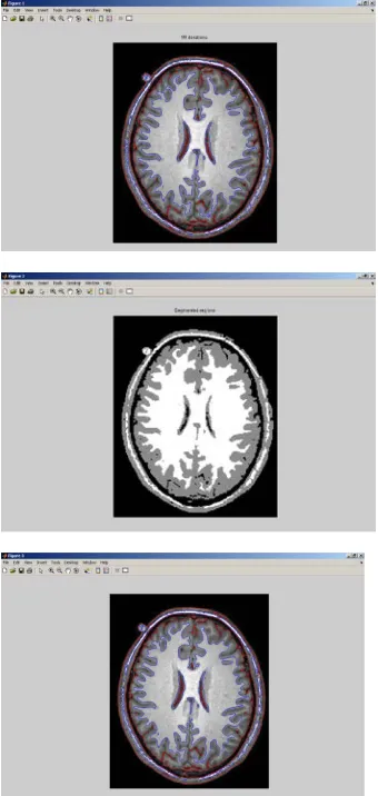
Fig 1 .Basic steps employed for new medical image segmentation technique based on level set method
Proposed Work:
Fig 2 Steps employed for Image segmentation of Intensity Homogeneity based on level set method
3. Results:
As a level set method, our method provides a contour as
the segmentation result. Therefore, we use the following contour-based metric for precise evaluation of the segmentation result.
Let C be a contour as a segmentation result, and S be the true object boundary, which is also given as a contour. For each point Pi; i=1,2,3,…..N. on the contour , we can compute the distance from the point Pi to the ground truth contour , denoted by dist(Pi,S). Then, we define the deviation from the contour C to the ground truth S by
4. Conclusions
It can be seen that the intensities within each tissue
become quite homogeneous in the bias corrected images.
The improvement of the image quality in terms of intensity
homogeneity can be also demonstrated by comparing the
histograms of the original images and the bias corrected
images. The histograms of the original images (left) and
the bias corrected images (right) are plotted. Based on a
generally accepted model of images with intensity
property, we define an energy of the level set functions
that represent a partition of the image domain and a bias
field that accounts for the intensity inhomogeneity.
Segmentation and bias field estimation are therefore
jointly performed by minimizing the proposed energy
functional. The slowly varying property of the bias field
derived from the proposed energy is naturally ensured by
the data term in our variational framework, without the
need to impose an explicit smoothing term on the bias
field.
REFERENCES
[1] G. Aubert and P. Kornprobst, Mathematical Problems in Image Processing: Partial Differential Equations and the Calculus of Variations. New York: Springer-Verlag, 2002.
[2] V. Caselles, F. Catte, T. Coll, and F. Dibos, “A geometric model for active contours in image processing,” Numer. Math., vol. 66, no. 1, pp. 1–31, Dec. 1993.
[3] V. Caselles, R. Kimmel, and G. Sapiro, “Geodesic active contours,” Int. J. Comput. Vis., vol. 22, no. 1, pp. 61–79, Feb. 1997.
[4] T. Chan and L. Vese, “Active contours without edges,” IEEE Trans. Image. Process., vol. 10, no. 2, pp. 266–277, Feb. 2001.
[5] D. Cremers, “A multiphase levelset framework for variational motion segmentation,” in Proc. Scale Space Meth. Comput. Vis., Isle of Skye, U.K., Jun. 2003, pp. 599–614.
[6] L. Evans, Partial Differential Equations. Providence, RI: Amer. Math. Soc., 1998.
[7] S. Kichenassamy, A. Kumar, P. Olver, A. Tannenbaum, and A. Yezzi,“Gradient flows and geometric active contour models,” in Proc. 5th Int. Conf. Comput. Vis., 1995, pp. 810–815.
[8] R. Kimmel, A. Amir, and A. Bruckstein, “Finding shortest paths on surfaces using level set propagation,” IEEE Trans. Pattern Anal. Mach. Intell., vol. 17, no. 6, pp. 635–640, Jun. 1995.
[9] C. Li, R. Huang, Z. Ding, C. Gatenby, D. Metaxas, and J. Gore, “A variational level set approach to segmentation and bias correction of medical images with intensity inhomogeneity,” in Proc. Med. Image Comput. Comput. Aided Intervention, 2008, vol. LNCS 5242, pp. 1083–1091, Part II.
[10] C. Li, C. Kao, J. C. Gore, and Z. Ding, “Minimization of region-scalable fitting energy for image segmentation,” IEEE Trans. Image Process., vol. 17, no. 10, pp. 1940–1949, Oct. 2008.
[11] C. Li, C. Xu, C. Gui, and M. D. Fox, “Distance regularized level set evolution and its application to image segmentation,”
IEEE Trans. Image Process., vol. 19, no. 12, pp. 3243–3254, Dec. 2010.
[12] R. Malladi, J. A. Sethian, and B. C.Vemuri, “Shape modeling with front propagation: A level set approach,” IEEE Trans. Pattern Anal. Mach. Intell., vol. 17, no. 2, pp. 158–175, Feb. 1995.
[13] D. Mumford and J. Shah, “Optimal approximations by piecewise smooth functions and associated variational problems,” Commun. Pure Appl. Math., vol. 42, no. 5, pp. 577–685, 1989
[14] Rafael C. Gonzalez and Richard E. Woods, Digital Image Processing, 3rd edition, Published by Pearson Prentice Hall, 2009. [15] S.K. Weeratunga and C. Kamath, An Investigation of Implicit Active Contours for Scientific Image Segmentation, Visual Communications and Image Processing Conference, IS&T/SPIE Symposium Electronic Imaging, San Jose, CA, January 18-22, 2004
[16] M. Khelif, F. Derraz and M. Beldagham, Application of Active Contour Models in Medical Image Segmentation. [17] M. Droskey et al, An adaptive level set method for medical image segmentation.
[18] J. A. Sethian, Level Set Methods: An Act of Violence, Evolving Interfaces in Geometry, Fluid Mechanics, Computer Vision and Material Science.
[19] Tony F. Chan and Luminita A. Vese, Active Contours Without Edges, IEEE Transaction On Image Processing, Vol. 10, No. 2, FEBRUARY 2001.
[20] V. Caselles, F. Catte, T. Coll, and F. Dibos, A geometric model for active contours in image processing, Numer. Math., Vol. 66, pp. 1-31, 1993.
[21] M. Airouche et al, Image Segmentation Using Active Contour Model and Level Set Method Applied to Detect Oil Spills, Proceedings of the World Congress on Engineering, Vol. I, July 1-3, 2009, London, U.K.