1071-412X/05/$08.00
⫹
0
doi:10.1128/CDLI.12.1.68–75.2005
Copyright © 2005, American Society for Microbiology. All Rights Reserved.
Development of Capture Assays for Different Modifications of Human
Low-Density Lipoprotein
Gabriel Virella,
1* M. Brooks Derrick,
1,2Virginia Pate,
1,2Charlyne Chassereau,
1,2Suzanne R. Thorpe,
3and Maria F. Lopes-Virella
2Department of Microbiology and Immunology, Medical University of South Carolina,
1and Department of Medicine, Division of
Endocrinology-Metabolism-Nutrition, Medical University of South Carolina, and Ralph H. Johnson VA Medical Center,
2Charleston, and Department of Chemistry and Biochemistry, University of South Carolina, Columbia,
3South Carolina
Received 15 June 2004/Returned for modification 18 August 2004/Accepted 8 September 2004
Antibodies to malondialdehyde (MDA)-modified low-density lipoprotein (LDL), copper-oxidized LDL (oxLDL),
N
(carboxymethyl) lysine (CML)-modified LDL, and advanced glycosylation end product (AGE)-modified LDL
were obtained by immunization of rabbits with in vitro-modified human LDL preparations. After absorption
of apolipoprotein B (ApoB) antibodies, we obtained antibodies specific for each modified lipoprotein with
unique patterns of reactivity. MDA-LDL antibodies reacted strongly with MDA-LDL and also with oxLDL.
CML-LDL antibodies reacted strongly with CML-LDL and also AGE-LDL. oxLDL antibodies reacted with
oxLDL but not with MDA-LDL, and AGE-LDL antibodies reacted with AGE-LDL but not with CML-LDL.
Capture assays were set with each antiserum, and we tested their ability to capture ApoB-containing
lipopro-teins isolated from precipitated immune complexes (IC) and from the supernatants remaining after IC
precipitation (free lipoproteins). All antibodies captured lipoproteins contained in IC more effectively than free
lipoproteins. Analysis of lipoproteins in IC by gas chromatography-mass spectrometry showed that they
contained MDA-LDL and CML-LDL in significantly higher concentrations than free lipoproteins. A significant
correlation (
r
ⴝ
0.706,
P
< 0.019) was obtained between the MDA concentrations determined by chemical
analysis and by the capture assay of lipoproteins present in IC. In conclusion, we have developed capture
assays for different LDL modifications in human ApoB/E lipoprotein-rich fractions isolated from precipitated
IC. This approach obviates the interference of IC in previously reported modified LDL assays and allows
determination of the degree of modification of LDL with greater accuracy.
Human lipoproteins are known to undergo spontaneous
modification in humans. Several chemical processes are
in-volved in this modification. Oxidation affects both the lipid and
protein components of low-density lipoprotein (LDL).
Reac-tive aldehyde products formed during the oxidation of
polyun-saturated fatty acids, such as malondialdehyde (MDA) and
4-hydroxynonenal (HNE), are capable of attaching covalently
to the
ε
-amino groups of lysine residues of apolipoprotein B
(ApoB) (21, 22, 33). Advanced glycosylation also affects LDL
(19). This process involves a chain of chemical reactions that
starts with the covalent, nonenzymatic addition of reducing
sugars to protein amino groups (Schiff base and Amadori
ad-ducts). Additional reactions take place leading to the
forma-tion of a heterogeneous family of sugar-amino acid adducts
collectively known as “advanced glycosylation end products”
(AGE) (31), including AGE-modified LDL (AGE-LDL).
Both oxidized LDL (oxLDL) and AGE-LDL have been
shown to have proatherogenic and proinflammatory properties
(11). This has led to a burst of interest in the development of
techniques for their assay in human sera. The immunogenicity
of modified lipoproteins first reported by Steinbrecher et al.
has been well documented in studies involving immunization
of laboratory animals with in vitro-modified lipoproteins (23).
The immunogenicity of these modifications in experimental
animals allowed the production of monoclonal antibodies
spe-cific for MDA and HNE-lysine which reacted with oxLDL
prepared in vitro, as well as with LDL isolated from
athero-sclerotic plaques (16, 33). AGE-modified proteins including
LDL (AGE-LDL) have also been shown to be immunogenic
(6). Antibodies raised in laboratory animals have been used for
the detection of AGE-modified proteins in serum (15) and
tissues (14, 15). Human autoantibodies to modified LDL have
also been extensively characterized (28) and shown to
recog-nize primarily MDA-modified LDL and
N
ε(carboxymethyl)
ly-sine (CML)-modified LDL (29). Some groups have proposed
the assay of such autoantibodies as a surrogate measurement
for modified LDL, but this approach is riddled with
inaccura-cies because of the interference caused by the formation of
antigen-antibody complexes involving modified forms of LDL
and the corresponding antibodies (28). We have shown in
previous studies that LDL-containing immune complexes
(LDL-IC) can be precipitated with 4% polyethylene glycol
(PEG), and that while modified forms of LDL are easily
mea-surable in PEG precipitates, very little modified LDL remains
soluble after IC precipitation. The enzymoimmunoassay
tech-niques developed by different groups for the detection of
mod-ified forms of LDL and AGE-modmod-ified proteins (7, 9, 15, 24,
25) attempt to avoid interference of IC and other confounding
factors by a variety of means. While most groups will trust the
ability of immobilized antibodies to preferentially capture and
retain modified LDL at high sample dilutions, this may not be
necessarily the case when monoclonal antibodies are used for
capture of modified LDL. Other groups have proposed
differ-* Corresponding author. Mailing address: Medical University of
South Carolina, Department of Microbiology and Immunology, 173
Ashley Ave., PO Box 250504, Charleston, SC 29425. Phone: (843)
792-4339. E-mail: [email protected].
68
on August 17, 2020 by guest
http://cvi.asm.org/
ent approaches to this issue, such as using competition
be-tween bound modified LDL and the unknown LDL in test
samples to which antibody is added (5), using purified LDL
instead of whole serum or plasma (8) or adding sodium
dode-cyl sulfate (32) or 4% PEG (26) to the samples. However, none
of the groups provided evidence for the effective dissociation
of IC as a consequence of the assay conditions.
We have demonstrated that rabbits immunized with
MDA-LDL, copper-oxidized LDL (oxLDL), AGE-MDA-LDL, and
CML-LDL produce antibodies that recognize each one of these
modifications individually. For example, rabbit oxLDL
anti-bodies show only a marginal degree of reactivity with
MDA-LDL, rabbit MDA-LDL antibodies react only weakly with
oxLDL, and rabbit AGE-LDL antibodies do not react with
CML-LDL (29). Using these antibodies, we have developed
capture assays whose specificity and ability to capture in
vivo-modified human LDL, isolated from circulating IC, have been
demonstrated.
MATERIALS AND METHODS
Isolation of human LDL.Blood for lipoprotein isolation was collected from four healthy volunteers in EDTA at 0.4 mmol/liter after 12 h of fasting. The donors used for this purpose were normolipemic healthy volunteers not receiving prescription medications for any acute or chronic condition and without family history of coronary artery disease, peripheral vascular disease, or stroke. None of the volunteers was receiving antioxidant therapy. All volunteers gave permission with informed consent for blood collection. LDL was isolated from pooled
plasma, after density adjustment (1.019⬍density⬍1.063 g/ml) with potassium
bromide, by preparative ultracentrifugation at 50,000 rpm for 17 h on a Beckman L-80 ultracentrifuge with a type 70 Ti rotor (19). After isolation, LDL was washed by ultracentrifugation, dialyzed against a 0.15-mol/liter sodium chloride
solution containing 300mol of EDTA (pH 8.0) per liter, passed through an
Acrodisc filter (0.22-m pore size) in order to sterilize and remove aggregates,
and stored under nitrogen in the dark at 4°C.
Preparation of modified proteins. oxLDL was prepared according to our modification (10) of the protocol described by Steinbrecher (22). LDL was diluted in phosphate-buffered saline (PBS) to a protein concentration of 1.5
mg/ml and incubated with 40mol of copper chloride (CuCl2) per liter. The
degree of oxidation was monitored continuously by fluorescence emission with a luminescence spectrophotometer (SLM-AMINCO Series 2; Spectronic Instru-ments, Rochester, N.Y.). LDL oxidation was stopped 4 to 6 h after the
fluores-cence values reached the peak (ⱖ1.1 fluorescence units). On average, this
cor-responds to the following degree of modification: 4 to 7 mmol of MDA per mol of lysine (0.4 to 0.7% modification of lysine residues), 0.8 mmol of CML per mol of lysine (0.08% modification of lysine residues), and 0.25 mmol of
Nε(carboxyethyl) lysine (CEL) per ml of lysine (0.025% modification of lysine
residues).
AGE-LDL was prepared by a modification of the method described by Schmidt et al. (20). Freshly isolated LDL (1.5 mg/ml) was sterilized by passage
through a 0.2-m-pore filter, added to 150 mM glucose-6-phosphate in 200 mM
phosphate buffer, pH 8.0, containing 40M butylated hydroxytoluene and 400
M EDTA, filter sterilized a second time, and incubated for 8 weeks at 37°C. At
the end of the incubation, LDL was dialyzed for 24 h against three changes of 4 liters of 0.15 M NaCl–0.3 mM EDTA, pH 8.0. Our AGE-LDL modifications contained 4.6 mmol of CML per mol of lysine (0.5% modification of lysine residues) and 0.45 mmol of CEL per mol of lysine, corresponding to 0.05% modification of lysine residues). The MDA content of AGE-LDL was usually below the detection limit, but in some preparations MDA was detectable in small amounts, similar to those measured for CEL.
MDA modification of proteins was performed according to Haberland et al. (4) by incubating equal volumes of freshly isolated LDL and 0.2 M MDA for 3 h at 37°C, followed by extensive dialysis against 0.15 M NaCl with 0.3 mM EDTA, pH 8.0. The degree of modification of the MDA-LDL preparations used to immunize rabbits was 67 mmol of MDA per mol of lysine, corresponding to the modification of 6.7% of lysine residues.
CML-modified proteins were prepared by incubation of the protein with
glyoxylic acid and NaBH3CN in phosphate buffer at 37°C, as described previously
for the preparation of CML in bovine serum albumin (17). The degree of
modification of the CML-LDL used in our studies was113 mmol of CML per mol of lysine (11.3% modification of lysine residues).
The characteristics of the different modified lipoproteins used in this study are summarized in Table 1.
Isolation of ApoB-containing lipoproteins from circulating IC.Human sera obtained as part of the ongoing Diabetes Control and Complications Trial/ Epidemiology of Diabetes Interventions and Complications (DCCT/EDIC) study aimed at assessing the development of macrovascular disease in type 1
diabetes (1, 30) were used in this part of the study. Sera were kept at⫺70°C until
our studies were performed. Isolation of soluble IC by precipitation with 4% PEG was performed as previously described (30). ApoB-rich lipoproteins (LDL and other ApoB-containing lipoproteins) were isolated from the supernatant and from the resuspended PEG-precipitated IC. The supernatants were split: one-half was fractionated on an immobilized oxLDL column, and the other one-half was fractionated on an immobilized AGE-LDL column. The washout fractions from both oxLDL and AGE-LDL columns were combined and then chromatographed on heparin-agarose columns (Sigma-Aldrich Corp., St. Louis, Mo.) that retain ApoB/E-containing lipoproteins. The PEG-precipitated IC were first submitted to affinity chromatography on protein G-Sepharose, and the washout fractions, containing all precipitated proteins other than immunoglobulin G (IgG), were later fractionated on heparin-agarose columns. The lipoprotein-containing sam-ples obtained from the heparin-agarose columns were dialyzed against saline containing 0.3 mM EDTA, pH 8.0, and stored at 4°C until tested in the capture assays.
Analysis of LDL modifications.Analysis of modified lipoproteins for their content of CML, CEL, and the advanced lipoxidation end products MDA-lysine and HNE-lysine was carried out by selected ion monitoring-gas chromatography-mass spectrometry (SIM-GC/MS) as described by Requena et al. (18). The degree of modification of MDA-LDL was also estimated by the thiobarbituric acid reactive substances assay (13), using MDA as a standard.
Rabbit antibodies.Antibodies to modified LDL were obtained by immuniza-tion of New Zealand White female rabbits with different modificaimmuniza-tions of human LDL (AGE-LDL, CML-LDL, oxLDL, and MDA-LDL) as previously described (29). The resulting rabbit antiserum was first fractionated by affinity chromatog-raphy with immobilized protein G, and the IgG fractions were absorbed in a column of immobilized native LDL. Depending on the protocol, the washout from the column of immobilized native LDL, containing antibodies to modified LDL and irrelevant IgG, was used as purified to capture modified LDL or as a peroxidase conjugate, prepared in our laboratory with the reagents and protocol obtained from Roche Diagnostics, Manheim, Germany, to detect captured LDL. The eluate, containing purified IgG antibodies to native LDL, predominantly reactive with ApoB, was used either to capture LDL or as a peroxidase conjugate to detect captured LDL.
Capture assays.Capture assays were carried out in two basic protocols. To capture MDA-LDL, oxLDL, and AGE-LDL, purified rabbit antibodies of the different specificities were absorbed to the plates at a protein concentration of
approximately 35g/ml, chosen as optimal based on the signal/noise ratio, from
three different concentrations (87.5, 35, and 17.5g/ml). After washing off the
unabsorbed antibodies, the plates were blocked with 5% bovine serum albumin. Serial dilutions of modified LDL preparations were added to the plates, and
rabbit peroxidase-conjugated anti-human ApoB, at a concentration of 0.67g/ml
(chosen as optimal between two concentrations, 0.22 and 0.67g/ml), was used
to detect bound LDL. To capture CML-LDL, we immobilized purified rabbit
anti-human ApoB at a protein concentration of 22g/ml to capture LDL from
the patient’s samples and used rabbit peroxidase-conjugated anti-CML-LDL at a
concentration of 0.5g/ml to detect bound CML-LDL. The capture assays for
TABLE 1. Characteristics of the different types of modified LDL
used in this study
Form of LDL
LDL modificationa
mmol of MDA/ mol of lysine
mmol of CEL/ mol of lysine
mmol of CML/ mol of lysine
oxLDL
4–7
0.25
0.8
MDA-LDL
67
ND
bND
AGE-LDL
ND
0.45
4.6
CML-LDL
ND
ND
113
aData obtained by SIM-GC/MS.
bND, not detectable (below the detection limit of SIM-GC/MS [0.005 mmol/
mol of lysine]).
V
OL. 12, 2005
CAPTURE ASSAYS FOR DIFFERENT MODIFICATIONS OF LDL
69
on August 17, 2020 by guest
http://cvi.asm.org/
MDA-LDL and CML-LDL were calibrated with reference preparations of oxLDL and CML-LDL with known concentrations of MDA and CML, deter-mined by SIM-GC/MS. The capture assay for oxLDL was calibrated in micro-grams per milliliter, taking advantage of the fact that we have determined a reproducible method for the preparation of oxLDL with a consistent degree of
modification (10). The assay of AGE-LDL could not be calibrated by any of the methods described above because AGE-LDL antibodies do not react with CML-LDL and because the degree of modification of different AGE-CML-LDL prepara-tions is variable. Therefore, we calibrated the assay in arbitrary units, defined as the reciprocal of the dilution of the reference AGE-LDL preparation that results
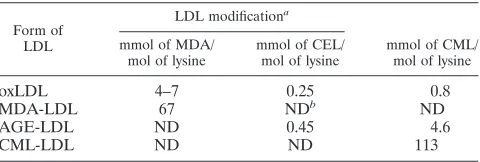
FIG. 1. Diagrammatic representation of the results of specificity testing for the capture assays for MDA-LDL (A) and oxLDL (B). n-LDL,
native LDL.
on August 17, 2020 by guest
http://cvi.asm.org/
in an optical density (OD) lower than 0.5 with a standard time for the reaction of the conjugate antibody with its substrate. In all assays, the concentration of
LDL added to the antibody-containing wells was kept constant (5g/ml) and the
final values were expressed as millimoles of MDA or CML per mole of lysine, micrograms of oxLDL per microgram of protein, or units of AGE-LDL per microgram of protein.
Technical validation and recovery studies.Intra-assay reproducibility was de-termined from six replicates of one in vitro-modified LDL preparation and from six replicates of an unknown LDL isolated from a PEG precipitate, both adjusted
at a protein concentration of 5g/ml. Interassay reproducibility was determined
by assaying the same samples in sextuplicate in six different plates.
Recovery experiments were performed by mixing equal volumes of native and
modified LDL at 5g/ml and determining the total amounts measured by the
capture assay by the calibration methods described above.
PEG precipitation of IC and isolation of free and IC-bound ApoB-rich li-poproteins.Fractionation of human sera with 4% PEG was performed as pre-viously described (30). ApoB/E-containing lipoproteins (native and modified) were isolated from the supernatant and from the resuspended PEG precipitates. The supernatants were fractionated directly on heparin-agarose columns (Sigma-Aldrich Corp., St. Louis, Mo.) that retain ApoB/E-containing lipoproteins (2). The PEG precipitates were first submitted to affinity chromatography on protein G-Sepharose and the washout, containing all precipitated proteins other than IgG, was later fractionated on heparin-agarose columns. The lipoprotein-con-taining samples were pooled and dialyzed against saline conlipoprotein-con-taining 0.3 mM EDTA, pH 8.0, and tested by SIM-GC/MS and by the capture assays.
RESULTS
The capture assays for MDA-LDL and oxLDL (Fig. 1)
showed specificity for the corresponding modification.
How-ever, while MDA-LDL antibodies captured oxLDL with low
efficiency (Fig. 1A), oxLDL antibodies did not capture
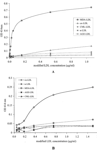
MDA-LDL (Fig. 1B). The capture assay for CML-MDA-LDL (Fig. 2)
showed great specificity and efficiency for CML-LDL (Fig.
2A). At high protein concentrations, AGE-LDL was captured
with greater efficiency than oxLDL and native LDL (Fig. 2B).
Trying different conditions, we determined that the best
dis-crimination between the capture of AGE-LDL versus the
cap-ture of oxLDL and native LDL with CML-LDL antibody was
obtained when ApoB antibody was used for capture and
per-oxidase-labeled CML-LDL antibody was used for detection.
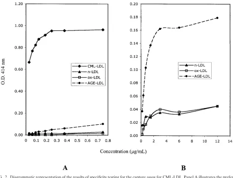
The capture assay with LDL antibodies captured
AGE-LDL with high efficiency and oxAGE-LDL with low efficiency (Fig.
3). The capture of CML-LDL was only marginally higher than
the capture of native LDL.
The sensitivity, reproducibility, and recovery data for the
four capture assays are summarized in Table 2. The intra-assay
coefficients of variation (CV) were under 10%, and the
inter-assay CV were under 11%. The recovery data were equally
excellent, ranging from 94% (AGE-LDL assay) to 103%
(MDA-LDL assay).
Previous observations have demonstrated that ApoB/E-rich
lipoproteins isolated from PEG-precipitated IC are enriched in
MDA-LDL and CML-LDL, using isotope dilution
SIM-FIG. 2. Diagrammatic representation of the results of specificity testing for the capture assay for CML-LDL. Panel A illustrates the preferential
capture of CM-LDL at low protein concentrations. Panel B illustrates the preferential capture of AGE-LDL relatively to oxLDL and native LDL
(nLDL) at high protein concentrations.
V
OL. 12, 2005
CAPTURE ASSAYS FOR DIFFERENT MODIFICATIONS OF LDL
71
on August 17, 2020 by guest
http://cvi.asm.org/
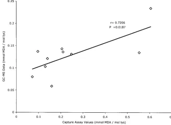
GC/MS for their measurement (30). We compared the capture
assay’s ability to distinguish the LDL isolated from
PEG-pre-cipitated IC from that remaining free in the supernatants and
compared the concentrations of MDA-LDL determined by
GC/MS and by capture assay. The ApoB-rich lipoproteins
iso-lated from precipitated IC were enriched in all the different
LDL modifications tested by our capture assays relative to
ApoB-rich lipoproteins isolated from the supernatants after
centrifugation of precipitated IC (Table 3). The comparison of
MDA-LDL levels assayed by SIM-GC/MS and the MDA-LDL
capture assay in 10 different ApoB-rich lipoprotein
prepara-tions isolated from precipitated IC showed a good correlation
between both assays (
r
⫽
0.706,
P
⫽
0.0187), although, in
general, the absolute values calculated from the capture assay
were greater than those obtained by GC/MS (Fig. 4).
DISCUSSION
There is great interest in the assay of modified lipoproteins
in serum or plasma samples because of their potential
patho-genic role in atherosclerosis (11). Given the immunopatho-genicity of
modified lipoproteins, several groups have developed
immu-FIG. 3. Diagrammatic representation of the results of specificity testing for the capture assay for AGE-LDL. nLDL, native LDL.
TABLE 2. Sensitivity, reproducibility, and recovery data for the capture assays of MDA-LDL, oxLDL, CML-LDL, and AGE-LDL
Sensitivity
MDA-LDL (0.042 mmol/mol of
lysine)
oxLDL (0.03
g/g of protein)
CML-LDL (0.086 mmol/mol of
lysine)
AGE-LDL
(0.037 U/g of
protein)
Intra-assay
CV
(%)
aIn vitro-modified LDL
5.96
6.76
5.03
5.30
LDL from IC
9.69
8.56
5.29
5.47
Interassay
CV
(%)
bIn vitro-modified LDL
7.94
9.48
6.53
5.61
LDL from IC
10.17
10.55
8.86
6.18
% Recovery
c103
⫾
2
100
⫾
3
98
⫾
3
94
⫾
1
aThe intra-assay coefficient of variation was determined using six replicates of both an in vitro-modified LDL and an LDL sample purified from LDL-IC on one plate.
Data are expressed as the mean CV obtained in six separate assays of both samples.
bThe interassay CV was determined over six separate assays, using six replicates of both an in vitro-modified LDL and an LDL sample purified from LDL-IC
complex. The CV for each sample was calculated from a total of 36 determinations in six different runs.
cRecovery experiments were performed by adding a known amount of in vitro-modified LDL specific for the given assay into native LDL and measuring the
concentration of modified LDL in the supplemented sample. Values are expressed as mean⫾1 standard deviation.
on August 17, 2020 by guest
http://cvi.asm.org/
noassays, particularly for MDA-LDL and AGE-LDL (7, 9, 15,
24, 25). Previous studies conducted in our laboratory showed
that rabbits immunized with MDA-LDL, oxLDL, AGE-LDL,
and CML-LDL produced antibodies that recognized epitopes
unique to those different in vitro modifications of LDL (29).
We have now demonstrated that the same antibodies are able
to capture modified LDL isolated from human sera and that
they can be used to develop capture assays for different
mod-ifications of LDL with excellent accuracy and reproducibility.
The recovery rates were close to 100%, except in the case of
the MDA-LDL assay, in which it exceeded 100%. At 103%, the
recovery value for MDA-LDL is within the range of variation
of the assay but could also reflect the recognition of
sponta-neously modified LDL in the native LDL preparation (27).
The specificity of our rabbit MDA-LDL antibodies could be
verified by comparison with the results of GC/MS assays of
MDA in ApoB-rich lipoproteins obtained from PEG
precipi-tates. A significant correlation existed between the two assays.
Similar validations were not possible for the other assays. In
the case of CML-LDL, the chemical assay of CML appeared
less sensitive than the capture assay and we did not obtain
sufficient data to compare the two assays. In the cases of the
oxLDL and AGE-LDL capture assays, the unknown nature of
the epitopes recognized by the rabbit antibodies makes any
such comparative analysis impossible.
The four antibodies used in the assay recognize different
epitopes of modified LDL. Although human antibodies to
oxLDL react primarily with MDA epitopes, rabbit antibodies
to oxLDL recognize a different epitope, also present in
spon-taneously modified human LDL (29). Similarly, human
anti-bodies to AGE-LDL react primarily with CML-LDL (30), but
rabbit antibodies to AGE-LDL recognize epitope(s) unrelated
to CML, which have been previously described by Ikeda et al.
(6). Our data suggest that these epitopes are expressed, at
lower levels, by oxLDL. As such, our rabbit AGE-LDL
anti-body does not differentiate well between oxLDL and
AGE-LDL.
All our antibodies captured significantly higher levels of
modified LDL in the ApoB/E-rich lipoproteins isolated from
IC. The highest level of discrimination between LDL isolated
from IC (apparently more modified) and LDL that remains
soluble after IC precipitation was obtained with the antibodies
to oxLDL and CML-LDL. The results obtained with
MDA-LDL and oxMDA-LDL antibodies suggest that the majority of
ox-LDL molecules in circulation are part of circulating IC.
Although we have previously proven that IC are precipitated
with PEG and native LDL is not (1, 12, 30), we cannot state
FIG. 4. Linear regression analysis of the correlation between assays for MDA-LDL in LDL isolated from PEG precipitates by the capture assay
and by chemical analysis by GC/MS. lys, lysine.
TABLE 3. Comparison of capture values obtained with equal
concentrations of ApoB/E-rich lipoproteins purified from
PEG-precipitated IC and the corresponding supernatants from sera
collected from 12 patients with type 1 diabetes
Capture assay
Value for:
Supernatant
PEG-precipitated IC
(mean⫾SD)
Pa
MDA-LDL (mmol of MDA/ mol of lysine)
0.031⫾0.013b
0.502⫾0.278 ⬍0.0001
oxLDL (g/g of protein) 0.001⫾0.005 0.648⫾0.361 ⬍0.0001 CML-LDL (mmol of CML/
mol of lysine)
0.259⫾0.140 0.576⫾0.134 ⬍0.0001
AGE-LDL (U/g of protein) 0.008⫾0.015 0.112⫾0.067 0.0003
aStatistical analysis was performed with the two-tailed pairedttest.
bMean⫾1 standard deviation (SD).
V
OL. 12, 2005
CAPTURE ASSAYS FOR DIFFERENT MODIFICATIONS OF LDL
73
on August 17, 2020 by guest
http://cvi.asm.org/
that all modified LDL is precipitated because of its
involve-ment in IC formation. Other possible causes for precipitation
in the presence of low concentrations of PEG, such as
aggre-gation, cannot be easily ruled out, although the presence of
LDL aggregates in circulation has never been proven. On the
other hand, there was a significant difference in the data
ob-tained with CML-LDL and AGE-LDL antibodies, because
while the AGE-LDL antibodies did not react with free LDL,
CML-LDL antibodies did. Therefore, CML-LDL seems to
exist in circulation in both IC-bound and free forms.
The formation of circulating IC containing modified LDL
and the corresponding antibodies has significant implications
for the assay of modified LDL in whole serum. During the
development of the capture assays, we observed that, when we
tried to perform them with whole serum samples, the OD
versus dilution curves were rather complex and the ranking of
different samples by their apparent contents of modified LDL
changed at different dilutions. This could be a consequence of
the interaction of modified lipoproteins with autoantibodies of
different affinities, leading to complex dissociation curves that
interfere with their capture. The interference of IC in the
modified LDL assays has been recognized by Ehara et al. (3),
who used isolated LDL in their capture assay. However, it is
highly unlikely that sequential ultracentrifugation will result in
dissociation of IC, and the IC themselves are likely to sediment
at a different rate from native LDL. The addition of 4% PEG
to the samples (26) is likely to enhance IC formation, as this is
the concentration that we use to precipitate IC from serum
samples. Addition of sodium dodecyl sulfate to the samples
(32) may help dissociate IC but may also negatively affect the
antigen-antibody reaction. Our protocol, involving the
isola-tion of antibody-free LDL from serum fracisola-tions, can certainly
avoid the interference of IC in the assay but has as its main
drawback the addition of several preparative steps that
signif-icantly complicate the screening of large numbers of samples.
Future work will focus on two areas: the clinical significance of
the detection of different types of modified LDL in PEG
pre-cipitates and the simplification of our assay protocol to allow
clinical studies in large groups of patients.
ACKNOWLEDGMENTS
The research reported in this publication was supported by a
pro-gram project grant funded by the National Institutes of Health/NHLBI
(PO1-HL55782) (M.L.V. and G.V.), the Juvenile Diabetes Research
Foundation International (1-20002-812) (M.L.-V. and G.V.), the
Re-search Service of the Ralph H. Johnson Department of Veteran Affairs
Medical Center (M.L.-V.), and by grants from the USPHS DK19971
and the Juvenile Diabetes Research Foundation (JDRF-1-2000-663)
(S.R.T.). We also want to acknowledge the support of the DCCT/
EDIC investigators sponsored through research contracts from the
Division of Diabetes, Endocrinology and Metabolic Diseases of the
National Institute of Diabetes and Digestive and Kidney Diseases
(NIDDK), National Institutes of Health.
REFERENCES
1.Atchley, D. H., M. F. Lopes-Virella, D. Zheng, and G. Virella.2002. Oxidized LDL-anti-oxidized LDL immune complexes and diabetic nephropathy.
Dia-betologia45:1562–1571.
2.Bentzen, C. L., K. J. Acuff, B. Marechal, M. A. Rosenthal, and M. E. Volk.
1982. Direct determination of lipoprotein cholesterol distribution with
mi-cro-scale affinity chromatography columns. Clin. Chem.28:1451–1456.
3.Ehara, S., M. Ueda, T. Naruko, K. Haze, A. Itoh, M. Otsuka, R. Komatsu, T. Matsuo, H. Itabe, T. Takano, Y. Tsukamoto, M. Yoshiyama, K. Takeuchi, J. Yoshikawa, and A. E. Becker.2001. Elevated levels of oxidized low density
lipoprotein show a positive relationship with the severity of acute coronary
syndromes. Circulation103:1955–1960.
4.Haberland, M. E., A. M. Fogelman, and P. A. Edwards.1982. Specificity of receptor-mediated recognition of malondialdehyde-modified low density
li-poproteins. Proc. Natl. Acad. Sci. USA79:1712–1716.
5.Holvoet, P., J. M. Stassen, J. Van Cleemput, D. Collen, and J. Vanhaecke.
1998. Oxidized low density lipoproteins in patients with transplant-associated
coronary artery disease. Arterioscler. Thromb. Vasc. Biol.18:100–107.
6.Ikeda, K., R. Nagai, T. Sakamoto, H. Sano, T. Araki, N. Sakata, H. Na-kayama, M. Yoshida, S. Ueda, and S. Horiuchi.1998. Immunochemical approaches to AGE-structures: characterization of anti-AGE antibodies.
J. Immunol. Methods215:95–104.
7.Itabe, H., E. Takeshima, H. Iwasaki, J. Kimura, Y. Yoshida, T. Imanaka, and T. Takano.1994. A monoclonal antibody against oxidized lipoprotein rec-ognizes foam cells in atherosclerotic lesions. Complex formation of oxidized
phosphatidylcholines and polypeptides. J. Biol. Chem.269:15274–15279.
8.Itabe, H., H. Yamamoto, T. Imanaka, K. Shimamura, H. Uchiyama, J. Kimura, T. Sanaka, Y. Hata, and T. Takano.1996. Sensitive detection of oxidatively modified low density lipoprotein using a monoclonal antibody. J.
Lipid Res.37:45–53.
9.Kotani, K., M. Maekawa, T. Kanno, A. Kondo, N. Toda, and M. Manabe.
1994. Distribution of immunoreactive malondialdehyde-modified
low-den-sity lipoprotein in human serum. Biochim. Biophys. Acta1215:121–125.
10.Lopes-Virella, M. F., S. Koskinen, M. Mironova, D. Horne, R. Klein, C. Chasssereau, C. Enockson, and G. Virella.2000. The preparation of copper-oxidized LDL for the measurement of copper-oxidized LDL antibodies by EIA.
Atherosclerosis152:105–113.
11.Lopes-Virella, M. F., and G. Virella.2003. The role of immune and inflam-matory processes in the development of macrovascular disease in diabetes.
Front. Biosci.8:s750–s768.
12.Mironova, M., G. Virella, I. Virella-Lowell, and M. F. Lopes-Virella.1997. Anti-modified LDL antibodies and LDL-containing immune complexes in
IDDM patients and healthy controls. Clin. Immunol. Immunopathol.85:73–
82.
13.Morel, D. W., J. R. Hessler, and G. M. Chisolm.1983. Low density lipopro-tein cytotoxicity induced by free-radical peroxidation of lipid. J. Lipid Res.
24:1070.
14.Nakamura, Y., Y. Horii, T. Nishino, H. Shiiki, Y. Sakaguchi, T. Kagoshima, K. Dohi, Z. Makita, H. Vlassara, and R. Bucala.1993. Immunochemical localization of advanced glycosylation end products in coronary atheroma
and cardiac tissue in diabetes mellitus. Am. J. Pathol.143:1649–1656.
15.Onorato, J. M., S. R. Thorpe, and J. W. Baynes.1998. Immunohistochemical and ELISA assays for biomarkers of oxidative stress in aging and disease.
Ann. N. Y. Acad. Sci.854:277–290.
16.Palinski, W., M. E. Rosenfeld, S. Yla¨-Herttuala, G. C. Gurtner, S. S. Socher, S. W. Butler, S. Parthasarathy, T. E. Carew, D. Steinbergand, and J. L. Witztum.1989. Low density lipoprotein undergoes oxidative modification in
vivo. Proc. Natl. Acad. Sci. USA86:1372–1376.
17.Reddy, S., J. Bichler, K. J. Wells-Knecht, S. R. Thorpe, and J. W. Baynes.
1995. N epsilon-(carboxymethyl)lysine is a dominant advanced glycation end
product (AGE) antigen in tissue proteins. Biochemistry34:10872–10878.
18.Requena, J. R., M. X. Fu, M. U. Ahmed, A. J. Jenkins, T. J. Lyons, J. W. Baynes, and S. R. Thorpe.1997. Quantification of malondialdehyde and 4-hydroxynonenal adducts to lysine residues in native and oxidized human
low-density lipoproteins. Biochem. J.322:317–325.
19.Schmidt, A. M., O. Hori, J. X. Chen, J. F. Li, J. Crandall, J. Zhang, R. Cao, S. D. Yan, J. Brett, and D. Stern.1995. Advanced glycation endproducts interacting with their endothelial receptor induce expression of vascular cell adhesion molecule-1 (VCAM-1) in cultured human endothelial cells and in mice. A potential mechanism for the accelerated vasculopathy of diabetes.
J. Clin. Investig.96:1395–1403.
20.Schmidt, A. M., S. D. Yan, J. Brett, R. Mora, R. Nowygrod, and D. Stern.
1993. Regulation of human mononuclear phagocyte migration by cell sur-face-binding proteins for advanced glycation end products. J. Clin. Investig.
91:2155–2168.
21.Steinberg, D.1988. Metabolism of lipoproteins and their role in
pathogen-esis of atherosclerosis. Atheroscler. Rev.18:1–23.
22.Steinbrecher, U. P.1987. Oxidation of human low density lipoprotein results in derivatization of lysine residues of apolipoprotein B by lipid peroxide
decomposition products. J. Biol. Chem.262:3603–3608.
23.Steinbrecher, U. P., M. Eisher, J. L. Witztum, and L. K. Curtiss.1984. Immunogenicity of homologous low density lipoprotein after methylation, ethylation, acetylation, or carbamylation: generation of antibodies specific
for derivatized lysine. J. Lipid Res.25:1109–1116.
24.Takeuchi, M., Z. Makita, K. Yanagisawa, Y. Kameda, and T. Koike.1999. Detection of noncarboxymethyllysine and carboxymethyllysine advanced
gly-cation end products (AGE) in serum of diabetic patients. Mol. Med.5:393–
405.
25.Takeuchi, M., Y. Yanase, N. Matsuura, S. Yamagishi Si, Y. Kameda, R. Bucala, and Z. Makita.2001. Immunological detection of a novel advanced
glycation end-product. Mol. Med.7:783–791.
26.Toshima, S., A. Hasegawa, M. Kurabayashi, H. Itabe, T. Takano, J. Sugano,
on August 17, 2020 by guest
http://cvi.asm.org/
K. Shimamura, J. Kimura, I. Michishita, T. Suzuki, and R. Nagai.2000. Circulating oxidized low density lipoprotein levels. A biochemical risk
marker for coronary heart disease. Arterioscler. Thromb. Vasc. Biol.20:
2243–2247.
27.Virella, G., S. Koskinen, G. Krings, J. M. Onorato, S. R. Thorpe, and M. Lopes-Virella.2000. Immunochemical characterization of purified human
oxidized low-density lipoprotein antibodies. Clin. Immunol.95:135–144.
28.Virella, G., and M. F. Lopes-Virella.2003. Lipoprotein autoantibodies:
mea-surement and significance. Clin. Diagn. Lab. Immunol.10:499–505.
29.Virella, G., S. Thorpe, N. L. Alderson, M. B. Derrick, C. Chassereau, J. M. Rhett, and M. F. Lopes-Virella.2004. Definition of the immunogenic forms of modified human LDL recognized by human autoantibodies and by rabbit
hyperimmune antibodies. J. Lipid Res.45:1859–1867.
30.Virella, G., S. R. Thorpe, N. L. Alderson, E. M. Stephan, D. H. Atchley, F.
Wagner, M. F. Lopes-Virella et al.2003. Autoimmune response to advanced glycosylation end-products of human low density lipoprotein. J. Lipid Res.
443:487–493.
31.Vlassara, H., R. Bucala, and L. Striker.1994. Pathogenic effects of advanced glycosylation: biochemical, biologic, and clinical implications for diabetes
and aging. Lab. Investig.70:138–151.
32.Yamazaki, K., H. Bujo, K. Taira, N. Itou, M. Shibasaki, K. Takahashi, and Y. Saito.2004. Increased circulating malondialdehyde-modified LDL in the patients with familial combined hyperlipidemia and its relation with the
hepatic lipase activity. Atherosclerosis172:181–187.
33.Yla-Herttuala, S., W. Palinski, M. E. Rosenfeld, S. Parthasarathy, T. E. Carew, S. Butler, J. L. Witztum, and D. Steinberg.1989. Evidence for the presence of oxidatively modified low density lipoprotein in atherosclerotic
lesions of rabbit and man. J. Clin. Investig.84:1086–1095.