C
LINICAL ANDV
ACCINEI
MMUNOLOGY, July 2008, p. 1067–1075
Vol. 15, No. 7
1556-6811/08/$08.00
⫹
0
doi:10.1128/CVI.00258-07
Copyright © 2008, American Society for Microbiology. All Rights Reserved.
Effects of Age and Oral Disease on Systemic Inflammatory and
Immune Parameters in Nonhuman Primates
䌤
J. L. Ebersole,
1* M. J. Steffen,
1J. Gonzalez-Martinez,
2and M. J. Novak
1Center for Oral Health Research, College of Dentistry, University of Kentucky, Lexington, Kentucky,
1and
Caribbean Primate Research Center, University of Puerto Rico, Sabana Seca, Puerto Rico
2Received 26 June 2007/Returned for modification 5 November 2007/Accepted 18 April 2008
This report evaluated systemic inflammatory and immune biomarkers in a cohort of
Macaca mulatta
(rhesus monkeys) maintained as a large family social unit, including an age range from <1 year to >24
years. We hypothesized that the systemic host responses would be affected by the age, gender, and clinical
oral presentation of the population, each contributing to inflammatory and immune responses that would
reflect chronic oral infections. The results demonstrated that the prevalence and severity of periodontitis,
including missing teeth, increased significantly with age. Generally, minimal differences in clinical
pa-rameters were noted between the genders. Systemic inflammatory mediators, including acute-phase
reac-tants, prostaglandin E
2(PGE
2), cytokines/chemokines, and selected matrix metalloproteinases (MMP),
demonstrated significant differences among the various age groups of animals. Levels of many of these
were increased with age, although PGE
2, RANTES, bactericidal permeability-inducing factor (BPI),
MMP-1, and MMP-9 levels were significantly increased in the young group (
⬃
1 to 3 years old) relative to
those for the older animals. We observed that in the adult and aged animals, levels of the systemic
inflammatory mediators related to gingival inflammation and periodontal tissue destruction were
signif-icantly elevated. Serum antibody levels in response to a battery of periodontal pathogens were generally
lower in the young animals, <50% of those in the adults, and were significantly related to aging in the
cohort. The levels of antibodies, particularly those to
Porphorymonas gingivalis
,
Fusobacterium nucleatum
,
and
Tannerella forsythia
, were most significantly elevated in animals with periodontal disease, irrespective
of the age of the animal. These results provide a broad description of oral health and host responses in
a large cohort of nonhuman primates from very young animals to the aged of this species. The findings
afford a base of data with which to examine the ontogeny of host responses at mucosal sites, such as the
gingival tissues.
Periodontal disease is the predominant chronic
inflamma-tory disease of humanity (37, 38, 78, 82) and has been noted to
occur naturally with increasing age in humans and nonhuman
primates (36, 63, 69, 88). This oral disease is an outcome of
complex oral infections, chronic immunoinflammatory
re-sponses, and resulting destruction of soft and hard tissues of
the periodontium (37, 78, 80, 82, 84). In both humans and
nonhuman primates, the extent of disease is predicted to be
controlled by the quality and quantity of the host response and
likely is modulated by systemic disease (48), environmental
stressors (6, 76, 85), and the genetic backgrounds of the
indi-viduals (3, 70, 84).
The oral microbial characteristics of subgingival biofilms in
younger and older individuals demonstrate differences in
com-position and complexity, which have been suggested to
con-tribute directly to the microbial infections that trigger the
de-structive disease of oral tissues that occurs during aging (4, 35,
49, 53, 67, 83). It is clear that levels of gram-negative
periodon-tal pathogens increase with age, although studies of young
humans and nonhuman primates demonstrate that many
mi-croorganisms associated with periodontal pathogenesis are
ac-quired early in life and become integrated into the commensal
autochthonous oral microbial ecology (9, 29, 30, 56). However,
it remains unclear how the age of the host impacts recognition
of and response to these oral microorganisms.
Increasing evidence also suggests that these microorganisms
can translocate from the oral cavity into the systemic
circula-tion, enabling routine stimulation of the reticuloendothelial
and immune systems, albeit generally in the absence of clinical
symptoms of bacteremia (17, 19, 58, 65, 74, 77). Recent studies
have provided clear data that the oral cavity can function as a
nidus for a variety of potential medical problems (33, 42, 75).
Bacterial infections frequently provide a strong stimulus for a
systemic acute-phase response manifested by the increased
production of some 25 plasma proteins (18, 22). Increased
levels of acute-phase proteins have been identified in adult
periodontitis patients and appear to reflect both the infection
and the acute and chronic inflammation that exists in the
periodontium (18, 39, 55). At the same time, it is clear that a
serum antibody response to these localized infections exists
and that it results from specific elicitation of antibody to an
infecting microorganism (19, 24, 40, 41, 46, 79).
Periodontal disease has been effectively used as a model of
host-bacterium interactions, inflammation, and chronic
inflam-matory diseases, particularly for the ability to longitudinally
describe bacterial and host factors in the oral cavity and to
correlate changes in these factors with pathological changes in
* Corresponding author. Mailing address: Center for Oral Health
Research, HSRB422, College of Dentistry, University of Kentucky,
Lexington, KY 40526-0297. Phone: (859) 323-5357. Fax: (859)
257-6566. E-mail: [email protected].
䌤
Published ahead of print on 30 April 2008.
1067
on August 17, 2020 by guest
http://cvi.asm.org/
the juxtaposed host tissues. The nonhuman primate model has
provided a model with which to critically define the interaction
of the subgingival microbiota with the host
inflammatory/im-mune response in the maintenance of gingival homeostasis or
the exacerbation of a chronic inflammatory process, leading to
progression of the disease (20, 22, 59, 62, 68). This study
described the characteristics of systemic inflammatory
media-tors and serum antibody responses to oral bacteria in
nonhu-man primates as functions of age and in relation to clinical
measures of periodontitis. The accessibility of oral tissues and
the development of chronic inflammation in the oral cavity in
response to microbial biofilms will provide tools for examining
the ontogeny of inflammatory/immune processes as related to
disease expression in this animal model.
MATERIALS AND METHODS
Animals and diet.Rhesus monkeys (Macaca mulatta) (n⫽208), housed at the Caribbean Primate Research Center (CPRC) at Sabana Seca, Puerto Rico, were used in these studies; 112 of these animals were females, and 66 were males. The age
of the animals ranged from⬃0.75 to⬎25 years, and they have been housed in a large
community representing 3 or 4 generations with many individual family units based on a matriarchal family lineage. An additional group of 30 animals (age range, 0.8 to 2.8 years) raised under specific-pathogen-free (SPF) conditions was also evaluated and included 24 female and 6 male monkeys. The CPRC’s SPF Program is a source of rhesus monkeys free of B virus (herpesvirus simiae or cercopithecine herpesvirus type 1), simian type D retrovirus, simian immunodeficiency virus, and simian T-lymphotropic virus 1. The monkeys are fed a 20% protein, 5% fat, and 10% fiber commercial monkey diet (diet 8773, Teklad NIB primate diet modified; Harlan Teklad). The diet is supplemented with fruits and vegetables, and water is provided ad libitum in an enclosed corral setting. This protocol was approved by the Institu-tional Animal Care and Use Committee of the University of Puerto Rico.
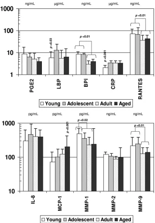
FIG. 1. Acute-phase reactants and inflammatory mediators in serum samples from nonhuman primates of different age groups: young (
⬍
3
years) (
n
⫽
76), adolescent (
ⱖ
3 to 8 years) (
n
⫽
61), adult (8 to 15 years) (
n
⫽
32), and aged (
⬎
15 years) (
n
⫽
10). Bars represent group means;
error bars, 1 standard deviation. Statistical differences among the groups are displayed.
on August 17, 2020 by guest
http://cvi.asm.org/
Oral clinical parameters.All animals were examined, while anesthetized, by the same periodontal investigator in this study. The periodontal examination was con-ducted using a Maryland probe (William’s markings) on the facial, mesiobuccal, and distobuccal aspects of all teeth, excluding the canines and third molars. The exam-ination included probing pocket depth (PD), clinical attachment level (CAL), plaque index, and gingival bleeding index (bleeding on probing [BOP]) (13). The plaque index characterizes the extent and quantity of tooth-associated bacterial plaque. PD and CAL measures were made using a calibrated probe and are measures of the extent and severity of periodontal pathology. The presence and degree of bleeding upon gentle periodontal probing provided a measure of the presence of inflamma-tion within the periodontal tissues.
Serum analyses.Blood was collected from all animals, serum was prepared, and levels of immunoglobulin G (IgG) antibodies to seven oral bacteria were evaluated using an enzyme-linked immunosorbent assay (ELISA) as we have
described previously (14, 15). Briefly, Campylobacter rectus, Fusobacterium
nucleatum,Aggregatibacter actinomycetemcomitans,Prevotella intermedia,
Trepo-nema denticola,Tannerella forsythia, andPorphyromonas gingivaliswere grown under anaerobic conditions in mycoplasma broth base with the addition of appropriate additives as we have reported previously (23). The bacteria were
harvested by centrifugation, formalin killed, washed, and stored at⫺20°C for use
as antigens (14, 15).
Selected systemic inflammatory biomarkers were quantified using ELISA pro-cedures developed in our laboratory (C-reactive protein [CRP] [21]). Luminex Beadlyte technology was used for interleukin-8 (IL-8), monocyte chemoattrac-tant protein 1 (MCP-1), and RANTES (Upstate, Temecula, CA) and for matrix metalloproteinase 1 (MMP-1), MMP-2, and MMP-9 (R&D Systems,
Minneap-olis, MN). Commercial ELISA kits were used for prostaglandin E2 (PGE2)
(Assay Design, Ann Arbor, MI), lipopolysaccharide binding protein (LBP; Cell Sciences, Canton, MA), and bactericidal permeability-inducing factor (BPI; Cell Sciences, Canton, MA) in serum samples from all animals.
Statistical analyses.An analysis of variance (ANOVA) with post hoc testing was used for the various continuous variables, including clinical parameters and
FIG. 2. Acute-phase reactants (A), inflammatory mediators (B), and antibodies (C) in serum samples from nonhuman primates
⬍
3 years old
raised under standard housing conditions (young) (
n
⫽
76) or under SPF conditions (
n
⫽
30). Bars represent mean levels of each mediator; error
bars, 1 standard deviation. Statistical differences between the groups are shown. Aa,
Aggregatibacter actinomycetemcomitans
; Cr,
Campylobacter
rectus
; Fn,
Fusobacterium nucleatum
; Pg,
Porphyromonas gingivalis
; Pi,
Prevotella intermedia
; Td,
Treponema denticola
; Tf,
Tannerella forsythia
. For
panels A and B, PGE
2, BPI, RANTES, MMP-2, and MMP-9 are measured in ng/ml; LBP and CRP are measured in
g/ml; and IL-8, MCP-1, and
MMP-1 are measured in pg/ml (as in Fig. 1). For panel C, antibody levels are presented in ng/ml.
V
OL. 15, 2008
AGE, ORAL DISEASE, AND IMMUNITY IN NONHUMAN PRIMATES
1069
on August 17, 2020 by guest
http://cvi.asm.org/
serum analytes. This was accomplished using a one-way ANOVA and a Holm-Sidak test for parametric values, and a Kruskal-Wallis ANOVA with Dunn’s
method for multiple comparisons of variables not normally distributed. An␣
value of⬍0.05 was accepted as the level of significant difference in comparing the
various parameters.
RESULTS
Systemic inflammatory mediators.
The levels of various
sys-temic inflammatory mediators were determined in serum
sam-ples from each animal and segregated based on the age of the
animal: young (
⬍
3 years), adolescent (3 to 8 years), adult (8 to
15 years), or aged (
⬎
15 years). Figure 1 summarizes the levels
of the various inflammatory mediators with aging in this cohort
of animals. CRP, LBP, and MCP-1 levels were significantly
decreased in the young animals, and MCP-1 levels were
ele-vated in the aged group. BPI, RANTES, MMP-1, and MMP-9
levels were all significantly elevated in the young and
adoles-cent animals compared to adult and aged monkeys.
Figure 2A and B show comparisons of the levels of these
inflammatory mediators in the sera of young animals within the
large group cohort versus those detected in the sera of animals
of similar ages maintained under SPF conditions. The results
showed elevated levels of PGE
2, CRP, BPI, MMP-1, and
MMP-9 in the sera of the SPF animals compared to the young
animals raised under standard housing conditions.
Systemic antibody responses to oral bacteria.
Figure 3
shows the levels of serum IgG antibodies to a group of oral
bacteria commonly associated with periodontal disease (1, 34,
44) in the various age groups of nonhuman primates. The
results show significantly lower antibody levels in the group of
young animals. The adult and aged animals routinely
demon-strated significantly elevated levels of antibodies to the
indi-vidual species, with minimal differences between these age
groups.
Figure 2C provides an analysis of the serum antibody levels
in the young animals housed under standard conditions
com-pared to the young SPF animals. There were few differences in
serum antibody levels between these groups; the SPF animals
had levels at least as high as those of the standard group of
animals.
Systemic responses and clinical parameters.
No clinical
dif-ferences were observed between the male and female animals,
although significant increases in disease parameters of BOP
(0.75
⫾
0.2 versus 0.95
⫾
0.15 units;
P
⬍
0.05), PD (2.50
⫾
0.10
versus 3.15
⫾
0.40 mm;
P
⬍
0.04), and CAL (0.25
⫾
0.05 versus
0.55
⫾
0.15 mm;
P
⬍
0.05) were noted in the aged animals. The
younger groups of animals demonstrated negligible plaque,
inflammation, or gingival tissue changes. We stratified the
adult and aged animals into two groups based on the bleeding
index (mean,
ⱖ
0.9 or
⬍
0.9 units) or on mean pocket depth
(
ⱖ
3 mm or
⬍
3 mm). When the animals were grouped based
on these clinical parameters, the differences in systemic
re-sponses were greater in animals with poorer oral health.
Consistent with the lack of differences in clinical
presenta-tion related to gender, no differences in serum inflammatory
mediators or serum antibody levels were observed between the
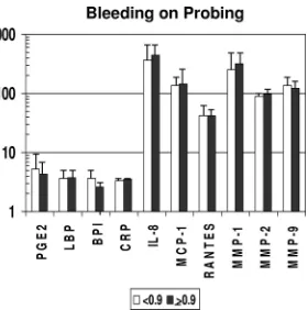
genders (data not shown). The results in Fig. 4 show the
dif-ferences in inflammatory mediator levels between the groups
stratified by gingival bleeding. No significant differences in
these serum response analytes were observed between the
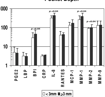
groups. However, in Fig. 5, it can be noted that levels of
antibody to
P. gingivalis
and the sum of antibodies to the seven
target bacteria were significantly increased with greater
gingi-val bleeding. In contrast, Fig. 6 and 7 depict significant
differ-FIG. 3. Serum IgG antibodies to individual oral bacteria and total antibodies to this battery (SUM) in nonhuman primates categorized on the
basis of age (see the legend to Fig. 1). Bars represent mean antibody levels; error bars, 1 standard deviation. Statistical differences are depicted
on the graph. For bacterium abbreviations, see the legend to Fig. 2.
on August 17, 2020 by guest
http://cvi.asm.org/
ences in multiple inflammatory mediators (BPI, IL-8, MMP-1,
MMP-2) and serum antibodies to
F. nucleatum
,
P. gingivalis
,
T.
forsythia
, and the sum of antibodies with more-severe
destruc-tive disease, i.e., greater pocket depth.
DISCUSSION
Evaluation of inflammatory and immune responses has
pro-vided evidence of ontogenic development of the immune
sys-tem (2, 5, 52, 61, 64, 71, 81), as well as alterations in various
host response parameters that are affected by aging (12, 28, 60,
87). However, while changes in oral health with aging similar
to those reported for humans (88) have been reported to occur
in various nonhuman primate species (7), little information on
the biologic underpinnings of these clinical differences has
been provided. While systemic inflammation, often resulting
from bacterial sepsis, is frequently related to negative clinical
outcomes with both morbidity and mortality, the systemic
in-flammatory response can also accomplish at the
whole-organ-ism level what the local inflammatory response is designed to
do: that is, utilize disparate, nonspecific effector molecules to
ameliorate potential tissue damage by noxious agents,
includ-ing infectinclud-ing bacteria (18). However, the characteristics of this
ancient response system have generally been evaluated in adult
individuals and have been related to sepsis, neoplastic changes,
and responses to chronic diseases (18). This study
demon-strated the patterns of selected systemic inflammatory
mole-cules in young individuals and demonstrated specific changes
in these levels with aging. Of particular note were the
signifi-cantly elevated levels of BPI, RANTES, and both MMP-1 and
MMP-9 in the younger groups of animals. This was
unex-FIG. 4. Acute-phase reactants and inflammatory mediators in
se-rum samples from nonhuman primates stratified on the basis of a low
(
⬍
0.9) (
n
⫽
32) or high (
ⱖ
0.9) (
n
⫽
9) mean index score for mouth
bleeding upon probing. Bars represent group means; error bars, 1
standard deviation. PGE
2, BPI, RANTES, MMP-2, and MMP-9 are
measured in ng/ml; LBP and CRP are measured in
g/ml; and IL-8,
MCP-1, and MMP-1 are measured in pg/ml (as in Fig. 1).
FIG. 5. Serum IgG antibodies to individual oral bacteria and total antibodies to this battery (SUM) in nonhuman primates stratified on the basis
of a low (
⬍
0.9) or high (
ⱖ
0.9) index score for mean bleeding upon probing. Bars represent mean antibody levels; error bars, 1 standard deviation.
Statistical differences are depicted on the graph. For bacterium abbreviations, see the legend to Fig. 2.
V
OL. 15, 2008
AGE, ORAL DISEASE, AND IMMUNITY IN NONHUMAN PRIMATES
1071
on August 17, 2020 by guest
http://cvi.asm.org/
pected, with minimal previous evidence of this type of response
except that reported in obese children and adolescents (86)
and a general concept of a lack of systemic challenge in
chil-dren in the absence of clinical changes, but it is consistent with
the maturation of host responses in young animals that have to
cope with a range of environmental challenges to naı¨ve
muco-sal surfaces. Testing this concept, we compared these systemic
responses in young animals housed under standard conditions
with those in comparably aged SPF monkeys. The results
showed that the SPF animals often had elevated levels of the
mediators compared to normal nonhuman primates. While
there are likely various explanations for these differences, one
existing theory, the “hygiene hypothesis,” suggests that the
increasing incidence of asthma and other allergic diseases in
the human population results from a lack of sufficient
onto-genic development or “training” of the immune system in the
young, who are then less able to effectively distinguish a
nox-ious challenge later in their development (27, 50, 66, 89).
Irrespective of the basis, these data demonstrate significant
differences in response profiles of the inflammatory and innate
immune systems during aging.
Nonhuman primates have historically been utilized to
eval-uate infectious agents (8, 32, 47, 54) and biologic processes (10,
16, 31, 51) associated with various human diseases. This is
related to homologies in a range of host responses between
humans and the other primate species, as well as species
tro-pisms for infectious agents that cross human and nonhuman
primate lines (26, 43, 73). These similarities extend to the
microbial ecology and host responses in the oral cavity related
to microbial biofilms that trigger periodontal disease (20, 22,
45, 62, 68). We observed that levels of serum antibody to
various bacteria associated with periodontal-disease biofilms
were significantly lower in the youngest animals. As is noted in
humans and nonhuman primates, the extent of disease was
increased in the aged group, although the antibody levels were
similar for the adult and aged animals. This is consistent with
the early acquisition and accumulation of these species as part
of the commensal microbiota of the oral cavity and an
associ-ation of these bacteria as etiologic triggers of periodontal
pa-thology related to aging. We have also characterized the effects
of aging on naturally occurring periodontitis, and we use a
specific ligature-induced model of specific challenge to the oral
cavity to describe acute responses of mucosal tissues during
aging. In addition, this model has allowed us to document
gender and diet effects on local and systemic inflammatory and
immune responses that are altered with aging (25).
FIG. 6. Acute-phase reactants and inflammatory mediators in serum samples from nonhuman primates stratified on the basis of a low (
⬍
3 mm)
(
n
⫽
25) or high (
ⱖ
3 mm) (
n
⫽
16) mean pocket depth in the mouth. Bars represent group means; error bars, 1 standard deviation. Statistical
differences are depicted on the graph. PGE
2, BPI, RANTES, MMP-2, and MMP-9 are measured in ng/ml; LBP and CRP are measured in
g/ml;
and IL-8, MCP-1, and MMP-1 are measured in pg/ml (as in Fig. 1).
on August 17, 2020 by guest
http://cvi.asm.org/
We then addressed specific questions regarding these
sys-temic responses and the expression of chronic periodontal
infections and inflammation in the oral cavities of adult and
aged animals. As is noted in humans and nonhuman
pri-mates, the extent of disease was increased in the aged group,
with no gender differences in expression of disease. This is
in contrast to our findings with
M. mulatta
raised in
individ-ual housing with ad libitum feeding. Our previous results
demonstrated that aged males exhibited significant weight
gain, demonstrated various biologic parameters of
un-healthy aging, and exhibited significantly greater
periodon-tal disease than similarly aged females (unpublished data).
Placing the males on a calorie-restricted diet decreased the
disease to a level similar to that of the female cohort. The
group of animals in this study was housed in a large corral
that permitted constant exercise, required food-scavenging
behaviors, and permitted natural competition among the
various strata of colony members. Thus, the older males
were much more physically fit than the sedentary singly
housed animals, which may have translated into a
preserva-tion of oral health for aging males comparable to that for
females.
We also stratified the animals, irrespective of age, based on
the clinical presentation of gingival bleeding, a measure of
local mucosal inflammation, and on pocket depth as an
indi-cator of local challenge resulting in destructive disease. These
subgroups were then evaluated for the patterns of systemic
inflammatory and antibody responses. The results
demon-strated rather minimal differences in the systemic responses
related to the level of gingival inflammation in these adult and
aged monkeys. In contrast, multiple significant differences
were observed in both the inflammatory mediators and the
levels of antibodies to oral bacteria in the animals with more
severe periodontitis. These data for the nonhuman primates
support similar data from humans indicating that the tissue
destruction associated with chronic periodontitis enhances
challenge of the systemic circulation with the potential to
alter the function of the vascular and/or distant tissues (11,
48, 57, 72).
The literature is generally lacking on the use of nonhuman
primates to elucidate the ontogenic development of the
inflam-matory, innate, and adaptive immune system. The oral cavity
provides a readily accessible model of these host response
changes at mucosal surfaces that interface with an evolving
microbial ecology. This study described oral clinical findings
and systemic responses in nonhuman primates and described
differences in these measures that occurred from young
through aged animals. The results from these initial studies of
this primate colony provide a basis for the use of this robust
resource to test hypotheses regarding the local and systemic
ontogeny of innate and adaptive immune responses in
rela-tionship to the acquisition, adaptation, and evolution of the
microbial ecology at this mucosal surface.
ACKNOWLEDGMENTS
This work was supported by CPRC grant 5P40 RR003640 from the
NCRR (National Institutes of Health) and a UKRF grant from the
University of Kentucky.
FIG. 7. Serum IgG antibodies to individual oral bacteria and total antibodies to this battery (SUM) in nonhuman primates stratified on the basis
of a low (
⬍
3 mm) or high (
ⱖ
3 mm) mean pocket depth in the mouth. Bars represent mean levels; error bars, 1 standard deviation. Statistical
differences are depicted on the graph. For bacterium abbreviations, see the legend to Fig. 2.
V
OL. 15, 2008
AGE, ORAL DISEASE, AND IMMUNITY IN NONHUMAN PRIMATES
1073
on August 17, 2020 by guest
http://cvi.asm.org/
We extend our gratitude to the entire technical support group from
the Caribbean Primate Research Center facility, especially Edmundo
Kraiselburd, for providing support for the conduct of this study.
REFERENCES
1.Aas, J. A., B. J. Paster, L. N. Stokes, I. Olsen, and F. E. Dewhirst.2005. Defining the normal bacterial flora of the oral cavity. J. Clin. Microbiol.
43:5721–5732.
2.Abraham, C. M., and D. R. Ownby.2005. Ontogeny of the allergic
inflam-matory response. Immunol. Allergy Clin. N. Am.25:215–229.
3.Agrawal, A. A., A. Kapley, R. K. Yeltiwar, and H. J. Purohit.2006.
Assess-ment of single nucleotide polymorphism at IL-1A⫹4845 and IL-1B⫹3954 as
genetic susceptibility test for chronic periodontitis in Maharashtrian
ethnic-ity. J. Periodontol.77:1515–1521.
4.Albandar, J. M., L. J. Brown, and H. Loe.1997. Putative periodontal patho-gens in subgingival plaque of young adults with and without early-onset
periodontitis. J. Periodontol.68:973–981.
5.Annacker, O., R. Pimenta-Araujo, O. Burlen-Defranoux, and A. Bandeira.
2001. On the ontogeny and physiology of regulatory T cells. Immunol. Rev.
182:5–17.
6.Apatzidou, D. A., M. P. Riggio, and D. F. Kinane.2005. Impact of smoking on the clinical, microbiological and immunological parameters of adult
pa-tients with periodontitis. J. Clin. Periodontol.32:973–983.
7.Avery, B. E., and D. M. Simpson.1973. The baboon as a model system for the study of periodontal disease: clinical and light microscopic observations. J.
Periodontol.44:675–686.
8.Barry, P. A., K. M. Lockridge, S. Salamat, S. P. Tinling, Y. Yue, S. S. Zhou, S. M. Gospe, Jr., W. J. Britt, and A. F. Tarantal.2006. Nonhuman primate
models of intrauterine cytomegalovirus infection. ILAR J.47:49–64.
9.Bimstein, E., S. Sapir, Y. Houri-Haddad, S. Dibart, T. E. Van Dyke, and L. Shapira.2004. The relationship betweenPorphyromonas gingivalisinfection
and local and systemic factors in children. J. Periodontol.75:1371–1376.
10.Black, A., and M. A. Lane.2002. Nonhuman primate models of skeletal and
reproductive aging. Gerontology48:72–80.
11.Bobetsis, Y. A., S. P. Barros, and S. Offenbacher.2006. Exploring the rela-tionship between periodontal disease and pregnancy complications. J. Am.
Dent. Assoc.137(Suppl. 2):7S–13S.
12.Candore, G., G. Colonna-Romano, C. R. Balistreri, D. Di Carlo, M. P. Grimaldi, F. Listi, D. Nuzzo, S. Vasto, D. Lio, and C. Caruso.2006. Biology
of longevity: role of the innate immune system. Rejuvenation Res.9:143–
148.
13.Cappelli, D., S. C. Holt, R. E. Singer, H. M. Pickrum, and J. L. Ebersole.
2000. Effects of 0.12% chlorhexidine gluconate on experimental gingivitis in non-human primates: clinical and microbiological alterations. Oral Dis.
6:124–131.
14.Celenligil, H., and J. L. Ebersole.1998. Analysis of serum antibody responses to periodontopathogens in early-onset periodontitis patients from different
geographical locations. J. Clin. Periodontol.25:994–1002.
15.Celenligil-Nazliel, H., E. Kansu, and J. L. Ebersole.1999. Periodontal
find-ings and systemic antibody responses to oral microorganisms in Behc¸et’s
disease. J. Periodontol.70:1449–1456.
16.Coughlin, S. R.2005. Protease-activated receptors in hemostasis, thrombosis
and vascular biology. J. Thromb. Haemost.3:1800–1814.
17.Craig, R. G., R. Boylan, J. Yip, D. Mijares, M. Imam, S. S. Socransky, M. A. Taubman, and A. D. Haffajee.2002. Serum IgG antibody response to peri-odontal pathogens in minority populations: relationship to periperi-odontal
dis-ease status and progression. J. Periodontal Res.37:132–146.
18.Ebersole, J. L., and D. Cappelli.2000. Acute-phase reactants in infections
and inflammatory diseases. Periodontol. 200023:19–49.
19.Ebersole, J. L., D. Cappelli, and S. C. Holt.2001. Periodontal diseases: to
protect or not to protect is the question? Acta Odontol. Scand.59:161–166.
20.Ebersole, J. L., D. Cappelli, S. C. Holt, R. E. Singer, and T. Filloon.2000. Gingival crevicular fluid inflammatory mediators and bacteriology of gingi-vitis in nonhuman primates related to susceptibility to periodontitis. Oral
Microbiol. Immunol.15:19–26.
21.Ebersole, J. L., D. Cappelli, E. C. Mathys, M. J. Steffen, R. E. Singer, M. Montgomery, G. E. Mott, and M. J. Novak.2002. Periodontitis in humans and non-human primates: oral-systemic linkage inducing acute phase
pro-teins. Ann. Periodontol.7:102–111.
22.Ebersole, J. L., D. Cappelli, G. Mott, L. Kesavalu, S. C. Holt, and R. E. Singer.1999. Systemic manifestations of periodontitis in the non-human
primate. J. Periodontal Res.34:358–362.
23.Ebersole, J. L., and K. S. Kornman.1991. Systemic antibody responses to oral microorganisms in the cynomolgus monkey: development of methodol-ogy and longitudinal responses during ligature-induced disease. Res.
Immu-nol.142:829–839.
24.Ebersole, J. L., M. J. Steffen, and D. Cappelli.1999. Longitudinal human
serum antibody responses to outer membrane antigens ofActinobacillus
actinomycetemcomitans. J. Clin. Periodontol.26:732–741.
25.Ebersole, J. L., M. J. Steffen, M. A. Reynolds, G. L. Branch-Mays, D. R. Dawson, K. F. Novak, J. C. Gunsolley, J. A. Mattison, D. K. Ingram, and M. J. Novak.Differential gender effects of a reduced calorie diet on systemic
inflammatory and immune parameters in nonhuman primates. J. Periodontal Res., in press.
26.Engel, G., L. L. Hungerford, L. Jones-Engel, D. Travis, R. Eberle, A. Fuentes, R. Grant, R. Kyes, and M. Schillaci.2006. Risk assessment: a model for predicting cross-species transmission of simian foamy virus from
ma-caques (M. fascicularis) to humans at a monkey temple in Bali, Indonesia.
Am. J. Primatol.68:934–948.
27.Folkerts, G., G. Walzl, and P. J. Openshaw.2000. Do common childhood infections ‘teach’ the immune system not to be allergic? Immunol. Today
21:118–120.
28.Fulop, T., A. Larbi, N. Douziech, I. Levesque, A. Varin, and G. Herbein.
2006. Cytokine receptor signalling and aging. Mech. Ageing Dev.127:526–
537.
29.Gafan, G. P., V. S. Lucas, G. J. Roberts, A. Petrie, M. Wilson, and D. A. Spratt.2004. Prevalence of periodontal pathogens in dental plaque of
chil-dren. J. Clin. Microbiol.42:4141–4146.
30.Hayashi, F., M. Okada, Y. Soda, K. Miura, and K. Kozai.2006. Subgingival
distribution ofCampylobacter rectusandTannerella forsythensisin healthy
children with primary dentition. Arch. Oral Biol.51:10–14.
31.Henson, M. C., and V. D. Castracane.2002. Leptin: roles and regulation in
primate pregnancy. Semin. Reprod. Med.20:113–122.
32.Herodin, F., P. Thullier, D. Garin, and M. Drouet.2005. Nonhuman pri-mates are relevant models for research in hematology, immunology and
virology. Eur. Cytokine Netw.16:104–116.
33.Hu, S. W., C. H. Huang, H. C. Huang, Y. Y. Lai, and Y. Y. Lin.2006.
Transvascular dissemination ofPorphyromonas gingivalisfrom a sequestered
site is dependent upon activation of the kallikrein/kinin pathway. J.
Peri-odontal Res.41:200–207.
34.Jervoe-Storm, P. M., M. Koltzscher, W. Falk, A. Dorfler, and S. Jepsen.
2005. Comparison of culture and real-time PCR for detection and quantifi-cation of five putative periodontopathogenic bacteria in subgingival plaque
samples. J. Clin. Periodontol.32:778–783.
35.Kamma, J. J., M. Nakou, and F. A. Manti.1995. Predominant microflora of severe, moderate and minimal periodontal lesions in young adults with
rapidly progressive periodontitis. J. Periodontal Res.30:66–72.
36.Katancik, J. A., S. Kritchevsky, R. J. Weyant, P. Corby, W. Bretz, R. O. Crapo, R. Jensen, G. Waterer, S. M. Rubin, and A. B. Newman.2005.
Periodontitis and airway obstruction. J. Periodontol.76:2161–2167.
37.Kilian, M., E. V. Frandsen, D. Haubek, and K. Poulsen.2006. The etiology of periodontal disease revisited by population genetic analysis. Periodontol.
200042:158–179.
38.Kinane, D. F., and P. M. Bartold. 2007. Clinical relevance of the host
responses of periodontitis. Periodontol. 200043:278–293.
39.Kinane, D. F., and T. C. Hart.2003. Genes and gene polymorphisms
asso-ciated with periodontal disease. Crit. Rev. Oral Biol. Med.14:430–449.
40.Kinane, D. F., J. Mooney, and J. L. Ebersole. 1999. Humoral immune
response toActinobacillus actinomycetemcomitansandPorphyromonas
gingi-valisin periodontal disease. Periodontol. 200020:289–340.
41.Kinane, D. F., M. Podmore, M. C. Murray, P. J. Hodge, and J. Ebersole.
2001. Etiopathogenesis of periodontitis in children and adolescents.
Peri-odontol. 200026:54–91.
42.Kinane, D. F., M. P. Riggio, K. F. Walker, D. MacKenzie, and B. Shearer.
2005. Bacteraemia following periodontal procedures. J. Clin. Periodontol.
32:708–713.
43.Kodama, M., K. Murakami, R. Sato, T. Okimoto, A. Nishizono, and T. Fujioka.2005.Helicobacter pylori-infected animal models are extremely suit-able for the investigation of gastric carcinogenesis. World J. Gastroenterol.
11:7063–7071.
44.Kumar, P. S., A. L. Griffen, J. A. Barton, B. J. Paster, M. L. Moeschberger, and E. J. Leys.2003. New bacterial species associated with chronic
peri-odontitis. J. Dent. Res.82:338–344.
45.Madden, T. E., and J. G. Caton.1994. Animal models for periodontal
disease. Methods Enzymol.235:106–119.
46.McArthur, W. P., C. Bloom, M. Taylor, J. Smith, T. Wheeler, and N. I. Magnusson.1995. Antibody responses to suspected periodontal pathogens
in elderly subjects with periodontal disease. J. Clin. Periodontol.22:842–849.
47.McMurray, D. N.2000. A nonhuman primate model for preclinical testing of
new tuberculosis vaccines. Clin. Infect. Dis.30(Suppl. 3):S210–212.
48.Mealey, B. L.2006. Periodontal disease and diabetes: a two-way street.
J. Am. Dent. Assoc.137(Suppl.):26S–31S.
49.Miura, M., T. Hamachi, O. Fujise, and K. Maeda.2005. The prevalence and
pathogenic differences ofPorphyromonas gingivalis fimAgenotypes in
pa-tients with aggressive periodontitis. J. Periodontal Res.40:147–152.
50.Moreels, T. G., and P. A. Pelckmans.2006. The hygiene hypothesis and inflammatory bowel diseases: role of helminths. Acta Gastroenterol. Belg.
69:413–417.
51.Morton, W. R., and K. Swindler.2005. Serendipitous insights involving
nonhuman primates. ILAR J.46:346–351.
52.Newburg, D. S., and W. A. Walker.2007. Protection of the neonate by the innate immune system of developing gut and of human milk. Pediatr. Res.
61:2–8.
53.Nonnenmacher, C., R. Mutters, and L. F. de Jacoby.2001. Microbiological
on August 17, 2020 by guest
http://cvi.asm.org/
characteristics of subgingival microbiota in adult periodontitis, localized juvenile periodontitis and rapidly progressive periodontitis subjects. Clin.
Microbiol. Infect.7:213–217.
54.O’Connor, C. M., and D. H. Kedes.2007. Rhesus monkey rhadinovirus: a
model for the study of KSHV. Curr. Top. Microbiol. Immunol.312:43–69.
55.Offenbacher, S., and J. D. Beck.2005. A perspective on the potential
car-dioprotective benefits of periodontal therapy. Am. Heart J.149:950–954.
56.Okada, M., F. Hayashi, Y. Soda, X. Zhong, K. Miura, and K. Kozai.2004. Intra-familial distribution of nine putative periodontopathogens in dental
plaque samples analyzed by PCR. J. Oral Sci.46:149–156.
57.Padilla, C., O. Lobos, E. Hubert, C. Gonzalez, S. Matus, M. Pereira, S. Hasbun, and C. Descouvieres.2006. Periodontal pathogens in atheromatous plaques isolated from patients with chronic periodontitis. J. Periodontal Res.
41:350–353.
58.Papapanou, P. N., A. M. Neiderud, A. Papadimitriou, J. Sandros, and G. Dahlen.2000. “Checkerboard” assessments of periodontal microbiota and
serum antibody responses: a case-control study. J. Periodontol.71:885–897.
59.Persson, G. R.2005. Immune responses and vaccination against periodontal
infections. J. Clin. Periodontol.32(Suppl. 6):39–53.
60.Prelog, M.2006. Aging of the immune system: a risk factor for
autoimmu-nity? Autoimmun. Rev.5:136–139.
61.Prescott, S.2004. Developmental immunology and vaccines: cellular im-mune development and future vaccine strategies. Expert Rev. Vaccines
3:339–342.
62.Roberts, F. A., L. S. Houston, S. A. Lukehart, L. A. Mancl, G. R. Persson, and R. C. Page.2004. Periodontitis vaccine decreases local prostaglandin E2
levels in a primate model. Infect. Immun.72:1166–1168.
63.Roth, G. S., J. A. Mattison, M. A. Ottinger, M. E. Chachich, M. A. Lane, and D. K. Ingram.2004. Aging in rhesus monkeys: relevance to human health
interventions. Science305:1423–1426.
64.Rumbo, M., and E. J. Schiffrin.2005. Ontogeny of intestinal epithelium immune functions: developmental and environmental regulation. Cell. Mol.
Life Sci.62:1288–1296.
65.Sakai, Y., H. Shimauchi, H. O. Ito, M. Kitamura, and H. Okada.2001.
Porphyromonas gingivalis-specific IgG subclass antibody levels as
immuno-logical risk indicators of periodontal bone loss. J. Clin. Periodontol.28:853–
859.
66.Schaub, B., R. Lauener, and E. von Mutius.2006. The many faces of the
hygiene hypothesis. J. Allergy Clin. Immunol.117:969–978.
67.Schlegel-Bregenzer, B., R. E. Persson, S. Lukehart, P. Braham, T. Oswald, and G. R. Persson.1998. Clinical and microbiological findings in elderly
subjects with gingivitis or periodontitis. J. Clin. Periodontol.25:897–907.
68.Schou, S., P. Holmstrup, and K. S. Kornman.1993. Non-human primates used in studies of periodontal disease pathogenesis: a review of the
litera-ture. J. Periodontol.64:497–508.
69.Seto, H., M. Ninomiya, and T. Nagata.2006. Alveolar bone resorption in
animal models of periodontitis. Clin. Calcium16:24–30. (In Japanese.)
70.Shapira, L., A. Wilensky, and D. F. Kinane.2005. Effect of genetic variability on the inflammatory response to periodontal infection. J. Clin. Periodontol.
32(Suppl. 6):72–86.
71.Smith, D. J., and M. A. Taubman.1992. Ontogeny of immunity to oral
microbiota in humans. Crit. Rev. Oral Biol. Med.3:109–133.
72.Spahr, A., E. Klein, N. Khuseyinova, C. Boeckh, R. Muche, M. Kunze, D. Rothenbacher, G. Pezeshki, A. Hoffmeister, and W. Koenig.2006. Periodon-tal infections and coronary heart disease: role of periodonPeriodon-tal bacteria and
importance of total pathogen burden in the Coronary Event and Periodontal
Disease (CORODONT) study. Arch. Intern. Med.166:554–559.
73.Stearns-Kurosawa, D. J., F. Lupu, F. B. Taylor, Jr., G. Kinasewitz, and S. Kurosawa. 2006. Sepsis and pathophysiology of anthrax in a nonhuman
primate model. Am. J. Pathol.169:433–444.
74.Takahashi, K., F. Nishimura, M. Kurihara, Y. Iwamoto, S. Takashiba, T. Miyata, and Y. Murayama.2001. Subgingival microflora and antibody re-sponses against periodontal bacteria of young Japanese patients with type 1
diabetes mellitus. J. Int. Acad. Periodontol.3:104–111.
75.Takai, S., T. Kuriyama, M. Yanagisawa, K. Nakagawa, and T. Karasawa.
2005. Incidence and bacteriology of bacteremia associated with various oral and maxillofacial surgical procedures. Oral Surg. Oral Med. Oral Pathol.
Oral Radiol. Endod.99:292–298.
76.Tanaka, H., N. Tanabe, M. Shoji, N. Suzuki, T. Katono, S. Sato, M. Mo-tohashi, and M. Maeno.2006. Nicotine and lipopolysaccharide stimulate the formation of osteoclast-like cells by increasing macrophage
colony-stimulat-ing factor and prostaglandin E2production by osteoblasts. Life Sci.78:1733–
1740.
77.Tanner, A. C., R. L. Kent, Jr., M. F. Maiden, P. J. Macuch, and M. A. Taubman.2000. Serum IgG reactivity to subgingival bacteria in initial
peri-odontitis, gingivitis and healthy subjects. J. Clin. Periodontol.27:473–480.
78.Tatakis, D. N., and P. S. Kumar.2005. Etiology and pathogenesis of
peri-odontal diseases. Dent. Clin. N. Am.49:491–516.
79.Taubman, M. A., A. D. Haffajee, S. S. Socransky, D. J. Smith, and J. L. Ebersole.1992. Longitudinal monitoring of humoral antibody in subjects
with destructive periodontal diseases. J. Periodontal Res.27:511–521.
80.Taubman, M. A., P. Valverde, X. Han, and T. Kawai.2005. Immune re-sponse: the key to bone resorption in periodontal disease. J. Periodontol.
76:2033–2041.
81.Teitelbaum, J. E., and W. Allan Walker.2005. The development of mucosal
immunity. Eur. J. Gastroenterol. Hepatol.17:1273–1278.
82.Timmerman, M. F., and G. A. van der Weijden.2006. Risk factors for
periodontitis. Int. J. Dent. Hyg.4:2–7.
83.Timmerman, M. F., G. A. Van der Weijden, F. Abbas, E. M. Arief, S. Armand, E. G. Winkel, A. J. Van Winkelhoff, and U. Van der Velden.2000. Untreated periodontal disease in Indonesian adolescents. Longitudinal clin-ical data and prospective clinclin-ical and microbiologclin-ical risk assessment. J. Clin.
Periodontol.27:932–942.
84.Van Dyke, T. E., and D. Sheilesh.2005. Risk factors for periodontitis. J. Int.
Acad. Periodontol.7:3–7.
85.van Winkelhoff, A. J., C. J. Bosch-Tijhof, E. G. Winkel, and W. A. van der Reijden.2001. Smoking affects the subgingival microflora in periodontitis. J.
Periodontol.72:666–671.
86.Visser, M., L. M. Bouter, G. M. McQuillan, M. H. Wener, and T. B. Harris.
2001. Low-grade systemic inflammation in overweight children. Pediatrics
107:E13.
87.Weng, N. P.2006. Aging of the immune system: how much can the adaptive
immune system adapt? Immunity24:495–499.
88.Willershausen-Zo¨nnchen, B., and C. Gleissner.1998. Periodontal disease in
elderly patients. Eur. J. Med. Res.3:55–64.
89.Zavos, C., D. Vini, J. Kountouras, N. Zavos, and E. Trivara.2007. Hygiene hypothesis and protection against asthma in infants: spending time in the countryside encountering natural allergens may boost maternal immunity.
Med. Hypotheses68:914–915.