The first phase IIb safety and efficacy trial of a new tuberculosis vaccine since that for BCG was completed in October 2012.
BCG-vaccinated South African infants were randomized to receive modified vaccinia virus Ankara, expressing the
Mycobacterium
tu-berculosis
antigen 85A (MVA85A), or placebo. MVA85A did not significantly boost the protective effect of BCG. Cryopreserved
samples provide a unique opportunity for investigating the correlates of the risk of tuberculosis disease in this population. Due
to the limited amount of sample available from each infant, preliminary work was necessary to determine which assays and
con-ditions give the most useful information. Peripheral blood mononuclear cells (PBMC) were stimulated with antigen 85A
(Ag85A) and purified protein derivative from
M. tuberculosis
in an
ex vivo
gamma interferon (IFN-
␥
) enzyme-linked
immu-nosorbent spot assay (ELISpot) and a Ki67 proliferation assay. The effects of a 2-h or overnight rest of thawed PBMC on ELISpot
responses and cell populations were determined. Both the ELISpot and Ki67 assays detected differences between the MVA85A
and placebo groups, and the results correlated well. The cell numbers and ELISpot responses decreased significantly after an
overnight rest, and surface flow cytometry showed a significant loss of CD4
ⴙand CD8
ⴙT cells. Of the infants tested, 50% had a
positive ELISpot response to a single pool of flu, Epstein-Barr virus (EBV), and cytomegalovirus (CMV) (FEC) peptides. This pilot
work has been essential in determining the assays and conditions to be used in the correlate study. Moving forward, PBMC will be
rested for 2 h before assay setup. The ELISpot assay, performed in duplicate, will be selected over the Ki67 assay, and further work is
needed to evaluate the effect of high FEC responses on vaccine-induced immunity and susceptibility to tuberculosis disease.
D
isease caused by
Mycobacterium tuberculosis
continues to be a
major global health problem. In 2012, there were 8.6 million
new cases of tuberculosis (TB) worldwide and 1.3 million people
died of the disease (
1
). Bacille Calmette-Guérin (BCG), the only
licensed TB vaccine, has variable efficacy, ranging from 0 to 80%,
depending on the geographical location and population (
2
). A
vaccine which is able to provide universal protection is urgently
needed. The lack of a known correlate of protection against disease
caused by infection with
M. tuberculosis
continues to be a major
obstacle for the TB vaccine field, making it difficult to select which
vaccines should progress to large-scale efficacy trials and to
pre-dict how successful those vaccines will be. Since 2002, more than a
dozen candidate vaccines have entered into clinical testing (
3
),
with the aim of boosting the efficacy of BCG or replacing it
alto-gether. Only a few of these candidate vaccines have progressed to
large-scale efficacy trials (
3
). The results of the most advanced of
these, a phase IIb safety and efficacy trial of modified vaccinia virus
Ankara, expressing the
M. tuberculosis
antigen 85A (MVA85A) in
BCG-vaccinated South African infants, were published in
Febru-ary 2013 (
5
). MVA85A did not significantly improve the efficacy
of BCG in this population, despite promising preclinical data
from animal models (6
⫺
9) and the induction of potent and
du-rable T cell responses in earlier phase I/IIa clinical trials (10 –13).
Although enhanced protection was not achieved in this
popula-tion, peripheral blood mononuclear cells (PBMC) stored pre- and
postvaccination provide a unique opportunity to investigate
im-munological differences between those infants who went on to
develop TB disease and those who did not. With limited PBMC
available to do this, careful planning was needed in order to select
the assays which give the most relevant and diverse information.
Prior work using the mycobacteria growth indicator tube (MGIT)
assay and gene expression analysis demonstrated that high-quality
data can be obtained using samples from the same population of
South African infants, and these two assays (our unpublished
data) will be included in the correlate analysis. The aim of this
work was to determine which other assays have utility for
inclu-sion. Here we describe some pilot work carried out to evaluate the
optimum time that thawed PBMC should be rested before setting
up immunological assays and compare antigen-specific responses
to antigen 85A (Ag85A) and purified protein derivative (PPD)
from
M. tuberculosis
in the gamma interferon (IFN-
␥
)
enzyme-linked immunosorbent spot assay (ELISpot) and the Ki67
prolif-Received6 March 2014Returned for modification21 March 2014
Accepted9 May 2014
Published ahead of print14 May 2014
Editor:D. L. Burns
Address correspondence to Stephanie A. Harris, [email protected], or Helen McShane, [email protected].
Copyright © 2014 Harris et al. This is an open-access article distributed under the
terms of theCreative Commons Attribution 3.0 Unported license.
doi:10.1128/CVI.00128-14
on August 17, 2020 by guest
eration assays. The utility of cell surface flow cytometry was also
evaluated. This process of assay selection and optimization, prior
to the processing of the valuable correlate samples, has relevance
for all trials of new vaccines, where the amount of sample available
for analysis will always be limited.
MATERIALS AND METHODS
Origin of samples. (i) Infant samples.Samples used in these pilot exper-iments were cryopreserved PBMC from a double-blind, randomized,
pla-cebo-controlled phase IIb efficacy trial of the candidate TB vaccine, MVA85A, in BCG-vaccinated, HIV-negative South African infants (South African National Clinical Trials Register DOH-27-0109-2654,
ClinicalTrials.govregistration no. NCT00953927). Collection of these trial samples was approved by the University of Cape Town Faculty of Health Sciences Human Research Ethics Committee, the Oxford Univer-sity Tropical Research Ethics Committee, and the Medicines Control Council of South Africa. The samples used were selected from a subgroup of infants on whom gene expression analysis had already been planned, and the remainder were from those infants who were lost to follow-up.
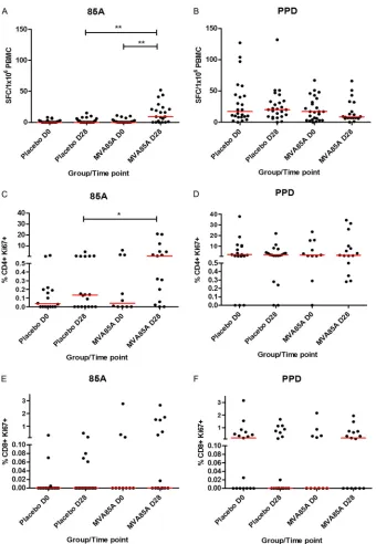
FIG 1Detection of antigen-specific responses in infants vaccinated with MVA85A. IFN-␥ELISpot responses (A and B) and percentages of proliferating CD4⫹ Ki67⫹(C and D) and CD8⫹Ki67⫹(E and F) in PBMC after stimulation with a single pool of antigen 85A peptides (A, C, and E) and purified protein derivative (PPD) fromM. tuberculosis(B, D, and F). Red lines show median responses. Significant differences (Mann-Whitney test): *,P⬍0.05; **,P⬍0.01.n⫽10 to 27 per time point.
on August 17, 2020 by guest
http://cvi.asm.org/
They were not from cases (infants who went on to develop TB disease) nor from matched controls.
(ii) Adult samples.Samples used in this study were cryopreserved PBMC from BCG-vaccinated, United Kingdom adults, 28 days after MVA85A vaccination (1⫻108PFU) (ClinicalTrials.govregistration no.
NCT01194180).
Experimental design.Infants enrolled in this trial were 4 to 6 months old and had received BCG (Danish 1331; Statens Serum Institute [SSI], Denmark) within 7 days of birth. They were randomized to receive an intradermal vaccination with either MVA85A (1⫻108PFU) or placebo
(Candidaskin test antigen) (5). Two subsets of samples were used in this work. The first set consisted of PBMC taken at 0 to 7 days prevaccination and 28 days postvaccination from 30 infants given MVA85A and 30 given placebo. These samples were used for the Ki67 proliferation assay, to quantify the proliferative potential of PBMC when stimulated with my-cobacterial antigens, and for theex vivoIFN-␥ELISpot assay, to quantify the magnitude of the response to mycobacterial and nonmycobacterial antigens. The ELISpot responses were also used to assess the intra-assay variation, comparing responses across either duplicate or triplicate anti-gen wells. The correlation between the two assays was investigated.
The second set of samples consisted of pre- and postvaccination PBMC from 15 infants from each group (MVA85A andCandidaplacebo). Due to the logistics of processing the large number of case and control samples in the TB correlates of risk study, it is desirable to thaw and rest PBMC overnight. This set of samples was used to compareex vivoIFN-␥ ELISpot responses when PBMC were rested for 2 h or overnight. A subset of 10 of these samples was used for cell surface flow cytometry to identify cell populations and determine differences between a 2-h and an over-night rest. As a comparison to the infant samples, PBMC from 12 adults at 28 days after MVA85A vaccination were subjected to the same thawing and resting conditions and were used for cell surface flow cytometry and IFN-␥ELISpot assays.
Cell thawing.Two vials of cryopreserved PBMC from each sample were rapidly thawed in a 37°C water bath and transferred to a 15-ml Falcon tube containing 10 ml R10 (RPMI medium, 10% fetal calf serum, 1% L-glutamine, 1% penicillin-streptomycin, 1% sodium pyruvate). PBMC were pelleted, supernatants were discarded, and the samples were resuspended in 10 ml R10 with 20l Benzonase (25 U/l) (Merck Chem-icals Ltd.) and rested overnight at 37°C and 5% CO2. For the 2-h versus
overnight comparison, PBMC were split into equal volumes; one set was rested for 2 h, and one set was rested overnight. PBMC were counted on a Casy counter (Roche) and split into the appropriate volumes for each assay. In some cases, there were inadequate numbers of cells to perform all assays and use all conditions.
Ex vivoIFN-␥ELISpot assay.Theex vivoIFN-␥ELISpot assay was performed on both subsets of samples, thawed PBMC from prevaccina-tion and 28 days postvaccinaprevaccina-tion as previously described (13). In a 96-well ELISpot plate (Millipore), triplicate wells containing 3⫻105PBMC were
stimulated with a single pool of Ag85A peptides, consisting of 66 15-mer peptides, overlapping by 10 amino acids (2g/ml/peptide) (Peptide Pro-tein Research), BCG from pooled SSI vaccine vials (2⫻105CFU/ml),
purified protein derivative (PPD) fromM. tuberculosis(20g/ml) (SSI), FEC, consisting of a single pool of flu, Epstein-Barr virus (EBV), and
cytomegalovirus (CMV) peptides (10g/ml/peptide) (Peptide Protein Research), and combined TB10.3 and TB10.4 peptides (10 g/ml/pep-tide) (Peptide Protein Research). Phytohemagglutinin (PHA) (Sigma) was used as a positive control, and unstimulated wells were used as a measure of background IFN-␥production. The results are reported as spot-forming cells (SFC) per million PBMC, calculated by subtracting the mean of the unstimulated wells from the mean of triplicate antigen wells and correcting for the numbers of PBMC in the wells. A response was considered positive if the mean number of spots in the antigen well was at least twice the mean of the unstimulated wells and at least 5 spots greater.
Ki67 proliferation assay.PBMC were counted and plated in 24-well plates at 1⫻106PBMC/1 ml R10. Plates were incubated for 6 days at 37°C
and 5% CO2with either 2g/ml PPD or 1g/ml/peptide of a single pool
of antigen 85A peptides. Two wells of PBMC were left unstimulated in R10. After 3 days, PHA was added to one of the unstimulated wells at 0.3
g/ml. Three days later, the cells were washed in phosphate-buffered saline (PBS) and stained with amine-reactive viability dye (Live/Dead fixable red dead cell stain; Invitrogen). Cells were then permeabilized with Perm/Wash (BD Biosciences) and incubated with monoclonal antibodies: anti-CD3-AF700, anti-CD4-PB, anti-Ki67-phycoerythrin (PE) (BioLegend), and anti-CD8-allophycocyanin (APC)/AF750 (Beck-man Coulter). Cells were then washed and acquired on a BD LSR II flow cytometer (BD Biosciences, San Jose, CA). Data were analyzed using FlowJo software (Tree Star Inc.). Dead cells were excluded from the anal-ysis, while singlet CD3⫹T cells, CD3⫹CD4⫹T cells, and CD3⫹CD8⫹T cells were included. Proliferating cells are presented as the percentage of Ki67⫹T cells out of the total CD4⫹or CD8⫹T cells. Background (un-stimulated) values were subtracted from all data.
Cell surface flow cytometry.PBMC were washed and stained with Live/Dead fixable red dead cell stain (Invitrogen) followed by surface staining with the following antibodies: anti-CD3-AF700, anti-CD4-PB, anti-CD14-PE/Cy7, anti-CD16-AF488, anti-CD19-PE/Cy5, anti-CD25-APC/Cy7,␥␦T cell receptor (TCR)-APC, anti-cytotoxic-T-lymphocyte-as-sociated antigen (CTLA)-PE (all from BioLegend), anti-CD8-efluorNC605, and anti-CD127-efluorNC650 (eBioscience). Fluorescence minus one (FMO) controls were used to identify boundaries of gates for CD25,
␥␦TCR, CD127, and CTLA. Samples were acquired on a BD LSR II flow cytometer. Results are presented as percentages of cells after gating out of dead cells and doublets. CD4⫹and CD8⫹T cells were identified as CD3⫹ cells, while CD14⫹/⫺and CD16⫹/⫺cells were identified as CD3⫺and CD19⫺populations. CTLA⫹and CD25⫹CD27⫺populations were gated on the CD4⫹cells.
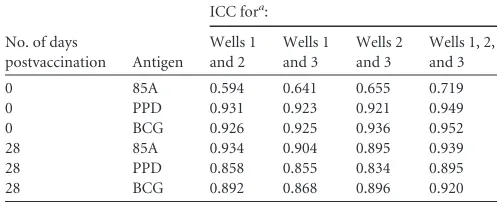
Statistical analysis.Statistical analyses were performed using GraphPad Prism and SPSS. The Mann-Whitney test was used to compare differences between groups. The Wilcoxon matched-pairs signed-rank test was used to compare responses after a 2-h or an overnight rest. Spearman’s rho was used to determine correlations between the ELISpot and Ki67 prolifera-tion assays. The intraclass correlaprolifera-tion (ICC) with a two-way mixed-effects 28 PPD 0.858 0.855 0.834 0.895 28 BCG 0.892 0.868 0.896 0.920
a
Intraclass correlation (ICC) values: 0 to 0.2, poor; 0.3 to 0.4, fair; 0.4 to 0.6, good; 0.6 to 0.8, very good; and 0.8 to 1.0, excellent.
on August 17, 2020 by guest
http://cvi.asm.org/
model was used to assess the reproducibility between duplicate and trip-licate wells in the IFN-␥ELISpot assay.
RESULTS
Ki67 proliferation assay results correlate with IFN-
␥
ELISpot
responses.
Both the IFN-
␥
ELISpot and Ki67 assays detected
sig-nificant differences (
P
⫽
0.001 and 0.03, respectively) between the
MVA85A and placebo groups at 28 days postvaccination in
re-sponse to stimulation with a single pool of antigen 85A peptides
(
Fig. 1A
and
C
). A significant difference between the baseline and
postvaccination responses in the MVA85A group was also
de-tected using the ELISpot assay (
P
⫽
0.001) but was not detected in
the Ki67 assay, possibly due to the low number of samples
avail-able for this assay in the MVA85A group on day 0 (D0). Neither
assay detected any differences between groups or time points in
response to stimulation with PPD (
Fig. 1B
,
D
, and
F
). Significant
correlations were observed between the two assays for PPD
re-sponses at baseline and for both PPD and antigen 85A rere-sponses at
D28 (vaccination groups combined) (
Table 1
).
Intra-assay variation.
The intraclass correlation (ICC) has
been reported as an improved measure of reliability for
quantita-tive data, where reliability is the reproducibility of a biological
measurement when it is repeated for the same study subject (
14
).
The ICC was used in this study to measure the reproducibility of
responses detected in replicate wells of the ELISpot assay. The ICC
improved as the magnitude of the detected IFN-␥
ELISpot
re-sponse increased. ICC values for duplicate (all combinations) and
triplicate ELISpot wells indicated a good (ICC of 0.4 to 0.6) or very
good (ICC of 0.6 to 0.8) level of agreement for baseline responses
to antigen 85A and an excellent (ICC of 0.8 to 1.0) level of
agree-ment for all other antigens and time points (
Table 2
).
Cell counts are significantly lower overnight.
PBMC were
counted on a Casy counter (Roche) after both the 2-h and
over-night rests (
Fig. 2
). Infant 2-h counts ranged from a total of 1.1
⫻
10
6to 2.3
⫻
10
7cells, with overnight counts ranging from a total of
1.4
⫻
10
6to 2.5
⫻
10
7cells. A significant reduction in the median
of the cell counts was observed, with counts of 7.7
⫻
10
6and 6.9
⫻
10
6for the 2-h and overnight rests, respectively (
P
⫽
0.02) (
Fig.
2A
). The median adult PBMC count also showed a significant
reduction after the overnight rest (
P
⫽
0.03) (
Fig. 2B
).
IFN-
␥
ELISpot responses are significantly lower after an
overnight rest.
Figure 3
shows IFN-
␥
ELISpot responses after a
2-h and an overnight rest in both South African infants (groups
and time points combined) and United Kingdom adults.
Re-sponses to all five antigens tested in the infant population were
significantly lower after the overnight rest than after the 2-h rest
(Wilcoxon matched-pairs signed-rank test), despite equivalent
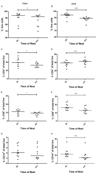
FIG 2Cell viability is reduced in both adult and infant PBMC following overnight resting of cells. Infant (A) and adult (B) PBMC counts from a Casy counter (Roche) after a 2-h or overnight rest. Lines show median and interquartile ranges. An asterisk indicates significant difference (P⬍0.05, Wilcoxon matched-pairs signed rank test). Infants,n⫽56; adults,n⫽12.
FIG 3IFN-␥ELISpot responses are reduced in both adult and infant PBMC following overnight resting of cells. Infant (pre- and postvaccination) (A) and adult (postvaccination) (B) IFN-␥ELISpot responses after a 2-h or overnight rest. Red lines show median responses. Asterisks indicate significant differences (Wilcoxon matched-pairs signed-rank test): *,P⬍0.05; **,P⬍0.01; ***,P⬍0.001. Infants,n⫽45 to 55; adults,n⫽12.
on August 17, 2020 by guest
http://cvi.asm.org/
numbers of PBMC in the ELISpot wells (
Fig. 3A
). The same was
true for the adult responses to the two antigens tested (
Fig. 3B
).
Loss of T cell populations is significant after an overnight
rest.
Cell surface flow cytometry of 10 infant samples and 12 adult
samples showed a significant loss of viability overnight: a drop in
median viability of 93.9 to 92.0% in the infants (
P
⫽
0.02) (
Fig.
4A
) and 94.3 to 89.2% in the adults (
P
⫽
0.0005) (
Fig. 4B
). The
infant samples showed a significant loss of CD4
⫹and CD8
⫹T
FIG 4Changes in lymphocyte and monocyte cell frequencies following overnight resting of cells. Infant (left) and adult (right) cell viability (A and B) and percentages of total live CD4⫹(C and D), CD8⫹(E and F), and CD14⫹(G and H) cells after a 2-h or overnight rest. Lines show median values. Asterisks indicate significant differences (Wilcoxon matched-pairs signed-rank test): *,P⬍0.05; ***,P⬍0.001. Infants,n⫽10; adults,n⫽12.
on August 17, 2020 by guest
http://cvi.asm.org/
cells after an overnight rest compared to that after a 2-h rest (
P
⫽
0.01) (
Fig. 4C
and
E
) and a nonsignificant loss of CD14
⫹mono-cytes (
Fig. 4G
). In adults, a significant loss of CD8
⫹and CD14
⫹cells was observed (
P
⫽
0.0005) (
Fig. 4F
and
H
). While the
per-centage of CD4
⫹T cells significantly increased after an
over-night rest (
P
⫽
0.0005) (
Fig. 4D
), the total number of CD4
⫹T
cells did not.
Proportion of infants responding on IFN-
␥
ELISpot assay to
the FEC peptides was higher than expected.
A single pool of FEC
peptides was included in the IFN-␥
ELISpot assay for 30 infants.
After a 2-h rest of PBMC, 15 out of 30 infants had a positive
baseline or D28 response to these peptides, ranging from 27 to 783
SFC/million PBMC (median, 102 SFC/million PBMC) (
Fig. 3A
).
DISCUSSION
Here we have presented preliminary work on the down-selection
of assays and methods to be carried forward for investigating a
correlate of infection/risk of tuberculosis. This work used pre- and
postvaccination PBMC from a phase IIb efficacy trial of MVA85A
in BCG-vaccinated South African infants and showed that
high-quality data can be obtained from these cryopreserved samples.
Cell counts were variable, ranging from 1.1 to 23.3 million
(median, 7.7 million) for the 2-h rest and dropping significantly
overnight (median, 6.9 million). For most subjects, ample PBMC
were available to carry out the desired assays and conditions,
sug-gesting that it is worthwhile to plan to carry out multiple assays on
the case versus control samples, as, based on the cell counts in this
study, sample sizes will be large enough to make meaningful
com-parisons within each assay.
Both the
ex vivo
IFN-␥
ELISpot and Ki67 proliferation assays
were able to detect differences in responses to antigen 85A
be-tween the placebo and MVA85A-vaccinated groups at 28 days
postvaccination. The ELISpot assay was also able to detect a
dif-ference between pre- and postvaccination responses to antigen
85A in the MVA85A group, which the Ki67 assay did not, possibly
due to a lower number of samples available for the Ki67 assay in
the MVA85A group at D0. The baseline responses to the PPD were
high due to all infants receiving BCG vaccination at birth, but
neither assay detected an increase in response to the PPD after
vaccination with MVA85A. This is in contrast to previous findings
in BCG-vaccinated United Kingdom adults and South African
adults, adolescents, and children, where we see significant
in-creases in responses to both Ag85A and PPD after vaccination
with MVA85A (
11
,
13
,
15
). The lack of an increase in PPD
re-sponses in infants may be due to the recency of the BCG
vaccina-tion and the fact that the corresponding Ag85A responses are
much lower than those we see in adults. There was good
agree-ment between the two assays, with significant correlations for PPD
responses at both the baseline and D28 and for antigen 85A
re-sponses at D28. The lack of correlation between the assays for
antigen 85A responses at baseline is probably due to the very low
responses detected at this time point. As a result of these findings
and due to the cell-intensive nature and length of time involved in
the Ki67 assay, we decided that the IFN-␥
ELISpot assay would be
taken forward for the full correlate analysis and the Ki67
prolifer-ation assay would not be performed on further samples from this
cohort.
Intra-assay variability was determined by looking at both
du-plicate and tridu-plicate repeats in the ELISpot assay. Both numbers
of repeats produced excellent ICC values, suggesting that it is
suf-ficient to carry out the ELISpot with duplicate antigen wells, thus
saving a considerable number of valuable PBMC for use in other
assays.
Due to the logistical advantage, many trials of new vaccines
perform analyses on cryopreserved PBMC. However, the loss of
responses and therefore the loss of sensitivity of an assay with the
use of cryopreserved samples instead of fresh must be minimized.
Logistically, an overnight rest of thawed PBMC is desirable;
how-ever, a significant decrease in ELISpot responses for all of the
an-tigens tested for both infant and adult samples was observed when
PBMC were rested overnight compared with that when the same
samples were rested for only 2 h. Cell surface flow cytometry to
determine cell populations in the infant samples showed a
signif-icant loss of percentages of CD4
⫹and CD8
⫹T cells overnight,
which might explain the loss of antigen-specific IFN-
␥
production
in the ELISpot. A loss of monocytes, although not significant,
might also result in lower levels of antigen presentation. As a
con-sequence of this result, future assays will be set up after a rest of
only 2 h as this will minimize the loss of antigen-specific
re-sponses.
The inclusion of FEC, a nonmycobacterial pool of CD8
epitopes, in the ELISpot assay provided some interesting results.
While 50% of infants had no response to these peptides at either
the baseline or D28, the remaining 50% had positive responses,
with a median of 102 SFC/million PBMC. Infants in this trial were
4 to 6 months old when enrolled, so even at this very young age, a
noticeable proportion have immunological memories of exposure
to at least one of these viral pathogens. It will be of interest and
possibly of relevance to determine which of these pathogens the
infants are responding to. As a result, in the study of the correlate
samples, separate pools of EBV, CMV, and influenza peptides will
be included in the ELISpot panel.
An immune correlate analysis of the phase III HIV vaccine
efficacy trial with RV144 used 6 assays for the primary analysis,
selected on the basis of reproducibility, ability to detect
postvac-cine responses, and uniqueness of responses (
16
). The preliminary
work we have shown here has also demonstrated the importance
of selecting assays that are reproducible and sensitive enough to
detect differences between groups and, importantly, those that
measure different aspects of the immune response. This pilot
study has been essential in determining the optimal assays and
conditions to continue with for the TB immune correlate study.
ACKNOWLEDGMENTS
We thank the trial participants and their families and the MVA85A 020 trial study team.
This work was supported by the Wellcome Trust, Aeras, Oxford-Emergent Tuberculosis Consortium (OETC) Ltd., and NEWTBVAC (EC FP7). H.M. is a Wellcome Trust Senior Clinical Fellow and Jenner Insti-tute Investigator.
REFERENCES
1.World Health Organization. 2012. Global Tuberculosis Report 2012. World Health Organization, Geneva, Switzerland.
2.Colditz GA, Brewer TF, Berkey CS, Burdick E, Fineberg HV, Mosteller F.1994. Efficacy of BCG vaccine in the prevention of tuberculosis. Meta-analysis of the published literature. JAMA271:698 –702.
3.Brennan MJ, Thole J.2012. Tuberculosis vaccines: a strategic blueprint for the next decade. Tuberculosis92:S6 –S13.http://dx.doi.org/10.1016 /S1472-9792(12)70005-7.
4. Reference deleted.
5.Tameris MD, Hatherill M, Landry BS, Scriba TJ, Snowden MA,
on August 17, 2020 by guest
http://cvi.asm.org/
Thomas AW.2009. MVA.85A boosting of BCG and an attenuated, phoP deficientM. tuberculosisvaccine both show protective efficacy against tu-berculosis in rhesus macaques. PLoS One4:e5264.http://dx.doi.org/10 .1371/journal.pone.0005264.
8.Goonetilleke NP, Mcshane H, Hannan CM, Anderson RJ, Brookes RH, Hill AVS. 2003. Enhanced immunogenicity and protective efficacy against Mycobacterium tuberculosis of bacille Calmette-Guérin vaccine using mucosal administration and boosting with a recombinant modified vaccinia virus Ankara. J. Immunol.171:1602–1609.http://dx.doi.org/10 .4049/jimmunol.171.3.1602.
9.Williams A, Goonetilleke NP, Mcshane H, Simon O, Hatch G, Gilbert SC, Hill AVS, Clark SO.2005. Boosting with poxviruses enhances My-cobacterium bovisBCG efficacy against tuberculosis in guinea pigs. Infect. Immun. 73:3814 –3816. http://dx.doi.org/10.1128/IAI.73.6.3814-3816 .2005.
10. Scriba TJ, Tameris M, Mansoor N, Smit E, van der Merwe L, Mauff K, Hughes EJ, Moyo S, Brittain N, Lawrie A, Mulenga H, de Kock M, Gelderbloem S, Veldsman A, Hatherill M, Geldenhuys H, Hill AVS, Hussey GD, Mahomed H, Hanekom WA, McShane H. 2011. Dose-finding study of the novel tuberculosis vaccine, MVA85A, in healthy
BCG-13. Meyer J, Harris SA, Satti I, Poulton ID, Poyntz HC, Tanner R, Rowland R, Griffiths KL, Fletcher HA, McShane H.2013. Comparing the safety and immunogenicity of a candidate TB vaccine MVA85A administered by intramuscular and intradermal delivery. Vaccine31:1026 –1033.http://dx .doi.org/10.1016/j.vaccine.2012.12.042.
14. White E.2011. Measurement error in biomarkers: sources, assessment, and impact on studies. IARC Sci. Publ.163:143–161.
15. Scriba TJ, Tameris M, Mansoor N, Smit E, van der Merwe L, Isaacs F, Keyser A, Moyo S, Brittain N, Lawrie A, Gelderbloem S, Veldsman A, Hatherill M, Hawkridge A, Hill AVS, Hussey GD, Mahomed H, McShane H, Hanekom WA.2010. Modified vaccinia Ankara-expressing Ag85A, a novel tuberculosis vaccine, is safe in adolescents and children, and induces polyfunctional CD4⫹T cells. Eur. J. Immunol.40:279 –290.
http://dx.doi.org/10.1002/eji.200939754.
16. Haynes BF, Gilbert PB, McElrath MJ, Zolla-Pazner S, Tomaras GD, Alam SM, Evans DT, Montefiori DC, Karnasuta C, Sutthent R, Liao HX, DeVico AL, Lewis GK, Williams C, Pinter A, Fong Y, Janes H, DeCamp A, Huang Y, Rao M, Billings E, Karasavvas N, Robb ML, Ng J. 2012. Immune-correlates analysis of an HIV-1 vaccine efficacy trial. N. Engl. J. Med.366:
1275–1286.http://dx.doi.org/10.1056/NEJMoa1113425.