_____________________________________________________________________________________________________
www.sciencedomain.org
Effectiveness of Tuberculosis Smear Microscopy in
Laboratory Diagnosis of
Mycobacterium
tuberculosis
in Resource Constrained Settings
Ameh James
1*, Kingsley Ochei
1,2, Nnamdi Emenyonu
3and Lovett Lawson
31Family Health International, 360 Plot 1073-A1, J.S. Tarka Street, Area 3, Garki, Abuja, Nigeria. 2General Hospital, Wuse, Abuja, Nigeria.
3
Zankli Medical Centre, Abuja, Nigeria.
Authors’ contributions
This work was carried out in collaboration of all authors. Author AJ performed statistical analysis, managed literature searches and wrote the first draft. Author KO conceived the study and performed laboratory work. Author NE performed the laboratory work. Author LL co-ordinated the study.
All authors read and approved the final manuscript.
Article Information
DOI: 10.9734/BJMMR/2015/6714 Editor(s): (1)Sinan INCE, Department of Pharmacology and Toxicology, University of Afyon Kocatepe, TURKEY. (2)Divya Kesanakurti, Dept. of Cancer Biology and Pharmacology, University of Illinois College of Medicine, USA. (3)Gaetano Santulli, College of Physicians & Surgeons, Columbia University Medical Center, New York, NY, USA. Reviewers: (1)Anonymous, Ahvaz Jundi-Shapour University of Medical Sciences, Iran. (2)Anonymous, Case Western reserve university, USA. (3)Anonymous, USA. (4)Anonymous, Rajiv Gandhi Medical University, Karnataka, India. (5)Anonymous, Chung Shan Medical University, Taiwan. (6)Jann-Yuan Wang, Department of Internal Medicine, National Taiwan University Hospital, Taiwan. Complete Peer review History:http://sciencedomain.org/review-history/10256
Received 2nd September 2013 Accepted 5th December 2013 Published 21st July 2015
ABSTRACT
Aim: To determine the correlation of accuracy of direct smear microscopy compared with BACTEC MGIT 960.
Design: The study prospectively compare direct smear microscopy with BACTEC MGIT 960 using the reference standard, Lowenstein Jensen culture.
Place and Duration: The study was conducted in Zankli Medical Centre, Abuja, between November 2004 and July 2005.
Methodology: 340 suspected patients for Mycobacterium tuberculosis referred from direct observation therapy clinics located in six different government owned health facilities were referred to our facility. These patients; male (192) and female (148) were between the age of 10 and 64
years old. Three sputa samples were collected over two consecutive days and direct smear microscopy and culture were performed on these samples.
Results: When compared with the reference standard, BACTEC MGIT 960 has a sensitivity and specificity of 100.0% and 56.4% respectively, and a negative predictive value of 100.0%; indicating the proportion of AFB negative participants were actually not infected with M. tuberculosis when tested with BACTEC MGIT 960. The sensitivity of direct microscopy was significantly lower than BACTEC MGIT 960 (84.9% versus 100%, p<0.001) and the specificity was significantly higher (96.6% versus 56.4%, p<0.001).
Conclusions: For the purpose of effectiveness of tuberculosis program in developing countries, direct smear microscopy may still be relevant in the diagnosis of Mycobacterium tuberculosis.
Keywords: Tuberculosis; developing countries; direct smear microscopy; BACTEC MGIT 960.
1. INTRODUCTION
Pulmonary Mycobacterium Tuberculosis (MTB) continues to be one of the infectious diseases affecting low and medium income countries (LMIC), especially in sub-Saharan African countries. In order to successfully control the spread of MTB, cases must be detected and treated immediately and effectively. The detection of cases has always been direct smear microscopy in LMIC, where nearly 95 percent of TB cases and 98 percent of deaths occur as a result of MTB [1]. Direct smear microscopy is a simple, rapid and inexpensive method which is very specific in areas with high prevalence of tuberculosis [1]. This method also identifies the most infectious patients and it is widely applicable in various populations with different socio-economic levels [1]. However, the current quest for automated liquid culture like BACTEC Mycobacterium Growth Indicator Tubes (MGIT) 960 (Becton Dickinson, USA) has challenged the relevance of direct microscopy in LMIC.
In brief, the BACTEC MGIT 960 is an automated equipment that consists of liquid broth medium in a growth indicator tube and a device that detects the growth of the mycobacteria. The liquid medium is known to yield better recovery and faster growth of mycobacteria. This medium is made up of modified Middlebrook 7H9 broth base and reconstituted with a growth supplement (Oleic acid, Albumin, Dextrose and Catalase) and PANTA (Polymyxin B, Amphotericin B, Nalidixic acid, Trimethoprim, and Azolocillin). Also, present in the tube is an oxygen-quenched fluorochrome, tris 4, 7-diphenyl-1, 10-phenonthroline ruthenium chloride pentahydrate, embedded in a silicone. During bacterial growth within the tube, the free oxygen is utilized and is replaced with carbon dioxide. As the free oxygen depletes, the fluorochrome fluorescence within the tube. The intensity of fluorescence is directly proportional to the extent of oxygen depletion
and this is dependent on the growth of bacteria as well as mycobacteria (2). This system has been described as an essential for strengthening tuberculosis diagnoses, care and treatment in many countries [2]. It has been reported that a well funded clinical laboratory can effectively detect MTB cases within 7 to 14 days using automated liquid culture system like BACTEC MGIT 960 [3].
However, there are challenges associated with the introduction of this system in a setting like ours, as described in a systematic review [4]. The major challenge has been attributed to lack of funding of clinical laboratories. In LMIC, MTB laboratories lack the required funding and human resource to support case detection using sophisticated and expensive equipment, like the BACTEC MGIT 960 [4,3].
There have been varied reports of the sensitivity of direct smear microscopy in different settings, as documented in a review [5]. Also, there have been substantial researches into alternative methods and validation in the laboratory diagnosis of MTB for developing countries [6-15]. However, there seems to be less frequent reporting on the performance evaluation of direct microscopy when compared with the automated liquid culture system [16]. This is also similar to comparing direct microscopy with the solid (egg-based) medium (Lowenstein Jensen, LJ); few studies reported this comparison to the best of our knowledge [13,17]. However, there are well established reports on BACTEC MGIT 960 validation, when compared with LJ [18-21].
specificity, positive and negative predictive values between direct smear microscopy and BACTEC MGIT 960.
2. MATERIALS AND METHODS
2.1 Settings and Patient Recruitment
340 suspected TB patients from direct observation therapy clinics located in six different governments owned health facilities in the Federal Capital Territory (FCT), Abuja in Nigeria were sent for TB diagnosis at the Zankli Research laboratory, also based in Abuja, Nigeria. These hospitals are managed through the government agency, Hospital Management Board. Patients, 192 males and 148 females, aged between 10 and 64 years old were prospectively enrolled into the study between November 2004 and July 2005. A verbal informed consent was obtained from each participant. Ethical approvals were obtained from the Ethical Committees of FCT Hospital Management Board and Zankli Medical Centre. Participants that did not submit three specimens over 2 day’s period and participants receiving anti-tuberculosis treatment were excluded from the study.
2.2 Specimen Collection
All the 340 participants submitted three sputum specimens over two consecutive days. First sputum samples (1st spot) were collected from the patients during their first visits to the laboratory. They were then given sputum bottles to take home and bring back early morning samples (morning) the following day. The third samples (2nd spot) were collected when the patients submitted the early morning samples. Thus, a total of 1,020 specimens were collected. Instructions were given to patients on the appropriate method of sputum collection. The two specimens collected in the laboratory were produced by patients in an open and free ventilated area. We performed direct smear microscopy on all the specimens collected and randomly selected one specimen of the three from each patient for culture. All diagnostic tests gave conclusive results on 340 participants.
2.3 Direct Smear Microscopy
1 by 2 cm smears were made from the purulent part of the sputum, air-dried and heat fixed on a hot plate at 850C for 2-3 minutes and stained with Ziehl-Neelsen technique using standard
carbol-fuchsin for microscopy staining, and methylene blue (6); while sulphuric acid for decolourisation was used.
2.4 Microscopic Examination and
Interpretation
The smears were read using oil immersion lens (x100) of ordinary light microscope by experienced microscopist who were blinded to culture results. Positive and negative smears were defined accordingly (6). Briefly, a patient is reported smear positive for tuberculosis if at least 1–9 acid fast bacilli (AFB) are seen in 100 high power fields. For the purpose of this study, we only considered a definitive case detection and not necessarily the grading. Hence, we reported on number of patients diagnosed with active M. tuberculosis infection.
2.5 Sputum Decontamination (Modified Petroff Method), Culture and Isolation
of M. tuberculosis
Sputum for culture was randomly selected from the three specimens provided by every participant. To 5 ml of sputum was added an equal volume of 2% sodium hydroxide / N-acetyl-L-cysteine (NaOH/NALC) solution in a 50 ml screw-cap tube. This was capped tightly and shaken gently to ensure that NaOH-NALC solution enough contact to aid sputum digest; thereafter the solution was allowed to stand at room temperature for 15 minutes with occasional shaking. The sputum NaOH-NALC was then diluted with sterile phosphate buffer Ph 6.8 and centrifuged at 3,000 X g for 15 minutes. The supernatant was carefully decanted and the deposit was re-suspended with 1-2 ml of sterile phosphate buffer pH 6.8. The re-suspended deposit was used for both culture techniques.
Solid media–0.5ml of the re-suspended sediment from the processed sputa were inoculated onto LJ agar slope and incubated at 37±2oC and observed daily for the first three days for possible contamination and thereafter regularly examined at weekly interval for 6 - 10 weeks for the isolation of M. tuberculosis. Positive and negative growth controls were always included using wild strains of M. tuberculosis complex and sterile distilled water respectively.
monitored automatically every one hour for increased fluorescence. The cultures were tested until positive or for 42 days.
Identification of mycobacteria – Identification of mycobacteria was based on colony morphology, colony pigmentation, rate of growth on solid media, the results of biochemical tests; nitrate reductase test, catalase heat-labile test. M. tuberculosis complex was only differentiated from non-tuberculous mycobacteria and no further identification tests were done.
2.6 Definitions
1) Reference standard is positive and negative LJ culture. 2) Positive MGIT culture was considered as positive signal and positive AFB smear. 3) Negative MGIT culture was considered as negative signal at 42 days and negative AFB smear. 4) Specimens were considered contaminated when AFB smear of a positive MGIT culture shows growth of commensal bacteria or bacteria other than mycobacteria species within the 42 days protocol (16).
2.7 Statistical Analysis
Sensitivity, specificity, negative and positive predictive values were calculated using standard definitions [22]. Statistical analysis was performed with Stata SE software version 11 (Stata Corp LP, College station TX, USA). A p-value of <0.05 was considered statistically significant. For the purposes of analysis, all contaminated and negative cultures were considered negative for MTB.
3. RESULTS AND DISCUSSION
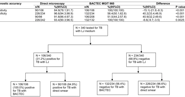
340 participants recruited during the 8 months period study profile, is shown in Fig. 1. When compared with the reference standard, BACTEC MGIT 960 has a sensitivity and specificity of 100.0% and 56.4% respectively, and a negative predictive value of 100.0% (Table 1) indicating the proportion of AFB negative participants were actually not infected with M. tuberculosis when tested with BACTEC MGIT 960. The sensitivity of direct microscopy was signifcantly lower than BACTEC MGIT 960 (84.9% versus 100%, p<0.001) and the specifcity was signifcantly higher (96.6% versus 56.4%, p<0.001).
Of the 340 positive sputa by MGIT, 102 specimens were contaminants representing 30% contamination rate.
One of the advantages of comparing one diagnostic method with another, especially with an identified reference standard, is to establish test performance characteristics [23]. This is helpful in determining diagnostic properties of the method like sensitivity and specificity before its introduction into the laboratory. This study therefore, compared direct microscopy with the BACTEC MGIT 960, using the solid medium as the reference standard. The choice of solid culture was simply because it was an existing method before the introduction of BACTEC MGIT 960 in our setting, but we are aware that the automated liquid culture is now an accepted gold standard. Our results indicate that in the absence of the automated liquid culture system, direct microscopy could be utilized, as indicated in the statistical analysis in the Table 1. In another study in Zimbabwe by Aper L et al. [16], direct microscopy was reported to be comparable with BACTEC MGIT 960, statistically. The study reported an agreement between the two diagnostic methods as 72% (p<0.0001). While the sensitivity and specificity of direct microscopy was reported as 67.5% and 95.5% respectively. Our study reported a higher sensitivity when compared with the study in Zimbabwe. This observation could have been as a result of increased sample size utilized in this study. However, there are limitations associated with this study. For example, we used direct microscopy to confirm mycobacterial growth from liquid culture and did not establish non-tuberculous mycobacteria. Also, the direct smear microscopy could have missed out 15.1% of the patients detected by BACTEC MGIT 960 since this method had a sensitivity of 100% (Table 1). It is also likely that true TB cases detected by BACTEC MGIT 960 were not detected by the LJ culture technique, contributing to the reported low specificity and PPV in this study. Furthermore, the study design did not compare smear microscopy to culture on a ‘per sample’ basis, since only one randomly selected sample was used for the culture techniques. Additionally, the HIV status of the patients was not evaluated.
5
Table 1. Diagnostic tests characteristics of BACTEC MGIT 960 and direct smear determined when compared with the reference standard. PPV – positive predictive value, NPV – negative predictive value, CI – confidence interval
Diagnostic accuracy Direct microscopy BACTEC MGIT 960 Difference
n/N %(95%CI) n/N %(95%CI) %(95%CI) P value
Sensitivity 90/106 84.9(78.1;91.7) 106/106 100(100;100) -15.1(-21.9;-8.3) <0.001
Specificity 226/234 96.6(94.3;98.9) 132/234 56.4(50.1;62.8) 40.2(33.4;46.9) <0.001
PPV 90/98 91.8(86.4;97.3) 106/208 51.0(44.2;57.8) 40.9(32.2;49.6) <0.001
NPV 226/242 93.4(90.3;96.5) 132/132 100(100;100) -6.6(-9.7;-3.5) 0.0025
Fig. 1. Enrolment and outcome. Study profile of the evaluation of BACTEC MGIT 960 and direct smear against the reference standard N = 340 tested for TB
with LJ medium
N = 234/340 (68.8%) negative for TB with LJ N = 106/340
(31.2%) positive for TB with LJ
N = 90/106 (84.9%) positive for TB with direct smear
N = 226/234 (96.6%) negative for TB with direct smear N = 106/106
(100.0%) positive for TB with BACTEC
This contamination rate could have contributed to the reduced specificity and it is a disadvantage of the MGIT system. However, this limitation cannot be compared with the advantage of a shorter detection time than any other culture method but it is not cost effective as already reported (16). Although, this study did not evaluate the cost effectiveness of the MGIT system but it is obvious that this diagnostic method is likely not realistic in a laboratory that lacks the human resource and infrastructure to support it.
However, direct smear microscopy may still remain the pillar for MTB detection in resource constraint settings. The reported variations in sensitivity range to more than 80% in some settings; reported ranged of 20 to 60% of this method (5) have not prevented tuberculosis control programs in LMIC countries from utilizing this approach. So, we could suggest that for the purpose of TB control, direct smear microscopy is still an essential method for the diagnosis of pulmonary tuberculosis. It is therefore not surprising, that the current guidelines of World Health Organization [29] and the International Union against Tuberculosis and Lung Disease [30] specify the importance of MTB diagnosis by microscopic examination of sputum samples. Though, we utilized three samples over two consecutive days but the WHO report recommends same day results using morning and spot samples. This approach, as recommended by WHO will reduce turn-around-time and reduce or eliminate diagnostic defaulters. These defaulters do not come back with morning samples or for results, and are lost to treatment and follow up [31].
4. CONCLUSION
In conclusion, direct smear microscopy is an important diagnostic tool in the control of TB in LMIC but efforts are still required to research a modified technique that could take into account the limitations associated with this method. Considering the high contamination rate associated with liquid culture, our data suggest that utilizing three sputum specimens for smear microscopy performs reasonably well, from a programmatic perspective, compared to a one-off specimen for liquid culture.
CONSENT
A verbal informed consent was obtained from the study participants.
ETHICAL APPROVAL
All authors hereby declare that the study have been examined and approved by the appropriate ethics committee and have therefore been performed in accordance with the ethical standards laid down in the 1964 Declaration of Helsinki.
ACKNOWLEDGEMENTS
The authors wish to thank all the patients recruited for the study.
COMPETING INTERESTS
Authors have declared that no competing interests exist.
REFERENCES
1. International Standards for Tuberculosis Care. Accessed 29 August 2013. Available:http://www.who.int/tb/publications /2006/istc_report.pdf
2. Siddiqi SH, Rusch-Gerdes S. MGIT procedure manual. Foundation for Innovative New Diagnostics, Geneva, Switzerland; 2006.
3. Luz C, Tien-Shun L, Robert HG, Patricia S, Emily S, Ellen HL, Douglas EB, Sonia MJ. Rapid efficient detection and drug susceptibility testing of Mycobacterium tuberculosis in sputum by microscopic observation of broth cultures. J Clin Microbiol. 2000;38(3):1203-1208.
4. Linda MP, Akos S, Cristina G, Evan L, Paramasivan CN, Alash’le A, Steven S, Giorgio R, John N. Laboratory diagnosis of tuberculosis in resource-poor countries: Challenges and opportunities. Clin Microbiol Rev. 2011;24(2):314-350. 5. Steingart KR, Vivienne N, Megan H,
Hopewell PC, Ramsay A, Cunningham J, Urbanczik R, Perkins MD, Aziz MA, Pai M. Sputum processing methods to improve the sensitivity of smear microscopy for tuberculosis: A systematic review. Lancet Infect Dis. 2006;(6):664-674.
7. Walid BS, Hedi H, Jalel B. Immunochromatographic IgG/IgM test for rapid diagnosis of active tuberculosis. Clin Vaccine Immunol. 2011;18(12):2090-2094. 8. Catharina CB, Pamela N, Doris H, Mark PN, Shubhada S, Fiorella K, Jenny A, Rasim T, Robert B, Roxana R, Ana M, Martin J, Sean MO, David HP, Sabine RG, Eduardo G, Camilla R, David A, Perkins MD. Rapid molecular detection of tuberculosis and rifampin resistance. N Engl J Med. 2010;363(11):1005-1015. 9. Rawat J, Biswas D, Sindhwani G, Masih V.
An alternative 1-day smear microscopy protocol for the diagnosis of pulmonary tuberculosis. Respirology. 2010;15:1127-1130.
10. Ongkhammy S, Amstutz, Barennes H, Buisson Y. The bleach method improves the detection of pulmonary tuberculosis in Laos. Int. J. Tuberc. Lung. Dis. 2009; 13(9):1124-1129.
11. Merid Y, Yassin MA, Yamuah L, Kumar R, Engers H, Aseffa A. Validaiton of bleach treated smears for the diagnosis of pulmonary tuberculosis. Int. J. Tuber. Lung Dis. 2009;13:136-141.
12. Maryline B, Andrew R, Willie G, Laramie G, Francis V, Philippe JG. Bleach sedimentation. An opportunity to optimize smear microscopy for tuberculosis diagnosis in settings of high prevalence of HIV CID. 2008;46:1710-1716.
13. Frimpong EH, Adukpo R, Owusu-Darko K. Evaluation of two novel Ziehl-Neelsen methods for tuberculosis diagnosis. West Afr J Med. 2005;24:316-320.
14. Farnia P, Mohammadi F, Zarifi Z, Tabatabee DJ, Ganavi J, Ghazisaeedi K, Farnia PK, Gheydi M, Bahadori M, Masjedi MR, Velayati AA. Improving sensitivity of direct microscopy for detection of acid-fast bacilli in sputum: Use of chitin in mucus digestion. J Clin Microbiol. 2002;40:508-511.
15. Wah WA, Mar MN, Ti T, Win M. Improved method of direct microscopy for detection of acid-fast bacilli in sputum. Southeast Asian J Trop Med Public Health. 2001; 32(2):390-393.
16. Apers L, Mutsvangwa J, Magwenzi J, Chigara N, Butterworth A, Mason P, Van der Stuyft P. A comparison of direct microscopy, the concentration method and the mycobacteria growth indicator tube for the examination of sputum for acid-fast
bacilli. Int J Tuberc Lung Dis.2003;7:376– 81.
17. Angeby KA, Alvarado-Galvez C, Pineda-Garcia L, Hoff ner SE. Improved sputum microscopy for a more sensitive diagnosis of pulmonary tuberculosis. Int J Tuberc Lung Dis.2000;4:684–87.
18. Lee JJ, Suo J, Lin CB, Wang JD, Lin TY, Tsai YC. Comparative evaluation of the BACTEC MGIT 960 system with solid medium for isolation of mycobacteria. Int J Tuberc Lung Dis. 2003;7(6):569-574. 19. Chien HP, Yu MC, Wu MH, Lin TP, Luh
KT. Comparison of the BACTEC MGIT 960 with Lowenstein-Jensen medium for recovery of mycobacteria from clinical specimens. Int J Tuberc Lung Dis. 2000; 4(9):866-870.
20. Hanna BA, Ebrahimzadeh A, Elliott LB, Morgan MA, Novak SM, Rusch-Gerdes S, et al. Multicentre evaluation of the BACTEC MGIT 960 system for recovery of mycobacteria. J Clin Microbiol. 1999;37: 748-752.
21. Tortoli E, Cichero P, Piersimoni C, Simonetti MT, Gesu G, Nista D. Use of BACTEC MGIT 960 for recovery of mycobacteria from clinical specimens: Multicentre study. J Clin Microbiol. 1999; 37:3578-3582.
22. Drewe JA, Tomlinson AJ, Walker NJ, Delahay RJ. Diagnostic accuracy and optimal use of three tests for tuberculosis in Live Badgers. PLoS One. 2010; 5(6):e11196.
23. Special program on tropical diseases and research. Evaluation of diagnostic tests for infectious diseases: General principles. Nature reviews. 2006;S21–22.
24. Kobayashi I, Toda H, Koyama E, Hasegawa M, Hashiguchi N, Arai H, Fukami T, Watanabe A. Evaluation of mycobacteria growth indicator tube (MGIT), an automated culture system for detection of mycobacteria from clinical specimens. Kansenshogaku Zasshi. 1999; 73:172–178.
25. Whyte T, Cormican M, Hanahoe B, Doran G, Collins T, Corbett-Feeney G. Comparison of BACTEC MGIT 960 and BACTEC 460 for culture of mycobacteria. Diagn Microbiol Infect Dis. 2000;38:123– 126.
specimens. Ann Clin Lab Sci. 2001; 31:279–283.
27. Chew WK, Lasaitis RM, Schio FA, Gilbert GL. Clinical evaluation of the mycobacteria growth indicator tube (MGIT) compared with radiometric (Bactec) and solid media for isolation of mycobacteria species. Med Microbiol. 1998;47:821–827.
28. Cornfield DB, Beavis KG, Greene JA, Bojak M, Bondi J. Mycobacterial growth and bacterial contamination in the mycobacteria growth indicator tube and BACTEC 460 culture systems. J Clin Microbiol. 1997;35:2068–2071.
29. World Health Organization, Global tuberculosis report, 2011. Accessed 15 August 2013.
Available:http://www.who.int/tb/publications /global_report/2011/gtbr11_full.pdf
30. Reider HL, Van Deun A, Kam KM, Kim SJ, Chonde TM, Trebucq A, Urbanczik R. International Union Against Tuberculosis and Lung Disease; 2007.
31. Prabha Desikan. Sputum smear
microscopy in tuberculosis: Is it still relevant? Indian J Med Res. 2013; 137:442-444.
© 2015 James et al.; This is an Open Access article distributed under the terms of the Creative Commons Attribution License (http://creativecommons.org/licenses/by/4.0), which permits unrestricted use, distribution, and reproduction in any medium, provided the original work is properly cited.
Peer-review history: