Spatially controlled expression of the
Drosophila pseudouridine synthase RluA-1
CHUNG-CHI WANG
1, JUN-CHIH LO
2, CHENG-TING CHIEN
3and MIN-LANG HUANG*
,11Department of Life Science, National Chung-Cheng University, 2Graduate Institute of Natural Healing Sciences, Nanhua University and 3Institute of Molecular Biology, Academia Sinica, Taiwan
ABSTRACT Pseudouridine () synthases function in the formation of , the most abundant of the modified RNA residues. All synthases in E. coli are classified into one of five families according to their sequences. Among them, members of the RluA synthase family catalyze certain formations in ribosomal RNA. RluA family members are required for ribosomal assembly and bacterial growth. None of the RluA in multicellular organisms has been studied. In the Drosophila peripheral nervous system, multiple dendritic (MD) neurons are recognized by their dendritic arbors. MD neurons can also be identified by using the enhancer trap line E7-2-36, which expresses the lacZ gene in MD neurons. Here, we show that the P-element of E7-2-36 inserts into the Drosophila RluA-1 gene. RluA-1 is homologous to E. coli RluA family members and is evolutionarily conserved in multicellular organisms. In situ hybridization and immunocytochemistry revealed that RluA-1 is expressed in MD neurons. We investigated the RluA-1 enhancer responsible for MD expression and found that the membrane-tethered green fluorescent protein driven by RluA-1-GAL4 was expressed in the dendritic arbors of MD neurons, confirming that RluA-1 is indeed expressed in MD neurons. Thus, the expression of RluA-1 is spatially controlled during develop-ment.
KEY WORDS: pseudouridine synthase, multiple dendritic neuron
Pseudouridine () is the most common modified nucleotide in cellular RNAs (Rozenski et al., 1999). is synthesized from uridine by synthases. According to the amino acid sequence, synthases have been classified into five families: TruD, TruA, TruB, RsuA, and RluA (Kaya and Ofengand, 2003, Koonin, 1996). All synthases share a conserved active site cleft containing catalytic aspartate (Hamma and Ferre-D’Amare, 2006). In E. coli and yeasts, it is known that each synthase functions in the synthesis of specific residues in tRNA or rRNA. For example, the RluA family member RluD in E. coli is responsible for the formation of residues in rRNA. Lack of RluD causes defects in ribosomal assembly and bacterial growth (Gutgsell et al., 2005). In addition, synthases may have functions other than synthe-sis, such as being an RNA chaperone (Grosshans et al., 2001). In humans, the TruB synthase dyskerin is involved in an RNP complex, which is required for rRNA processing and telomerase activity as well as synthesis (Hamma and Ferre-D’Amare, 2006). Mutation in dyskerin causes the human bone marrow failure syndrome dyskeratosis congenita (Heiss et al., 1998). No RluA family synthases in multicellular organisms have been
BIOLOGY
www.intjdevbiol.com*Address correspondence to: Min-Lang Huang. Department of Life Science, National Chung-Cheng University, Chia-Yi 621, Taiwan. Fax: +886-5-2722871. e-mail: [email protected] - web: http://admbio.ccu.edu.tw/new/teacher_hml.html
Accepted: 22 September 2010. Final author corrected PDF published online: 8 June 2011.
ISSN: Online 1696-3547, Print 0214-6282 © 2011 UBC Press
Printed in Spain
Abbreviations used in this paper: PNS, peripheral nervous system; MD, multiple dendritic; , pseudouridine.
investigated.
The Drosophila embryonic peripheral nervous system (PNS) consists of type I and type II neurons (Jan and Jan, 1993, Jan and Jan, 1994). Type I neurons locate within external sensory (ES) and chordotonal (CH) organs. Each of the type I neurons has one dendrite, whereas each of the type II multiple dendritic (MD) neurons possesses more than one dendrite. MD neurons can be recognized in the enhancer trap line E7-2-36, which bears a lacZ-containing P-element and expresses lacZ in all MD neurons (Brewster and Bodmer, 1995). The lacZ expression in E7-2-36 suggests the existence of a nearby gene expressed in MD neurons.
Fig. 1. Insertion of the E7-2-36 P-element. The P-element of E7-2-36
resides between RluA-1 and RluA-2. The transcripts of RluA-1 and RluA-2 are illustrated under the genomic DNA. Filled bars indicate exons. The 3 kb enhancer is shown in hatched bars.
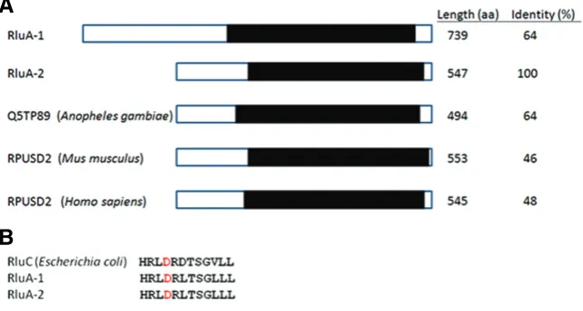
Fig. 2. Comparison of RluA and homologues.(A) Similarity among RluA-1, RluA-2, and the homologues in mosquito, mouse, and human. Q5TP89 is the accession number on Swiss-Prot. The filled boxes indicate the synthase domain. The identities of the synthase domain compared with that of RluA-2 are indicated. (B) Alignment of the residues in the active site cleft containing the catalytic aspartate (red).
spatially regulated during development.
Results
Insertion of E7-2-36 and evolutional conservation of RluA
synthases
The genomic sequence flanking the E7-2-36 P-element inser-tion site was identified by plasmid rescue and sequencing. We found the P-element inserts between two genes, RluA-1 and RluA-2 (Fig. 1), both of which encode synthases.
Sequence analyses revealed that Drosophila RluA-1 and RluA-2 contain the synthase domain (InterPro IPR006225), which is found in the RluC and RluD synthases in E. coli. RluA-1 and RluA-2 are highly similar in the synthase domain, which is evolutionarily conserved among RluA synthases (Fig. 2A). In addition, the synthase domain contains the catalytic aspartate (Fig. 2B), which suggests that RluA-1 and RluA-2 function in the formation. On the other hand, RluA-1 is longer than RluA-2. In RluA-1, the N-terminal sequence of amino acids 1–132 exhibits no functionally conserved domain and is absent in RluA-2.
Se-quence analyses revealed that RluA-1 and RluA-2 have one distinct homologue, respectively, in all Drosophila species se-quenced. However, in each of the other sequenced arthropods and mammals, only one gene homologous to RluA-1 and RluA-2 is found. The homologue is more similar to 2 than to RluA-1 because of the lack of a region similar to the RluA-RluA-1 N-terminus (Fig. 2). Thus, RluA-2 seems to be conserved across phyla, whereas RluA-1 may be produced by gene duplication only present in Drosophila.
Expression of RluA-1 mRNA
The insertion site of the P-element was 30 bp upstream of the RluA-1 transcription start site, suggesting that RluA-1 may be the candidate gene expressed in MD neurons. We performed in situ hybridization to analyze the expression of RluA-1 mRNA during embryogenesis. At stage 14, RluA-1 mRNA was expressed in the cells that appear iteratively in peripheral tissues (Fig. 3A). The signal of RluA-1 mRNA was also detected in the dorsal pharyn-geal musculature (Fig. 3B). At stage 16–17, RluA-1 mRNA was expressed in the cells that distribute as PNS neurons (Fig. 3C and 4C). In addition, RluA-1 mRNA was expressed in the dorsal vessel (Fig. 3D). No RluA-1 mRNA was detected in the embryonic central nervous system (CNS). RluA-2 mRNA was expressed ubiquitously (data not shown).
Comparison of the distribution of multiple dendritic (MD) neurons and RluA-1 expression
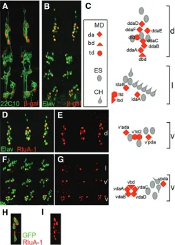
MD neurons are divided into three classes, da, td, and bd, according to the nature of their dendrites: da neurons exhibit large dendritic arbors, td neurons’ dendrites are associated with the trachea, and bd neurons have bipolar dendrites (Brewster and Bodmer, 1995). When all PNS and MD neurons were labeled using pan-neuronal markers and E7-2-36, respectively, the loca-tion of MD neurons among PNS neurons were observed (Fig. 4A– C). We generated RluA-1 antibody and examined its distribution in embryos. To avoid cross-reaction with RluA-2, we chose the 20–37 amino acids, which are not homologous to RluA-2, as the antigen. Immunocytochemistry studies using anti-RluA-1 and anti-Elav, which recognizes all neuronal nuclei, revealed that RluA-1 was ex-pressed in the subsets of PNS neurons (Fig. 4 D–G). The colocalization of their signals sug-gested that RluA-1 functions in the nuclei, where eukaryotic rRNA formations take place (Maden, 1990). The distribution of the RluA-1 expressing cells suggests that RluA-1 is ex-pressed in MD neurons.
Confirmation of the RluA-1 expression in MD neurons
We examined whether RluA-1 is indeed ex-pressed in MD neurons. In wild-type embryos, the dorsal part in one abdominal hemisegment consists of 12 ES and MD neurons (Fig. 4C). The scB57 allele lacks the proneural genes of the achaete-scute complex, which are required for ES and some MD neuron formation (Lindsley, 1992). Therefore, in scB57 embryos, only two MD neurons remain in the dorsal abdominal
Fig. 4. RluA-1 expression in multiple dendritic neurons. (A,B) MD neurons in stage 16 E7-2-36 embryos were marked by -galactosidase (red). (A) The MAb22C10 antibody (green) marks the morphology of PNS neurons. (B) Anti-Elav (green) is a pan-neuronal nuclear marker. (C) Schematic diagram of an abdominal hemisegment of the Drosophila
embryonic PNS. MD neurons are in red, ES (oval), and CH (drop shape) neurons are in gray. da, td, and bd neurons are indicated by diamonds, circles, and triangles, respectively. MD neurons are indicated according to the nomenclature described (Brewster and Bodmer, 1995, Grueber et al., 2002). (D, E, F, G) Stage 16 embryos stained with anti-Elav (green) and anti-RluA-1 (red). d, dorsal; l, lateral; v, ventral. (H,I) In stage 16
109(2)80>mCD8::GFP embryos, RluA-1 was expressed in the GFP-labeled MD neurons. Shown is the dorsal group.
hemisegment (Huang et al., 2000). We found that RluA-1 mRNA was expressed in two cells in the dorsal abdominal hemisegment of scB57 (Fig. 3E), suggesting that RluA-1 is expressed in MD neurons. In addition, we examined RluA-1 expression in the fly whose MD neurons were labeled by the MD neuron-specific marker GAL4 109(2)80 (Gao et al., 1999). This GAL4 line starts driving transgene expression at embryonic stage 15 (Sugimura et al., 2004). In stage 16 embryos bearing GAL4 109(2)80 and UAS-mCD8::GFP, most MD neurons are labeled by the membrane-tethered green fluorescent protein (GFP) (Fig. 4H). Later on, at stage 17 all MD neurons are recognized by GFP. Because the RluA-1 antibody cannot label MD neurons after embryonic stage 16, we examined RluA-1 expression in the stage 16 embryos. In the embryos we found that RluA-1 was expressed in the GFP-positive cells (Fig. 4 H–I), confirming the expression of RluA-1 in MD neurons.
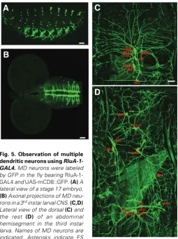
Observing MD neurons using an enhancer of RluA-1 One way to ensure that the RluA-1-expressing cells are MD neurons is to examine whether the cells exhibit multiple dendrites. We investigated the enhancer of RluA-1 and used the membrane-tethered GFP, mCD8::GFP (Lee and Luo, 1999), to label cellular morphology. If RluA-1 enhancer could drive the GFP expression in MD neurons, we would observe their dendritic arbors. Indeed, we found that the 3 kb genomic fragment including upstream sequences of RluA-1 (Fig. 1) was such an enhancer. We used the 3 kb fragment to generate 1-GAL4. In the fly carrying RluA-1-GAL4 and UAS-mCD8::GFP (RluA-RluA-1-GAL4>mCD8::GFP), some MD neurons began to be labeled by GFP at embryonic stage 16. At stage 17, all MD neurons were GFP-positive (Fig. 5A). The dendritic arbors of MD neurons were clearly seen at the third instar larvae (Fig. 5 C–D). By comparing the dendritic arbor
with that of the da neurons (Grueber et al., 2002), we identified all da neurons. db and td neurons were also recognized by the bipolar and trachea-associated dendrites, respectively. In each abdominal hemisegment, we detected all MD except ltd and lbd neurons. ltd and lbd neurons were observed occasionally possi-bly because they locate deeply in the body and exhibit low levels of GFP (data not shown). In addition, GFP signals were present in the cells that were negative for anti-RluA-1 in stage 16 em-bryos, such as v’td2 neurons, and subsets of ES neurons and muscles (Fig. 5, 4). In these cells, RluA-1 may be transcribed in a low level or later in embryogenesis, or only at larval stages. Consistently, RluA-1 mRNA appeared to be expressed in the
B
C
D
E
A
G
B
C
D
E
F
H
I
A
Fig. 3. RluA-1 mRNA expression in embryos. Whole mount in situ hybrid-ization of embryos using RluA-1 RNA probe. At stage 14, RluA-1 mRNA ap-peared in PNS-like cells (A) and the dor-sal pharyngeal musculature (red arrow) (B). At stage 17, RluA-1 mRNA is likely expressed in PNS neurons (C) and in the dorsal vessel (black arrow) (D). The parentheses in (C,D) indicate the cells similar to the ventral and dorsal neurons in Fig. 4C, respectively. Arrowheads indicate the cells similar to the dorsal most ES neurons in Fig. 4C. (E) Dorsal abdominal hemisegments of scB57embryos. All except (D) are in lateral view. Dorsal is up, anterior is
B
C
D
A
dorsal most ES neurons at stage 17 (Fig. 3D). In the larval CNS of RluA-1-GAL4>mCD8::GFP, axonal projections of MD neurons were observed by GFP in the ventral nerve cord (VNC) (Fig. 5B). Although there is a low expression of -galactosidase in the VNC of E7-2-36 (data not shown), RluA-1-GAL4 did not drive GFP in any cell body in the CNS and there is no expression in the CNS with the RluA-1 antibody, indicating that RluA-1 is not expressed in the CNS.
In summary, our data indicate that RluA-1 is expressed in MD neurons. Therefore, although synthases appear to function ubiquitously, the Drosophila RluA-1 expression is spatially con-trolled during development.
Materials and Methods
Fly stocks and transgenic flies
Flies were raised on a standard cornmeal–yeast–agar medium at 25C. The following flies were used: E7-2-36 (Brewster and Bodmer, 1995); UAS-mCD8::GFP (Lee and Luo, 1999); scB57 (Bloomington Stock
Center); Gal4 109(2)80 (Gao et al., 1999). To make RluA-1-GAL4, the 3 kb enhancer of the fly and the GAL4 gene of Saccharomyces cerevisiae were amplified from their genomic DNA by polymerase chain reaction. Primer sequences are available upon request. We removed the UAS sequence from the pUAST vector and used this modified vector as a backbone. The 3 kb enhancer and the GAL4 gene were thus cloned into the modified pUAST vector. Transgenic flies were generated by P-element mediated germ-line transformation techniques (Spradling, 1986).
In situ hybridization
The procedure used for performing in situ RNA hybridization of Drosophila embryos has been described (Tautz and Pfeifle, 1989). RluA-1 and RluA-2 riboprobes were made by using the EST clones RE33RluA-1RluA-13 and LD40728 (Berkeley Drosophila Genome Project), respectively. Em-bryos were visualized with a Zeiss Axioskop2 plus microscope under Normarski optics.
Immunocytochemistry
The rabbit polyclonal antibody against the RluA-1 peptide of 20–37 amino acids was generated (Quality Controlled Biochemicals). Standard embryo fixing and staining protocols (Patel, 1994) were used. The primary antibodies were: mouse MAb22C10 (1:250) and rat anti-Elav (1:100) (Developmental Studies Hybridoma Bank), rabbit anti--galactosidase (1:100) (Cappel), rabbit anti-RluA-1 (1:40). Images were collected on a Zeiss LSM 510 confocal microscope and merged in Adobe Photoshop.
Acknowledgements
We thank the Bloomington Stock Center for fly stocks and the Devel-opmental Studies Hybridoma Bank for antibodies. This work was sup-ported by grants from Academia Sinica and the National Science Council of Taiwan.
References
BREWSTER, R. and BODMER, R. (1995). Origin and specification of type II sensory neurons in Drosophila. Development 121: 2923-2936.
GAO, F.B., BRENMAN, J.E., JAN, L.Y. and JAN, Y.N. (1999). Genes regulating dendritic outgrowth, branching, and routing in Drosophila. Genes Dev 13: 2549-2561.
GROSSHANS, H., LECOINTE, F., GROSJEAN, H., HURT, E. and SIMOS, G. (2001). Pus1p-dependent tRNA pseudouridinylation becomes essential when tRNA biogenesis is compromised in yeast. J Biol Chem 276: 46333-46339. GRUEBER, W.B., JAN, L.Y. and JAN, Y.N. (2002). Tiling of the Drosophila
epidermis by multidendritic sensory neurons. Development 129: 2867-2878. GUTGSELL, N.S., DEUTSCHER, M.P. and OFENGAND, J. (2005). The
pseudouridine synthase RluD is required for normal ribosome assembly and function in Escherichia coli. RNA 11: 1141-1152.
HAMMA, T. and FERRE-D’AMARE, A.R. (2006). Pseudouridine synthases. Chem Biol 13: 1125-1135.
HEISS, N.S., KNIGHT, S.W., VULLIAMY, T.J., KLAUCK, S.M., WIEMANN, S., MASON, P.J., POUSTKA, A. and DOKAL, I. (1998). X-linked dyskeratosis congenita is caused by mutations in a highly conserved gene with putative nucleolar functions. Nat Genet 19: 32-38.
HUANG, M.L., HSU, C.H. and CHIEN, C.T. (2000). The proneural gene amos promotes multiple dendritic neuron formation in the Drosophila peripheral nervous system. Neuron 25: 57-67.
JAN, Y.N. and JAN, L.Y. (1993). Functional gene cassettes in development. Proc Natl Acad Sci USA 90: 8305-8307.
JAN, Y.N. and JAN, L.Y. (1994). Genetic control of cell fate specification in
Drosophila peripheral nervous system. Annu Rev Genet 28: 373-393. KAYA, Y. and OFENGAND, J. (2003). A novel unanticipated type of pseudouridine
synthase with homologs in bacteria, archaea, and eukarya. RNA 9: 711-721. KOONIN, E.V. (1996). Pseudouridine synthases: four families of enzymes
contain-ing a putative uridine-bindcontain-ing motif also conserved in dUTPases and dCTP deaminases. Nucleic Acids Res 24: 2411-2415.
LEE, T. and LUO, L. (1999). Mosaic analysis with a repressible cell marker for studies of gene function in neuronal morphogenesis. Neuron 22: 451-461. LINDSLEY, D.L., AND ZIMM, G.G. (1992). The genome of Drosophila
Melano-gaster. Academic press, San Diego.
MADEN, B.E. (1990). The numerous modified nucleotides in eukaryotic ribosomal RNA. Prog Nucleic Acid Res Mol Biol 39: 241-303.
PATEL, N.H.(1994). Imaging neuronal subsets and other cell types in whole-mount
Drosophila embryos and larvae using antibody probes. In Practical Uses in Cell
Fig. 5. Observation of multiple dendritic neurons using RluA-1-GAL4. MD neurons were labeled by GFP in the fly bearing RluA-1-GAL4 and UAS-mCD8::GFP. (A) A lateral view of a stage 17 embryo. (B)Axonal projections of MD neu-rons in a 3rd instar larval CNS. (C,D)
Lateral view of the dorsal (C) and the rest (D) of an abdominal hemisegment in the third instar larva. Names of MD neurons are indicated. Asterisks indicate ES
and Molecular Biology., (ed. E.A. Fyrberg, L S.B.G.A.). Academic Press, San Diego, California, pp.446-485.
ROZENSKI, J., CRAIN, P.F. and MCCLOSKEY, J.A. (1999). The RNA Modification Database: 1999 update. Nucleic Acids Res 27: 196-197.
SPRADLING, A.C. (1986). P element-mediated transformation in Drosophila: a practical approach. IRL, Oxford.
SUGIMURA, K., SATOH, D., ESTES, P., CREWS, S. and UEMURA, T. (2004). Development of morphological diversity of dendrites in Drosophila by the BTB-zinc finger protein abrupt. Neuron 43: 809-822.
TAUTZ, D. and PFEIFLE, C. (1989). A non-radioactive in situ hybridization method for the localization of specific RNAs in Drosophila embryos reveals translational control of the segmentation gene hunchback. Chromosoma 98: 81-85.
Further Related Reading, published previously in the
Int. J. Dev. Biol.
Mammalian hibernation: differential gene expression and novel application of epigenetic controls
Pier Morin Jr. and Kenneth B. Storey Int. J. Dev. Biol. (2009) 53: 433-442
Oyster sperm bindin is a combinatorial fucose lectin with remarkable intra-species diversity
Stevan A. Springer, Gary W. Moy, Daniel S. Friend, Willie J. Swanson and Victor D. Vacquier Int. J. Dev. Biol. (2008) 52: 759-768
Developmental expression of CagMdkb during gibel carp embryogenesis
Jun Yin, Jian-Hong Xia, Xin-Zheng Du, Jun Liu, Li Zhou, Yun-Han Hong and Jian-Fang Gui Int. J. Dev. Biol. (2007) 51: 761-769
Identification of a second Xenopus twisted gastrulation gene
Michael Oelgeschläger, Uyen Tran, Kristina Grubisic and Edward M De Robertis Int. J. Dev. Biol. (2004) 48: 57-61
Neurotrophic regulation of retinal ganglion cell synaptic connectivity: from axons and dendrites to synapses
Susana Cohen-Cory and Barbara Lom Int. J. Dev. Biol. (2004) 48: 947-956
Sequence and translation initiation properties of the xenopus TGFbeta5, PDGF-A, and PDGF-alpha receptor 5' untranslated regions
A W van der Velden, A Los, H O Voorma and A A Thomas Int. J. Dev. Biol. (2000) 44: 851-859
Nucleoskeleton and nucleo-cytoplasmic transport in oocytes and early development of Xenopus laevis
F Rudt, I Firmbach-Kraft, M Petersen, T Pieler and R Stick Int. J. Dev. Biol. (1996) 40: 273-278