Kidney regeneration in fish
THOMAS BATES
#,1, UTA NAUMANN
#,1, BEATE HOPPE
#,1and CHRISTOPH ENGLERT*
,1,2 1Leibniz Institute on Aging. Fritz Lipmann Institute (FLI) and2Institute of Biochemistry and Biophysics, Friedrich Schiller University Jena, Germany
ABSTRACT Age-related diseases, such as kidney diseases, are becoming more prevalent in aging societies. Currently, patients with reduced kidney function require dialysis or organ transplants. Those who suffer from kidney disease would benefit from regenerative therapies. Thus, one of the ultimate goals of regeneration research is to enhance an individual’s capacity of self-repairing dam-aged tissue; something that fish models can contribute towards. Kidney structures are conserved among vertebrates highlighting the opportunities for fish to act as human disease models. Here, different species can offer respective advantages. An understanding of the different modes of re-generation can help to visualize the differences in mammalian and fish regenerative capacity. The remarkable regenerative capacity of fish is well known, but kidney regeneration is an understud-ied area. The kinetics of kidney regeneration allows one to investigate early damage responses, as well as the initiation and completion of repair. Age-related reductions in regeneration are an additional societal problem; again an area where fish models can be of help. Age-matched experi-ments between varied vertebrate species will help us to learn from those that do or do not exhibit age-related phenotypes. The goal of such experiments is not only to outline important age-related factors and pathways, but, in addition, to see if age-related decreases in regenerative capacity can be reduced. Widening our knowledge of this very complex process will help to address many of the unanswered questions in the field.
KEY WORDS:
kidney, regeneration, fish, disease, aging
Introduction
The kidney is an indispensable organ that regulates metabolic waste removal and fluid balance. Mammalian kidneys have only limited proliferative and regenerative potential. Thus, kidney diseases can lead to life-threatening complications. In our aging society, the incidence of renal insufficiencies is predicted to increase (PHE, 2014) and highlights the need for new and efficient thera-peutic approaches. Modeling human diseases in other vertebrate species can contribute to a better understanding of the underlying mechanisms and support biomedical research.
The kidney is an organized labyrinth of tubes. Kidneys of adult humans filter approximately 180 liters of blood daily, with sub-sequent efficient reabsorption of the filtrate retrieving it into the circulation. Among vertebrates, the basic composition of the kidney is conserved. In particular, the structure and the development of the filtering units, the nephrons, show high similarities. Various fish models have been recognized as valuable systems to investigate
www.intjdevbiol.com
*Address correspondence to: Christoph Englert. Leibniz Institute on Aging – Fritz Lipmann Institute (FLI), Beutenbergstrasse 11, 07745 Jena, Germany.
Fax: +49 3641 656040. Tel: +49 3641 656042. E-mail: [email protected] - web: http://www.leibniz-fli.de/research/research-groups/englert - https://orcid.org/0000-0002-5931-3189
# These authors contributed equally to this work
Submitted: 8 December, 2017; Accepted: 12 December, 2017.
ISSN: Online 1696-3547, Print 0214-6282
© 2018 UPV/EHU Press Printed in Spain
Abbreviations used in this paper: AKI, acute kidney injury; CKD, chronic kidney disease.
renal function and development, but also to model renal diseases such as acute kidney injuries. This review will focus on models of kidney disease in fish and how these can impact future clinical and basic research.
Comparative kidney structure and function
kidney are named nephrons and are mainly organized in the renal cortex. They show a specific segmentation (Fig. 1) responsible for the filtration of the blood, with subsequent reabsorption of 99% of the filtered electrolytes and water, and 1% excretion into the urine (Zhuo and Li, 2013).
Each segment of the nephron is composed of different cell types. The most proximal part is the glomerulus that consists of a capillary loop bound by mesangial cells, enveloped in podocytes,
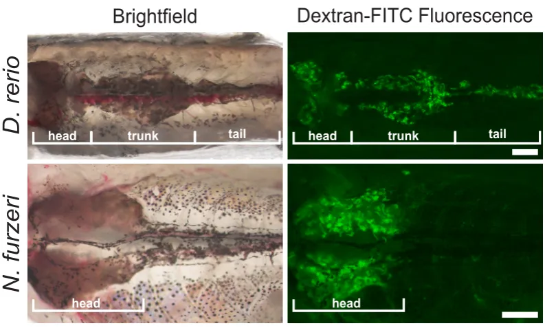
In both zebrafish (Danio rerio) and a distant relative the African Killifish (Nothobranchius furzeri) the kidney consists of a flattened structure located along the dorsal inner body wall. In Danio rerio there are morphologically separable head, trunk and tail kidney, in which nephrons can be detected. In contrast, in N. furzeri, there is only the head part of the kidney found, which is clearly enlarged compared to the zebrafish (Fig. 2) (Mccampbell et al., 2015; Sander and Davidson, 2014).
head
head
trunk tail
Brightfield
Dextran-FITC Fluorescence
D. rerio
N. furzeri
head trunk tail
head
Fig. 1. Organization of the mammalian kidney and nephron.(A) Fluorescent image of a two-month-old mouse kidney (Rosa26-flTomatofl-EGFP(B6);Pod-cre) where glomeruli are labeled in green and all other structures remain red. (B)
Diagrammatic representation of nephron compartments. G, glomerulus; PCT, proximal convoluted tubule (Segments 1 + 2); PST, proximal straight tubule; DTL, descending thin limb; ATL, ascending thin limb; TAL, thick ascending limb; MD, macula densa, DCT, distal convoluted tubule; CNT, connecting tubule. Scale bar in A, 1 mm. Panel (B) adapted from (Li and Wingert, 2013).
Fig. 2. Comparison of fish kidney structures. Adult fish were injected with a 40 kDa fluorescently labeled Dextran in order to visualize the proximal tubule. Signals are observed throughout the head-, trunk- and tail portions of the D. rerio kidney, while in N. furzeri, tubules are mainly detected in the head portion. Figure adapted from Hoppe et al. (2015).
and enclosed by Bowman’s capsule (Costantini and Kopan, 2010). Podocytes are specialized epithelial cells that possess a basket-like extension of cellular processes around the blood capillaries. The basement membrane between podocytes and capillary endothelial cells, together with specialized junctions between the podocyte processes form the blood filtration bar-rier. It allows passage of small molecules, ions and fluids, while retaining cells and macromolecules in the blood (Drummond and Davidson, 2010). The filtrate flows from the glomerular space into the joining nephron tubule. The tubule is composed as a pipe of mainly epithelial cells surrounded by a basement membrane (Li and Wingert, 2013) and is divided into multiple specialized segments. The epithelial cell populations that make up each tubule segment have a distinct cellular morphology and are characterized by an unique sets of solute transport-ers to perform discrete roles in modifying the glomerular filtrate (Davidson, 2008). In mammalian kidneys the ‘Loop of Henle’ (DTL and ATL see Fig. 1) is a specialized structure for concentration of urine.
When comparing the kidneys of mammals and fish species, the overall organization differs. In mammals, the kidneys are two bean-shaped organs found below the ribcage on either side of the spinal column. They display a high grade of orga-nization, with aligned nephrons situated in defined regions of medulla and cortex, while fish kidneys are less organized.
G
ATL CNT
DCT
DTL MD
PCT (S1)
PCT (S2)
PST (S3) TAL
Within all parts of the kidney the nephrons are homogeneously distributed, randomly oriented and interfused into hematopoietic tissue which forms a matrix around the nephrons. Although the kidney architecture varies between vertebrates, the specific seg-mentation of the nephrons is broadly conserved (Mccampbell and Wingert, 2014; Wingert and Davidson, 2008). As in mammals, zebrafish nephrons consist of a glomerulus, proximal and distal tubule segments and the collecting duct (Mccampbell et al., 2015; Mccampbell and Wingert, 2014). However, zebrafish nephrons do not develop a loop of Henle but show a branching of the distal tubules (Mccampbell and Wingert, 2014). Furthermore, special-ized cell types and orthologues of tubular segment marker genes are highly conserved between the species (Wingert and David-son, 2008). Based on these similarities in nephron composition, fish species have emerged as favorable models to study human kidney diseases.
Types of regeneration
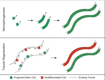
Vertebrate kidneys show specific regenerative responses after nephron damage. The first type of response to restore nephron functionality has been designated classically as “renal regenera-tion” or “tubular regeneraregenera-tion” (Mccampbell et al., 2015; Reim-schuessel, 2001). It describes a local self-repair of nephrons with intact basement membrane after a limited injury (Fig. 3). In this process, an initial phase of cell death and detachment of tubular cells from the basement membrane is followed by an initiation of cellular proliferation of either dedifferentiated cells or resident stem/
patterns as observed during nephrogenesis in developing vertebrate kidneys, eventually forming a new functional nephron (Davidson, 2014; Reimschuessel, 2001).
The regenerative capacity of the mammalian kidney
When trying to better understand human disease, animal models have proven incredibly informative. Understanding the onset and subsequent response to damage and repair in an organismal set-ting is necessary for basic research. Mammalian models of acute kidney injury (AKI) have been performed in murine models (Sho-skes et al., 1990; Wei and Dong, 2012) and have included surgical approaches to induce ischemia/reperfusion injuries (Duffield et al., 2006) and injection of nephrotoxins such as Cisplatin (Ramesh and Reeves, 2003). In addition, a more recent publication describes a renal cryo-injury model aimed at uncovering endogenous damage and repair processes (Abdulmahdi et al., 2015).
Mammalian kidneys have developed regenerative strategies to restore their integrity after damage, but also to manage cell turn-over to ensure continuous nephron maintenance. For mammals, it has never been reported that their kidneys have the capability to form new nephrons via “neo-nephrogenesis”, once nephrogenesis is completed in the embryo or neonate (Fine, 1986; Hartman et
al., 2007; Larsson et al., 1980). Instead, the answer to kidney
damage is a combination of “tubular regeneration” responses, to repair the nephric epithelium along the intact basement membrane (Humphreys et al., 2008), and “compensatory renal hypertrophy” to substitute for the loss of not repairable nephrons (Hayslett,
Tubular Regeneration
Neonephrogenesis
Progenitor/Stem Cell Dedifferentiated Cell Existing Tubule
Fig. 3. Visualization of kidney regeneration models. Neonephrogenesis is defined as the de novo
formation of tubules from a resident progenitor/stem cell. Tubular regeneration describes a process where either existing tubular cells dedifferentiate or resident progenitor/stem cells within existing tubules repopulate damaged tissue.
progenitor cells and migration along the tubule, leading to a repopulation of the existing nephron (Davidson, 2011; Reimschuessel, 2001).
A second strategy of the vertebrate kidney to restore nephron function is called “compensatory renal hypertro-phy”, meaning an enlargement of kidney size by cellular proliferation (Fine, 1986; Kaufman et al., 1975; Reimschuessel, 2001). After injury, surviving nephrons enhance the excretion of metabolic waste by increasing their single nephron filtration rate and undergo morphologi-cal changes, such as expanding their glomeruli diameter and proximal tubule cell volume (Davidson, 2011). This re-generative strategy mimics the enlarge-ment of nephrons seen during postnatal growth in mammals, suggesting that both processes may be induced by a common mechanism (Davidson, 2011). The last renal repair response des-ignated as “neo-nephrogenesis” is a process of generating entire nephrons
de novo after injury (Davidson, 2011,
1979; Mccrory, 1972).
Both processes require proliferative cells. Although the source of cells has been demonstrated to be nephron-intrinsic (Humphreys
et al., 2008), their identity remains still controversial. There is given
evidence, that reparative programs of the nephric tubules depend on surviving tubular cells that dedifferentiate and undergo epithelial to mesenchymal transition to become proliferative and migratory (Bonventre and Yang, 2011). Consistent with this, it was shown that the renal epithelium is not quiescent. Many tubular epithelial cells in uninjured nephrons are arrested in the G1 phase, waiting for a rapid recruitment into the cell cycle and allowing a renal repopulation after injury (Vogetseder et al., 2008). However, this mechanism does not exclude the coexistence of specialized stem or progenitor cells within the nephron. Several candidate populations have been proposed. Researchers have identified renal progeni-tor cells scattered along the tubular epithelium and located within the Bowman’s capsule of the glomerulus, that are suggested to give rise to tubular and glomerular cell types (Benigni et al., 2010; Bussolati et al., 2005; Hopkins et al., 2009). In addition, Lazzeri et
al., has demonstrated existence of bipotent progenitor populations
located at the urinary pole of the glomerulus that can give rise to both tubular epithelium as well as glomerular podocytes (Lazzeri
et al., 2010; Ronconi et al., 2009).
Taken together, there are several indications that mammalian nephrons harbor cells with a robust proliferation capacity sufficient to perform epithelial replacement. While “tubular regeneration” and “compensatory renal hypertrophy” processes enable for a partial rescue from nephron damage, kidney regeneration remains a limited process in mammals and can even lead to insufficiencies in kidney function. A continuous loss of nephrons as well as pro-longed hyperfiltration caused by extended hypertrophy eventually lead to glomerular injury, toxic proteinuria and kidney sclerosis (Brenner, 1985).
Kidney diseases
Humans are affected by a wide spectrum of renal diseases, ranging from minor dysfunctions to kidney failure, which requires organ replacement therapies by dialysis or kidney transplanta-tion. In general, a sudden damage of the kidney as induced by, e.g. ischemia reperfusion injury or nephrotoxic agents is termed “acute kidney injury” (AKI). Slow progressive loss of tubules and ongoing fibrosis can lead to “chronic kidney disease” (CKD). AKI is a life-threatening disease, characterized by an increase in serum creatinine levels and a decrease in urine volume. Due to secondary complications such as pulmonary edema, high potassium levels in the blood (hyperkalemia) and metabolic acidosis, AKI has a world-wide mortality rate of between 30% and 50% (Yaklin, 2011). In the United States of America the total number of deaths from AKI doubled from 18,000 to 39,000 between 2000 and 2009 (Hsu
et al., 2013). As such, AKI is a large socio-economical problem in
our increasingly populated and aging society.
Morphologically, AKI manifests in different, overlapping phases starting with the appearance of dedifferentiated cells at the injury site, followed by morphological changes of the cells in the proxi-mal tubule. As a consequence, cells detach from the basement membrane, undergo apoptosis or necrosis (in severe and rapid cases) and can be seen as cellular casts in the lumen causing tubular destruction downstream of the lesion site (Mccampbell et
al., 2014). Many molecular pathways are involved in the ensuing
damage response; such as activation of transcription factors like hypoxia induced factor (HIF) and NF-kB, Toll-like receptor (TLR) signaling and inflammatory processes (Basile, 2007; Eltzschig and Carmeliet, 2011; Havasi and Borkan, 2011; Leemans et al., 2005). When loss of renal function proceeds over a long period, CKD develops, which can lead to end-stage renal disease (Chawla and Kimmel, 2012). The complex pathophysiology of CKD by progressive scarring is usually too severe to be repaired. Due to chronic lesions, remaining nephrons try to increase functionally by intra-glomerular hypertension and hyper-filtration (Benigni et
al., 2010). This leads to further renal damage, such as excessive
accumulation of ultra-filtered proteins in the Bowman’s space and the lumen of the tubules. As a consequence, mitochondrial oxidative stress, expression of epithelial growth factor receptors (EGFR) and other factors can lead to glomerulosclerosis (Bollee
et al., 2011; Daehn et al., 2014). It is thought that one of the main
causes of progression from AKI to CKD is an increase of reactive oxygen species (ROS). ROS are formed as a consequence of increased oxygen consumption in the proximal tubules, because of increasing levels of nephrotoxic indoxyl sulfate (Barreto et al., 2009; Palm et al., 2010). The progression from AKI to CKD will be a very important aspect of future research. However, this review will concentrate on models of AKI in fish species, both at larval and adult stages.
The remarkable regenerative capacities of fish
Compared to kidney regeneration in mammals, adult fish kidneys have a different strategy to maintain and restore their functionality. It has been demonstrated that different fish species like goldfish (Reimschuessel et al., 1990b), trout (Reimschuessel et al., 1993), catfish (Cormier et al., 1995), toadfish (Brown and Reimschuessel, 1998), medaka (Watanabe et al., 2009) and zebrafish (Diep et al., 2011) have a remarkable regenerative ability to recover from an acute kidney injury. In response to nephrotoxins they undergo a two-phase repair mechanism (Reimschuessel et al., 1990c). Initially, fish kidneys perform a repair of existing nephric tubules via “tubular regeneration”, leading to the re-population of the basement mem-brane with epithelial cells consistent with what has been found in mammalian models (Augusto et al., 1996; Reimschuessel, 2001; Reimschuessel et al., 1990c, 1993). The origin of proliferating progenitor cells in this response has not yet been identified, but it is suggested that underlying mechanisms are similar to mammals (Kusaba and Humphreys, 2014).
The second repair response enables fish to compensate ef-ficiently for the loss of nephrons after an acute injury. A major difference between fish and mammalian kidneys is their potential for ongoing nephrogenesis in adults (Reimschuessel, 2001). Fish continue to add nephrons throughout their lifespan and have the abil-ity to significantly increase nephrogenesis after an injury (Augusto
et al., 1996; Brown and Reimschuessel, 1998; Elger et al., 2003;
Reimschuessel, 2001; Reimschuessel et al., 1990c; Watanabe
et al., 2009; Zhou et al., 2010). The capacity to generate entire
nephrons de novo has been traced to nephrogenic aggregates that appear during kidney regeneration and give rise to new nephrons in a process that recapitulates developmental nephrogenesis (Diep
et al., 2011; Zhou et al., 2010) but also follows the same pattern of
mammalian kidneys ((Reimschuessel, 2001). A major question that remains is about the origin and identity of cells that contribute to these cell clusters. Whether they arise from stem cells, progenitors cells or terminally differentiated cells, and if they are of intra- or extra-tubular origin has to be investigated.
Among animals that have been used for developmental and regenerative studies of the kidney, the zebrafish has become a favorable model organism (Mccampbell and Wingert, 2014). It has been shown that zebrafish embryos, larvae and adults can model kidney diseases of humans, including AKI caused by tubular or glomerular damage (Mccampbell and Wingert, 2014; Zhou and Hildebrandt, 2012), but also other common nephropathies like polycystic kidney disease (Drummond et al., 1998). This is based on the high conservation of nephron structures, cell types, devel-opment and functionality between mammals and fish (Drummond
et al., 1998; Sander and Davidson, 2014; Wingert and Davidson,
2008; Wingert et al., 2007). In addition zebrafish harbor an amazing regenerative potential. This includes the ability to restore nephron epithelia and to form entirely new nephrons. Zebrafish show de
novo formation of kidney structures after an injury, a major
differ-ence to mammals that can complement research in traditional AKI models such as the mouse and rat (Mccampbell and Wingert, 2014; Romagnani et al., 2013).
Zebrafish is a well-established genetic model with a steady increase of transgenic reporter lines and CRISPR/Cas9-mediated mutants. This allows studying of specific cell types and fates (Zhou and Hildebrandt, 2012) and supports transplantation assays to prove stem-cell capabilities (Diep et al., 2011). Analysis of kidney regeneration is much improved by newly established methods to determine the structure and functionality of nephrons (Mccampbell
et al., 2015). This comprises standard histological stains, such
as hematoxylin and eosin but also complex immunological stains with zebrafish specific antibodies, both on tissue section and in whole-mount. Furthermore, numerous solute transporters and renal transcription factors have been identified and assigned to in-dividual segments of the zebrafish nephron (Wingert and Davidson, 2008; Wingert et al., 2007). One of these, the sodium-dependent phosphate transporter slc20a1a, is specifically found in the proxi-mal tubule epithelium of zebrafish and mamproxi-malian kidneys, and
slc20a1a transcripts are transiently abrogated after gentamicin
administration (Diep et al., 2011). Similarly, alkaline phosphatase reactivity has been shown to be a specific feature of the luminal surface of proximal tubules in nephrons, meaning one can follow structural changes in this segment (Mccampbell et al., 2014, 2015). To visualize kidney functionality in the zebrafish, injections of 40
kDa Fluorescein-isothiocyanate-labeled dextran (Dextran-FITC) is a significant method. The fluorescent sugar is selectively reabsorbed in the proximal parts of the tubules. Functional destruction of this segment is indicated by an inability to reabsorb Dextran-FITC (Diep et al., 2011). Taken together, zebrafish represents a unique tool to investigate temporal and spatial progress of regeneration.
Modeling acute kidney injury in fish
In 1990, hexachlorobutadiene-induced nephrogenesis was used as a model of recovery in adult goldfish (Reimschuessel et
al., 1990a), with newly formed nephrons marked as basophilic
structures in H&E staining. Gentamicin, another injury model, is a nephrotoxic aminoglycoside (AG) antibiotic, which affects the proximal parts of the tubule leading to a dose-dependent apop-tosis (Laurent et al., 1983) or necrosis of tubular epithelial cells (Edwards et al., 2007). Traditionally, AGs are used in infections with gram-negative bacteria and bacterial endocarditis (Chen and Kaye, 2009) and are, despite their side effects, the most effective treatment against germs insensitive to other antibiotics. Classical clinical manifestations of AG toxicity are, among others, proteinuria, increase in plasma creatinine and electrolyte alterations (Parsons
et al., 1997), signs that are also observed during AKI.
The first published use of gentamicin’s efficacy as a nephrotoxin in adult fish was performed in goldfish (Reimschuessel and Wil-liams, 1995). Similarly to the earlier work with hexachlorobutadiene, gentamicin treatment induced an increase in developing tubules in adult fish. Gentamicin has also been used in larval zebrafish experiments (Cianciolo Cosentino et al., 2010; Hentschel et al., 2005). Injection into the larval bloodstream of zebrafish led to an increased occurrence of edema. In addition, with the application of a novel rhodamine-dextran clearance assay, it was shown that gentamicin reduced larval glomerular function (Hentschel et al., 2005). This approach also became the basis for a novel small molecule screen, searching for substances that could improve regeneration (Cianciolo Cosentino et al., 2013). It was not until 2010 that the regenerative response of the adult zebrafish to Gentamicin-induced kidney damage was described (Zhou et al., 2010). Utilizing two important zebrafish kidney markers, wt1b (developing tubules) and podocin (developed glomerulus), the authors showed that the number of tubules is positively correlated with body weight and age suggesting that neonephrogenesis is a life-long process in zebrafish (Zhou et al., 2010). In addition to the descriptive aspects of this work, functional AKI modeling with gentamicin in the wt1b and podocin reporter lines allowed certain populations of kidney cells to be investigated during adult kidney
Control
N.furzeri
Damaged
Recovered
D.rerio
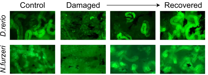
Fig. 4. Kidney regenerationki-netics in fish. Example pictures of Dextran uptake in control, dam-aged and recovering animals. One can see that uptake is lost upon damage and recovered following regeneration. Scale bar, 1 mm. Part of the figure is adapted from Hoppe
regeneration. With significantly higher numbers of wt1b-positive nephrons seen throughout regeneration, wt1b became an important marker of tubular regeneration in zebrafish.
With the gentamicin protocol established, a more mechanistic study uncovered further aspects of kidney regeneration in the zebrafish (Diep et al., 2011). With further data suggesting that new nephrons are created throughout life, the authors utilized a combination of methods to better describe in detail, the processes of regeneration. In addition, the presence of nephron progenitors was explored with transplantation studies. One very helpful tool was introduced in this publication; that the selective uptake of a 40kDa tagged sugar can be used as readout of regeneration (Fig. 4). Following this breakthrough discovery, additional publications have increased the tool kit of those investigating kidney regen-eration in zebrafish. In 2014, an atlas of markers was published (Mccampbell et al., 2014). Covering many aspects of histological staining of the adult kidney during regeneration, working markers for proliferation and cell death were combined with markers of both the proximal and distal tubules. In this readout of regeneration, an
in situ-based quantification of tubular recovery allows
research-ers to look for differences in regeneration at different time points and under different experimental conditions (Mccampbell et al., 2014). Another publication, which will contribute to the success of kidney regeneration experiments, was published in 2015. The work concentrates on how one can be sure of inducing AKI in the adult animal, without having to sacrifice the animal (Kamei et al., 2015). It is now clear that following AKI, adult zebrafish excrete extra waste into the water. Due to death of the kidney epithelial cells, tubular renal casts are formed. These accumulate in the collecting ducts of the fish and are passed into the water by the fish. This screening possibility avoids the further use of non-injured fish in an experiment, ensuring the use of animals with high-level, non-lethal AKI (Kamei et al., 2015).
Kinetics of regeneration
Shortly after application of the nephrotoxin gentamicin, epithelial cell death occurs in the proximal tubule segment of the zebrafish nephron (Verghese et al., 2008), followed at one-day post injury (dpi) by an accumulation of intraluminal cellular debris and a disorganized proximal tubular epithelium (Mccampbell et al., 2015; Zhou et al., 2010). Cellular and functional disruption results in a failure to take up Dextran-FITC, that persists until 3 dpi (Diep et al., 2011). Around 4 dpi, partial recovery of nephron function via “tubular regenera-tion” can be observed by appearance of few Dextran-FITC labeled nephrons (Diep et al., 2011). This is caused by a repopulation of the basement membrane probably by proliferating tubular cells, a process similar to mammalian nephron regeneration (Diep et al., 2011). At 5 dpi, cells within the proximal tubules start to display an epithelial organization as a single layer of cells and a restored luminal opening, leading to a complete recovery one week after gentamicin induced AKI (Mccampbell et al., 2015).
“Neo-nephrogenesis” occurs in a partially overlapping time frame in order to replace severely injured nephrons that cannot be repaired (Mccampbell et al., 2015). Beginning at approximately 5 dpi a small number of nephrogenic cellular aggregates begin to form by merging of nephron progenitors (Diep et al., 2011). By 7 dpi, the number of aggregates increases and begins to show signs of proliferation and differentiation (Mccampbell et al., 2015). The aggregates undergo a mesenchymal to epithelial transition to form
an epithelial ball also referred to as renal vesicle (Diep et al., 2011; Sander and Davidson, 2014). Eventually, the vesicles elongate showing comma- and S-shaped body stages and form a lumen within the second week after injury (Sander and Davidson, 2014). The immature tubules fuse into existing nephrons and mature in size and characteristics (Mccampbell et al., 2015; Sander and Davidson, 2014). At about 14 dpi the nascent nephrons exhibit functional brush borders, shown by positive Dextran-FITC uptake, indicating proximal tubule character (Mccampbell et al., 2015). The overall structure of the kidney is close to undamaged tissue, although immature nephrons can still be detected. At 21 dpi tissue staining reveals an absence of nephrogenic aggregates, suggest-ing that the functionality of the regenerated kidney in zebrafish is restored between 2 and 3 weeks following damage (Diep et al., 2011; Mccampbell et al., 2015).
The nature of progenitor/stem cells for kidney regeneration
The de novo formation of new nephrons is a feature, unique to fish, amphibians and reptiles. In zebrafish small cellular aggregates have been identified as the source of new nephrons. They are formed by the coalescence of multiple progenitor cells (Diep et al., 2011; Zhou et al., 2010). In goldfish, similar findings were made, identifying basophilic clusters of cells as source of new nephrons after kidney injury (Salice et al., 2001). These clusters display an enlargement, lumen formation and maturation of nephrons. Similar observations have been made in medaka as well, where first repair processes of the damaged nephrons took place, fol-lowed by development of new nephrons (Watanabe et al., 2009). Diep et al., (2011) addressed the question about the origin and stemness of the cell clusters by analyzing a transgenic zebrafish reporter line for lhx1a, a transcription factor active in “pre-tubular” aggregates during mammalian nephrogenesis. Studies of kidneys in this reporter line revealed three distinctive cell populations: (I) single cells with mesenchymal morphology that are suggested to be migratory, (II) homogeneous aggregates comprising few lhx1a expressing cells, and (III) renal vesicle-like bodies. Interestingly, in transplantation assays, only the cellular aggregates could engraft in recipient fish and generate new functional nephrons, suggesting that these comprise nephron progenitors that are long-lived and possess extensive proliferative potential, consistent with stem-cell activity (Diep et al., 2011). Because single cells failed to engraft, the question about the identity of the individual cells that form these nephrogenic clusters, and signals that regulates activation of lhx1a cells remains. It is also not clear whether lhx1a cells in zebrafish display an equivalent to mammalian nephron progeni-tors (Diep et al., 2011; Kobayashi et al., 2005).
that systemic signals play a role (Elger, 2003). A partial unilateral nephrectomy caused induction in nephrogenesis of the contralat-eral portion of the kidney. The remote site of the affect suggests a circulating substance that activates or initiates regenerative processes. Comparative metabolomics analysis between dam-aged and control kidneys in the hours following kidney damage could highlight differences. With many candidates, the strength of large-scale zebrafish screens could help with a kidney-specific functional screen (Cianciolo Cosentino et al., 2013).
Another aspect that should be considered is the ability of fish to detect changes in salt or fluid. Certain species of fish are able to survive in both fresh- and saltwater environments, as nephron numbers can adapt to the environments (Reviewed in (Davidson, 2011). Some authors discuss that although there are not many studies where the effect of the environment has been linked to kidney function, some species increase GFR when moved from fresh-to saltwater (Moriarty et al., 1978). Some hypothesise (Da-vidson, 2011) that low blood pressure requires fish to have neph-rogenic capacity in order to adapt. Measurements of glomerular filtration rates (GFR) are related to pressure (lower GFR = lower pressure) and in the river Lamprey (Lampetra fluviatilis), single nephron filtration rates were found to be roughly 4 times lower than the recordings from rat (Moriarty et al., 1978). It is suggested that this reduces the ability of fish to increase pressure to adapt to reduced kidney function. These ideas could potentially be tested experimentally with kidney damage performed in murine models with lower blood pressure or conversely seeing if mild increases in the blood pressure of fish reduce their need or ability to perform nephrogenesis.
A better understanding of the molecular and cellular events underlying de novo formation of kidney structures in fish could help to resolve limitations of kidney regeneration in humans.
Age-related regeneration
In almost all organisms one can observe aging, being defined as gradual decline in physical, physiological and mental abilities, with homeostatic imbalances and a decreased capacity to respond to stress. On a cellular level, aging is thought to be the result of several factors, such as accumulation of ROS due to a decline in mitochondrial function (Harman, 1972), accumulation of DNA damage and telomere shortening (Hasty et al., 2003), resulting in cellular senescence, increased inflammatory response and altered cellular response to injury. As our society ages, we are encounter-ing increases in age-related kidney disease in aged populations (reviewed in (Schmitt and Melk, 2017)); and as research into aging and associated clinical problems increases, we are finally able to ask important socially relevant questions. In regards to kidney regeneration specifically, what can we learn from animal models that either exhibit or don’t exhibit aging phenotypes, and how can we relate these questions to improve human health-span?
During aging a general decline of the regenerative response in tissues is observed, being the result of a decreased number of adult stem and progenitor cells. Next to their regenerative func-tions, these cells usually modulate maintenance and repair of tis-sues (Conboy and Rando, 2005). This age-dependent decrease is thought to be the result of changes in in the stem cell niche in addition to local and systemic factors, resulting in an inhibition and decline of regenerative activities. In 2005 Conboy and colleagues showed that parabiotic pairings of young and aged mice led to significantly improved regenerative capacity of muscle and liver in old mice. They could determine that pre-existing old muscle stem cells (satellite cells) are positively influenced by systemic factors of young mice (Conboy et al., 2005). Due to changed gene expression and telomere shortening, cells reach a finite lifespan, termed as
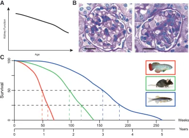
Fig. 5. Age-dependent changes in the kidney. (A) Human kidney function reduces with age as seen in (Davies and Shock, 1950). (B) Hu-man kidney diseases can be studied in animal models. Loss of Wt1 in the mouse can cause glomerulosclerosis as seen by an increase of sclerotic tissue (adapted from (Gebeshuber et al., 2013)). (C) Graphical estimation of lifespan data for N. furzeri (red),
M. musculus (green) and D. rerio
(blue). Median and 75% lifespans were determined (see colored dot-ted lines) to highlight the large dif-ferences between these lab-based animal models. This data serves as a rough estimation of lifespans based upon data from (Gerhard et al., 2002), C57BL/6J mouse lifespan from jax. org and (Wendler et al., 2015). Animal pictures adapted from zdmsociety. org, FLI fish facility, jax.org.
50 100 150 200 250
1 2 3 4 5
0
0
Weeks
Years
50
100
0
Survival
Kidney Function
Age
B
“cellular senescence” (Hayflick and Moorhead, 1961). Senescent fibroblasts for example are known to secrete epithelial growth fac-tors, matrix metalloproteinases and inflammatory cytokines, leading to an alteration in the tissue structure and causing local inflammation (Campisi, 2005). These effects foster a microenvironment, promot-ing age-associated diseases and neoplastic transformations in old organisms, like atherosclerotic and hyperplastic epithelial lesions. As aging effects the whole organism, clear signs of functional decrease can be observed in the kidney as well. In 1950, Davies and Shock reported an inverse relationship between glomerular filtration rate (GFR) and age (Davies and Shock, 1950) measuring the urinary clearance of inulin in individuals aged 24 to 89 years. More recently scientists have shown a significant reduction of the GFR by 22% in humans ≥55 years compared to younger individuals (Hoang et al., 2003). One of the biggest studies in humans is the Baltimore Longitudinal Study of Aging (BLSA), measuring GFR by creatinine clearance, next to other aging associated factors. Combining data from the Baltimore Longitudinal Study of Aging (BLSA), a large-scale study of human aging (Ferrucci, 2008), biopsies of kidney donor transplantations and healthy kidney tis-sue of nephrectomy patients, Wheeler and colleagues analyzed gene expression and identified 630 genes being changed during aging. 101 of these genes showed expression-associated single nucleotide polymorphisms (SNPs) correlating with GFR decline (Wheeler et al., 2009). Already in 2004, transcriptional profiling of the aging human kidney was done, identifying 985 genes that were changed with age, being associated with immune cell response and extracellular matrix (Rodwell et al., 2004). Aging affects the kidney globally as decline in size is observed in cortex and me-dulla (Lindeman, 1990). Implying a general decline of the repair and regeneration processes, several diseases of the kidney are associated with aging, like focal segmental glomerulosclerosis, interstitial nephritis or renal stones (Fig. 5 A,B) (Nitta et al., 2013).
Fish models of aging and regeneration
Only a few studies have tried to address the age-dependency of regeneration in the adult zebrafish, looking at the caudal fin and heart. Caudal fin amputation experiments showed that 28-month-old animals were able to regenerate to the same extent as both 4-month and 12-month-old animals (Shao et al., 2011). These results were backed up by a further study that showed when comparing young fish (6-12 months) to older animals (26-36 months) similar regenerative capacities were described following fin amputation but also ventricular resection (Itou et al., 2012). In contrast to zebraf-ish, Nothobranchius furzeri do show age-dependent differences.
Nothobranchius furzeri, also known as the turquoise killifish
belongs to the group of teleost fish and has, with 3-12 months, the shortest reported lifespan of a vertebrate in captivity (Valdesalici and Cellerino, 2003). The short lifespan of N. furzeri is an adapta-tion to the special environmental condiadapta-tions it has to deal with. N.
furzeri shows clear signs of aging, such as accumulation of the
aging markers lipofuscin in the liver and b-galactosidase in the skin (Terzibasi et al., 2008), impairment of mitochondrial function (Hartmann et al., 2011) and telomere shortening Hartmann et al., (2009). Additionally, changes in learning/behavioral capabilities, weakening of the color and muscle dysplasia are typical signs of aging in this fish. In a recent study, recovery from caudal amputation was reduced by more than 50% in aged animals. These reductions
are most likely due to reduction in proliferation and an increase in apoptosis, suggesting that the response to amputation is initiated earlier and is more efficient in young fish (Wendler et al., 2015).
These findings suggest that while zebrafish do not exhibit age-related losses in regenerative capacity, Nothobranchius furzeri shows clear signs of aging and loss of regenerative capacity with age, and could be of help to understand missing signals in those animals that do exhibit such phenotypes. Comparative studies with both fish species could lead to highly informative results, which could prove helpful for future clinical therapies. When comparing the lifespans of Nothobranchius furzeri (Killifish), Mus musculus (Mouse) and Danio rerio (Zebrafish) one can see a large difference (Fig. 5 C is a graphical reconstruction of data from (Gerhard et al., 2002), jax.org and (Wendler et al., 2015)). With a median lifespan roughly three times longer than it’s distant relative N. furzeri, aged or old zebrafish would ideally be older than 3 years (at least 36 months), for age-related experiments. And as this is not easy in a laboratory setting for time reasons, Nothobranchius furzeri can be used as an aged vertebrate in comparative studies. Age matched experiments in both fish species could help to understand the age-related regenerative capacities and help us to better understand increases in kidney disease.
Kidney regeneration is a fascinating but understudied research field. The source of the regenerative capacity in fish must be further investigated, in order to see whether the resident progenitor cells are sufficient, or if as yet non-described stem cells are important for the initiation of regeneration. Secondly, an in-depth analysis of the age-dependency of regeneration is necessary. Learning which changes cause animal models to exhibit age-related phenotypes, whilst also finding out what can allow other models to have life-long regeneration is a vital future direction. A field-wide definition of aged-fish will be important for further research. Fish models have the opportunity to be the basis for further human clinical treatments (Cao et al., 2009), with cell- and tissue-specific genome editing required in order to find the crucial signals for kidney regenera-tion. Additional molecular studies are needed to find important pathways and signaling, which can also give insights for future pharmacological targets for human therapies. In the coming years, kidney regeneration research will become increasingly important, as the kidney is one of the organs most affected by the increase in human lifespan.
Acknowledgements
Some of the work from this review is based upon thoughts and ideas from the doctoral theses of U.N. and B.H. In such a review, we are unable to include all of the excellent work from the field. To those authors whose work we have not included, we apologize. Birgit Perner (FLI) took the fluorescent picture in figure 1.
References
ABDULMAHDI, W, ZULLO, J, NESI, L, GOLIGORKSY, M S, RATLIFF, B B (2015). Charting the course of renal cryoinjury. Physiol Rep 3: pii: e12357.
AUGUSTO, J, SMITH, B, SMITH, S, ROBERTSON, J, REIMSCHUESSEL, R (1996). Gentamicin-induced nephrotoxicity and nephroneogenesis in Oreochromis nilotica, a tilapian fish. Diseases of Aquatic Organisms 26: 49-58.
for acute and chronic function. Kidney Int 72: 151-156.
BENIGNI, A, MORIGI, M, REMUZZI, G (2010). Kidney regeneration. Lancet 375: 1310-1317.
BOLLEE, G, FLAMANT, M, SCHORDAN, S, FLIGNY, C, RUMPEL, E, MILON, M, SCHORDAN, E, SABAA, N, VANDERMEERSCH, S, GALAUP, A, RODENAS, A, CASAL, I, SUNNARBORG, S W, SALANT, D J, KOPP, J B, THREADGILL, D W, QUAGGIN, S E, DUSSAULE, J C, GERMAIN, S, MESNARD, L, ENDLICH, K, BOUCHEIX, C, BELENFANT, X, CALLARD, P, ENDLICH, N, THARAUX, P L (2011). Epidermal growth factor receptor promotes glomerular injury and renal failure in rapidly progressive crescentic glomerulonephritis. Nat Med 17: 1242-1250. BONVENTRE, J V, YANG, L (2011). Cellular pathophysiology of ischemic acute kidney
injury. J Clin Invest 121: 4210-4221.
BRENNER, B M (1985). Nephron adaptation to renal injury or ablation. Am J Physiol 249: F324-337.
BROWN, M, REIMSCHUESSEL, R (1998). Nephron neogenesis in an aglomerular fish. FASEB J. 12: A800-A800.
BUSSOLATI, B, BRUNO, S, GRANGE, C, BUTTIGLIERI, S, DEREGIBUS, M C, CANTINO, D, CAMUSSI, G (2005). Isolation of renal progenitor cells from adult human kidney. Am J Pathol 166: 545-555.
CAMPISI, J (2005). Senescent cells, tumor suppression, and organismal aging: good citizens, bad neighbors. Cell 120: 513-522.
CAO, Y, SEMANCHIK, N, HUN LEE, S, SOMLO, S, EMILIO BARBANO, P, COIF-MAN, R, SUN, Z (2009). Chemical modifier screen identifies HDAC inhibitors as suppressors of PKD models. Proc Natl Acad Sci USA 106: 21819-21824. CHAWLA, L S, KIMMEL, P L (2012). Acute kidney injury and chronic kidney disease:
an integrated clinical syndrome. Kidney Int 82: 516-524.
CHEN, L F, KAYE, D (2009). Current use for old antibacterial agents: polymyxins, rifamycins, and aminoglycosides. Infect Dis Clin North Am. 23: 1053-1075. CIANCIOLO COSENTINO, C, ROMAN, B L, DRUMMOND, I A, HUKRIEDE, N A
(2010). Intravenous microinjections of zebrafish larvae to study acute kidney injury. J Vis Exp. 4: pii: 2079.
CIANCIOLO COSENTINO, C, SKRYPNYK, N I, BRILLI, L L, CHIBA, T, NOVITS-KAYA, T, WOODS, C, WEST, J, KOROTCHENKO, V N, MCDERMOTT, L, DAY, B W, DAVIDSON, A J, HARRIS, R C, DE CAESTECKER, M P, HUKRIEDE, N A (2013). Histone deacetylase inhibitor enhances recovery after AKI. J Am Soc
Nephrol 24: 943-953.
CONBOY, I M, CONBOY, M J, WAGERS, A J, GIRMA, E R, WEISSMAN, I L, RANDO, T A (2005). Rejuvenation of aged progenitor cells by exposure to a young systemic environment. Nature 433: 760-764.
CONBOY, I M, RANDO, T A (2005). Aging, stem cells and tissue regeneration: les-sons from muscle. Cell Cycle 4: 407-410.
CORMIER, S M, NEIHEISEL, T W, WERNSING, P, RACINE, R N, REIMSCHUES-SEL, R (1995). New nephron development in fish from polluted waters: a possible biomarker. Ecotoxicology 4: 157-168.
COSTANTINI, F, KOPAN, R (2010). Patterning a complex organ: branching morpho-genesis and nephron segmentation in kidney development. Dev Cell 18: 698-712. DAEHN, I, CASALENA, G, ZHANG, T, SHI, S, FENNINGER, F, BARASCH, N, YU, L,
D’AGATI, V, SCHLONDORFF, D, KRIZ, W, HARALDSSON, B, BOTTINGER, E P (2014). Endothelial mitochondrial oxidative stress determines podocyte depletion in segmental glomerulosclerosis. J Clin Invest 124: 1608-1621.
DAVIDSON, A J (2008). Mouse kidney development, StemBook, Cambridge (MA). DAVIDSON, A J (2011). Uncharted waters: nephrogenesis and renal regeneration in
fish and mammals. Pediatr Nephrol 26: 1435-1443.
DAVIDSON, A J (2014). Kidney regeneration in fish. Nephron Exp Nephrol 126: 45. DAVIES, D F, SHOCK, N W (1950). Age changes in glomerular filtration rate, ef-fective renal plasma flow, and tubular excretory capacity in adult males. J Clin
Invest 29: 496-507.
DIEP, C Q, MA, D, DEO, R C, HOLM, T M, NAYLOR, R W, ARORA, N, WINGERT, R A, BOLLIG, F, DJORDJEVIC, G, LICHMAN, B, ZHU, H, IKENAGA, T, ONO, F, ENGLERT, C, COWAN, C A, HUKRIEDE, N A, HANDIN, R I, DAVIDSON, A J (2011). Identification of adult nephron progenitors capable of kidney regeneration in zebrafish. Nature 470: 95-100.
DRUMMOND, I A, DAVIDSON, A J (2010). Zebrafish kidney development. Methods
Cell Biol 100: 233-260.
DRUMMOND, I A, MAJUMDAR, A, HENTSCHEL, H, ELGER, M, SOLNICA-KREZEL,
L, SCHIER, A F, NEUHAUSS, S C, STEMPLE, D L, ZWARTKRUIS, F, RANGINI, Z, DRIEVER, W, FISHMAN, M C (1998). Early development of the zebrafish pronephros and analysis of mutations affecting pronephric function.
Develop-ment 125: 4655-4667.
DUFFIELD, J S, HONG, S, VAIDYA, V S, LU, Y, FREDMAN, G, SERHAN, C N, BONVENTRE, J V (2006). Resolvin D Series and Protectin D1 Mitigate Acute Kidney Injury. J. Immunology 177: 5902-5911.
EDWARDS, J R, DIAMANTAKOS, E A, PEULER, J D, LAMAR, P C, PROZIALECK, W C (2007). A novel method for the evaluation of proximal tubule epithelial cellular necrosis in the intact rat kidney using ethidium homodimer. BMC Physiol 7: 1. ELGER, M (2003). Nephrogenesis Is Induced by Partial Nephrectomy in the
Elasmo-branch Leucoraja erinacea. J. Am. Soc. Nephrol. 14: 1506-1518.
ELGER, M, HENTSCHEL, H, LITTERAL, J, WELLNER, M, KIRSCH, T, LUFT, F C, HALLER, H (2003). Nephrogenesis is induced by partial nephrectomy in the elasmobranch Leucoraja erinacea. J Am Soc Nephrol 14: 1506-1518. ELTZSCHIG, H K, CARMELIET, P (2011). Hypoxia and inflammation. N Engl J Med
364: 656-665.
FERENBACH, D A, BONVENTRE, J V (2016). Kidney tubules: intertubular, vascular, and glomerular cross-talk. Curr Opin Nephrol Hypertens 25: 194-202. FERRUCCI, L (2008). The Baltimore Longitudinal Study of Aging (BLSA): A
50-Year-Long Journey and Plans for the Future. J Gerontol A Biol Sci Med Sci 63: 1416-1419. FINE, L (1986). The biology of renal hypertrophy. Kidney Int 29: 619-634. GEBESHUBER, C A, KORNAUTH, C, DONG, L, SIERIG, R, SEIBLER, J, REISS,
M, TAUBER, S, BILBAN, M, WANG, S, KAIN, R, BOHMIG, G A, MOELLER, M J, GRONE, H J, ENGLERT, C, MARTINEZ, J, KERJASCHKI, D (2013). Focal segmental glomerulosclerosis is induced by microRNA-193a and its downregula-tion of WT1. Nat Med 19: 481-487.
GERHARD, G, KAUFFMAN, E, WANG, X, STEWERT, R, MOORE, J, KASALES, C, DEMIDENKO, E, CHENG, K (2002). Life spans and senescent phenotypes in two strains of Zebrafish (Danio rerio). Exp Gerontol 37: 1055-1068.
HARMAN, D (1972). The biologic clock: the mitochondria? J Am Geriatr Soc 20: 145-147. HARTMAN, H A, LAI, H L, PATTERSON, L T (2007). Cessation of renal morphogenesis
in mice. Dev Biol 310: 379-387.
HARTMANN, N, REICHWALD, K, LECHEL, A, GRAF, M, KIRSCHNER, J, DORN, A, TERZIBASI, E, WELLNER, J, PLATZER, M, RUDOLPH, K L, CELLERINO, A, ENGLERT, C (2009). Telomeres shorten while Tert expression increases during age-ing of the short-lived fish Nothobranchius furzeri. Mech Ageage-ing Dev 130: 290-296. HARTMANN, N, REICHWALD, K, WITTIG, I, DROSE, S, SCHMEISSER, S, LUCK, C,
HAHN, C, GRAF, M, GAUSMANN, U, TERZIBASI, E, CELLERINO, A, RISTOW, M, BRANDT, U, PLATZER, M, ENGLERT, C (2011). Mitochondrial DNA copy number and function decrease with age in the short-lived fish Nothobranchius furzeri. Aging Cell 10: 824-831.
HASTY, P, CAMPISI, J, HOEIJMAKERS, J, VAN STEEG, H, VIJG, J (2003). Aging and genome maintenance: lessons from the mouse? Science 299: 1355-1359. HAVASI, A, BORKAN, S C (2011). Apoptosis and acute kidney injury. Kidney Int
80: 29-40.
HAYFLICK, L, MOORHEAD, P S (1961). The serial cultivation of human diploid cell strains. Exp Cell Res 25: 585-621.
HAYSLETT, J P (1979). Functional adaptation to reduction in renal mass. Physiol
Rev 59: 137-164.
HENTSCHEL, D M, PARK, K M, CILENTI, L, ZERVOS, A S, DRUMMOND, I, BON-VENTRE, J V (2005). Acute renal failure in zebrafish: a novel system to study a complex disease. Am J Physiol Renal Physiol 288: F923-929.
HOANG, K, TAN, J C, DERBY, G, BLOUCH, K L, MASEK, M, MA, I, LEMLEY, K V, MYERS, B D (2003). Determinants of glomerular hypofiltration in aging humans.
Kidney Int 64: 1417-1424.
HOPKINS, C, LI, J, RAE, F, LITTLE, M H (2009). Stem cell options for kidney disease.
J Pathol 217: 265-281.
HOPPE, B, PIETSCH, S, FRANKE, M, ENGEL, S, GROTH, M, PLATZER, M, EN-GLERT, C (2015). MiR-21 is required for efficient kidney regeneration in fish.
BMC Dev Biol 15: 43.
HSU, R K, MCCULLOCH, C E, DUDLEY, R A, LO, L J, HSU, C Y (2013). Temporal changes in incidence of dialysis-requiring AKI. J Am Soc Nephrol 24: 37-42. HUMPHREYS, B D, VALERIUS, M T, KOBAYASHI, A, MUGFORD, J W, SOEUNG,
cells repair the kidney after injury. Cell Stem Cell 2: 284-291.
ITOU, J, KAWAKAMI, H, BURGOYNE, T, KAWAKAMI, Y (2012). Life-long preservation of the regenerative capacity in the fin and heart in zebrafish. Biol Open 1: 739-746. KAMEI, C N, LIU, Y, DRUMMOND, I A (2015). Kidney Regeneration in Adult Zebrafish
by Gentamicin Induced Injury. J Vis Exp. e51912.
KAUFMAN, J M, HARDY, R, HAYSLETT, J P (1975). Age-dependent characteristics of compensatory renal growth. Kidney Int 8: 21-26.
KOBAYASHI, A, KWAN, K M, CARROLL, T J, MCMAHON, A P, MENDELSOHN, C L, BEHRINGER, R R (2005). Distinct and sequential tissue-specific activities of the LIM-class homeobox gene Lim1 for tubular morphogenesis during kidney development. Development 132: 2809-2823.
KUSABA, T, HUMPHREYS, B D (2014). Controversies on the origin of proliferating epithelial cells after kidney injury. Pediatr Nephrol 29: 673-679.
LARSSON, L, APERIA, A, WILTON, P (1980). Effect of normal development on compensatory renal growth. Kidney Int 18: 29-35.
LAURENT, G, MALDAGUE, P, CARLIER, M B, TULKENS, P M (1983). Increased renal DNA synthesis in vivo after administration of low doses of gentamicin to rats. Antimicrob Agents Chemother 24: 586-593.
LAZZERI, E, MAZZINGHI, B, ROMAGNANI, P (2010). Regeneration and the kidney.
Curr Opin Nephrol Hypertens 19: 248-253.
LEEMANS, J C, STOKMAN, G, CLAESSEN, N, ROUSCHOP, K M, TESKE, G J, KIRSCHNING, C J, AKIRA, S, VAN DER POLL, T, WEENING, J J, FLORQUIN, S (2005). Renal-associated TLR2 mediates ischemia/reperfusion injury in the kidney. J Clin Invest 115: 2894-2903.
LI, Y, WINGERT, R A (2013). Regenerative medicine for the kidney: stem cell prospects & challenges. Clin Transl Med 2: 11.
LINDEMAN, R D (1990). Overview: renal physiology and pathophysiology of aging.
Am J Kidney Dis 16: 275-282.
MCCAMPBELL, K K, SPRINGER, K N, WINGERT, R A (2014). Analysis of nephron composition and function in the adult zebrafish kidney. J Vis Exp, e51644. MCCAMPBELL, K K, SPRINGER, K N, WINGERT, R A (2015). Atlas of Cellular
Dy-namics during Zebrafish Adult Kidney Regeneration. Stem Cells Int 2015: 547636. MCCAMPBELL, K K, WINGERT, R A (2014). New tides: using zebrafish to study
renal regeneration. Transl Res 163: 109-122.
MCCRORY, W W (1972). Developmental nephrology. Harvard University Press, Cambridge, Mass. USA.
MORIARTY, R J, LOGAN, A, RANKIN, J C (1978). Measurement of single nephron filtration rate in the kidney of the river lamprey, Lampetra fluviatilis L. J Exp Biol 77: 57-71.
NITTA, K, OKADA, K, YANAI, M, TAKAHASHI, S (2013). Aging and chronic kidney disease. Kidney Blood Press Res 38: 109-120.
PALM, F, NANGAKU, M, FASCHING, A, TANAKA, T, NORDQUIST, L, HANSELL, P, KAWAKAMI, T, NISHIJIMA, F, FUJITA, T (2010). Uremia induces abnormal oxygen consumption in tubules and aggravates chronic hypoxia of the kidney via oxidative stress. Am J Physiol Renal Physiol 299: F380-386.
PARSONS, P P, GARLAND, H O, HARPUR, E S, OLD, S (1997). Acute gentamicin-induced hypercalciuria and hypermagnesiuria in the rat: dose-response relationship and role of renal tubular injury. Br J Pharmacol 122: 570-576.
PHE (Public Health England) (2014). Chronic kidney disease prevelence model. PHE
publications gateway number: 2014386
RAMESH, G, REEVES, W B (2003). TNFR2-mediated apoptosis and necrosis in cisplatin-induced acute renal failure. Am J Physiol Renal Physiol 285: F610-618. REIMSCHUESSEL, R (2001). A fish model of renal regeneration and development.
ILAR J 42: 285-291.
REIMSCHUESSEL, R, BENNETT, R, MAY, E, LIPSKY, M (1990a). Development of newly formed nephrons in the goldfish kidney following hexachlorobutadiene-induced nephrotoxicity. Toxicol Pathol 18: 32-38.
REIMSCHUESSEL, R, BENNETT, R O, MAY, E B, LIPSKY, M M (1990b). Development of newly formed nephrons in the goldfish kidney following hexachlorobutadiene-induced nephrotoxicity. Toxicol Pathol 18: 32-38.
REIMSCHUESSEL, R, BENNETT, R O, MAY, E B, LIPSKY, M M (1990c). Renal Tubular Cell Regeneration, Cell-Proliferation and Chronic Nephrotoxicity in the Goldfish Carassius-Auratus Following Exposure to a Single Sublethal Dose of Hexachlorobutadiene. Dis. Aquat. Organ. 8: 211-224.
REIMSCHUESSEL, R, BENNETT, R O, MAY, E B, LIPSKY, M M (1993). Pathological Alterations and New Nephron Development in Rainbow-Trout (Oncorhynchus-Mykiss) Following Tetrachloroethylene Contamination. J. Zoo Wildlife Med. 24: 503-507.
REIMSCHUESSEL, R, WILLIAMS, D (1995). Development of new nephrons in adult kidneys following gentamicin-induced nephrotoxicity. Ren Fail 17: 101-106. RODWELL, G E, SONU, R, ZAHN, J M, LUND, J, WILHELMY, J, WANG, L, XIAO,
W, MINDRINOS, M, CRANE, E, SEGAL, E, MYERS, B D, BROOKS, J D, DAVIS, R W, HIGGINS, J, OWEN, A B, KIM, S K (2004). A transcriptional profile of aging in the human kidney. PLoS Biol 2: e427.
ROMAGNANI, P, LASAGNI, L, REMUZZI, G (2013). Renal progenitors: an evolution-ary conserved strategy for kidney regeneration. Nat Rev Nephrol 9: 137-146. RONCONI, E, SAGRINATI, C, ANGELOTTI, M L, LAZZERI, E, MAZZINGHI, B,
BALLERINI, L, PARENTE, E, BECHERUCCI, F, GACCI, M, CARINI, M, MAGGI, E, SERIO, M, VANNELLI, G B, LASAGNI, L, ROMAGNANI, S, ROMAGNANI, P (2009). Regeneration of glomerular podocytes by human renal progenitors. J
Am Soc Nephrol 20: 322-332.
SALICE, C J, ROKOUS, J S, KANE, A S, REIMSCHUESSEL, R (2001). New neph-ron development in goldfish (Carassius auratus) kidneys following repeated gentamicin-induced nephrotoxicosis. Comp Med 51: 56-59.
SANDER, V, DAVIDSON, A J (2014). Kidney injury and regeneration in zebrafish.
Semin Nephrol 34: 437-444.
SCHMITT, R, MELK, A (2017). Molecular mechanisms of renal aging. Kidney Int. 92: 569-579.
SHAO, J, CHEN, D, YE, Q, CUI, J, LI, Y, LI, L (2011). Tissue regeneration after injury in adult zebrafish: the regenerative potential of the caudal fin. Dev Dyn 240: 1271-1277.
SHOSKES, D, PARFREY, N, HALLORAN, P (1990). Increased Major Histocompat-ability Complex Antigen Expression in Unilateral Ischemic Acute Tubular Necrosis in the Mouse. Transplantation 49: 201-207.
TERZIBASI, E, VALENZANO, D R, BENEDETTI, M, RONCAGLIA, P, CATTANEO, A, DOMENICI, L, CELLERINO, A (2008). Large differences in aging phenotype between strains of the short-lived annual fish Nothobranchius furzeri. PLoS One 3: e3866.
THATCHER, E J, PAYDAR, I, ANDERSON, K K, PATTON, J G (2008). Regula-tion of zebrafish fin regeneraRegula-tion by microRNAs. Proc Natl Acad Sci USA 105: 18384-18389.
VALDESALICI, S, CELLERINO, A (2003). Extremely short lifespan in the annual fish Nothobranchius furzeri. Proc Biol Sci 270 Suppl 2: S189-191.
VERGHESE, E, WEIDENFELD, R, BERTRAM, J F, RICARDO, S D, DEANE, J A (2008). Renal cilia display length alterations following tubular injury and are present early in epithelial repair. Nephrol Dial Transplant 23: 834-841.
VOGETSEDER, A, PICARD, N, GASPERT, A, WALCH, M, KAISSLING, B, LE HIR, M (2008). Proliferation capacity of the renal proximal tubule involves the bulk of differentiated epithelial cells. Am J Physiol Cell Physiol 294: C22-28.
WATANABE, N, KATO, M, SUZUKI, N, INOUE, C, FEDOROVA, S, HASHIMOTO, H, MARUYAMA, S, MATSUO, S, WAKAMATSU, Y (2009). Kidney regeneration through nephron neogenesis in medaka. Dev Growth Differ 51: 135-143. WEI, Q, DONG, Z (2012). Mouse model of ischemic acute kidney injury: technical
notes and tricks. Am J Physiol Renal Physiol 303: F1487-1494.
WENDLER, S, HARTMANN, N, HOPPE, B, ENGLERT, C (2015). Age-dependent decline in fin regenerative capacity in the short-lived fish Nothobranchius furzeri.
Aging Cell 14: 857-866.
WHEELER, H E, METTER, E J, TANAKA, T, ABSHER, D, HIGGINS, J, ZAHN, J M, WILHELMY, J, DAVIS, R W, SINGLETON, A, MYERS, R M, FERRUCCI, L, KIM, S K (2009). Sequential use of transcriptional profiling, expression quantitative trait mapping, and gene association implicates MMP20 in human kidney aging.
PLoS Genet 5: e1000685.
WINGERT, R A, DAVIDSON, A J (2008). The zebrafish pronephros: a model to study nephron segmentation. Kidney Int 73: 1120-1127.
WINGERT, R A, SELLECK, R, YU, J, SONG, H D, CHEN, Z, SONG, A, ZHOU, Y, THISSE, B, THISSE, C, MCMAHON, A P, DAVIDSON, A J (2007). The cdx genes and retinoic acid control the positioning and segmentation of the zebrafish pronephros. PLoS Genet 3: 1922-1938.
treat-ments. Nephrol Nurs J 38: 13-18; quiz 19.
YIN, V P, THOMSON, J M, THUMMEL, R, HYDE, D R, HAMMOND, S M, POSS, K D (2008). Fgf-dependent depletion of microRNA-133 promotes appendage regeneration in zebrafish. Genes Dev 22: 728-733.
ZHOU, W, BOUCHER, R C, BOLLIG, F, ENGLERT, C, HILDEBRANDT, F (2010).
Characterization of mesonephric development and regeneration using transgenic zebrafish. Am J Physiol Renal Physiol 299: F1040-1047.
ZHOU, W, HILDEBRANDT, F (2012). Inducible podocyte injury and proteinuria in transgenic zebrafish. J Am Soc Nephrol 23: 1039-1047.
Preformed Wolffian duct regulates Müllerian duct elongation independently of canonical Wnt signaling or Lhx1 expression
Masahiko Chiga, Tomoko Ohmori, Takashi Ohba, Hidetaka Katabuchi and Ryuichi Nishinakamura Int. J. Dev. Biol. (2014) 58: 663-668
https://doi.org/10.1387/ijdb.140261rn
Induction of intermediate mesoderm by retinoic acid receptor signaling from differentiating mouse embryonic stem cells
Shiho Oeda, Yohei Hayashi, Techuan Chan, Minoru Takasato, Yuko Aihara,Koji Okabayashi, Kiyoshi Ohnuma and Makoto Asashima Int. J. Dev. Biol. (2013) 57: 383-389
https://doi.org/10.1387/ijdb.130058ma
Characterization of an lhx1a transgenic reporter in zebrafish
Lisa M. Swanhart, Nobuhiro Takahashi, Rachel L. Jackson, Gregory A. Gibson, Simon C. Watkins, Igor B. Dawid and Neil A. Hukriede Int. J. Dev. Biol. (2010) 54: 731-736
https://doi.org/10.1387/ijdb.092969ls
5 yr ISI Impact Factor (2016) = 2.421
Isolation and expression analysis of foxj1 and foxj1.2 in zebrafish embryos
Emil Aamar and Igor B. Dawid Int. J. Dev. Biol. (2008) 52: 985-991 https://doi.org/10.1387/ijdb.072477ea
Cadherin-6 is required for zebrafish nephrogenesis during early development
Fumitaka Kubota, Tohru Murakami, Kenji Mogi and Hiroshi Yorifuji Int. J. Dev. Biol. (2007) 51: 123-129
https://doi.org/10.1387/ijdb.062200fk
The long and winding path to understanding kidney structure in amphioxus - a review
Nicholas D. Holland
Int. J. Dev. Biol. (2017) 61: 683-688 https://doi.org/10.1387/ijdb.170196nh
pdzrn3 is required for pronephros morphogenesis in Xenopus laevis
Silvia Marracci, Alberto Vangelisti, Vittoria Raffa, Massimiliano Andreazzoli and Luciana Dente Int. J. Dev. Biol. (2016) 60: 57-63