Michael Absoud, MD, PhD
Benjamin M. Greenberg, MD, MHS
Ming Lim, MD, PhD Tim Lotze, MD Terrence Thomas, MD Kumaran Deiva, MD,
PhD
Correspondence to Dr. Absoud:
Pediatric transverse myelitis
ABSTRACT
Pediatric acute transverse myelitis (ATM) is an immune-mediated CNS disorder and contributes to 20% of children experiencing a first acquired demyelinating syndrome (ADS). ATM must be dif-ferentiated from other presentations of myelopathy and may be the first presentation of relapsing ADS such as neuromyelitis optica (NMO) or multiple sclerosis (MS). The tenets of the diagnostic criteria for ATM established by the Transverse Myelitis Consortium Working Group can generally be applied in children; however, a clear sensory level may not be evident in some. MRI lesions are often centrally located with high T2 signal intensity involving gray and neighboring white matter. Longitudinally extensive ATM occurs in the majority. Asymptomatic lesions on brain MRI are seen in more than one-third and predict MS or NMO. The role of antibodies such as myelin oligodendro-cyte glycoprotein in monophasic and relapsing ATM and their significance in therapeutic ap-proaches remain unclear. ATM is a potentially devastating condition with variable outcome and presents significant cumulative demands on health and social care resources. Children generally have a better outcome than adults, with one-half making a complete recovery by 2 years. There is need for standardization of clinical assessment and investigation protocols to enable interna-tional collaborative studies to delineate prognostic factors for disability and relapse. There are no robust controlled trials in children or adults to inform optimal treatment of ATM, with one study currently open to recruitment. This review provides an overview of current knowledge of clinical features, investigative workup, pathogenesis, and management of ATM and suggests future directions.Neurology®2016;87 (Suppl 2):S46–S52
GLOSSARY
ADS5acquired demyelinating syndrome;AFM5acute flaccid myelitis;AQP45aquaporin-4;ASIA5American Spinal Injury Association;ATM5acute transverse myelitis;GBS5Guillain-Barré syndrome;IL5interleukin;IVIg5IV immunoglobulin; LETM5longitudinally extensive TM;MOG5myelin oligodendrocyte glycoprotein;MS5multiple sclerosis;NMO5 neuro-myelitis optica;PLEX5plasmapheresis;SLE5systemic lupus erythematosus;TMCWG5Transverse Myelitis Consortium Working Group.
Pediatric acute transverse myelitis (ATM) is an immune-mediated CNS disorder classically
described as demyelinating. ATM comprises a subgroup of the noncompressive transverse
mye-lopathies.
1,2It is a potentially devastating condition with variable outcome.
3ATM must be
differentiated from other, rarer presentations of noncompressive myelopathy.
4,5ATM may be
the first presentation of relapsing acquired demyelinating syndromes (ADS) such as
neuro-myelitis optica (NMO) or multiple sclerosis (MS). A PubMed search using the terms
“pediatric
or paediatric transverse myelitis”
revealed that more than 200 articles have been published in
English on the topic between 1976 and 2015. In this article we discuss current knowledge on
clinical features, pathogenesis, and investigative and management strategies in ATM and
pro-pose future directions.
DEMOGRAPHICS AND CLINICAL FEATURES Active surveillance studies from Canada and the UK have esti-mated that the incidence of ATM in children under 16 years of age is 2/million children/year. ATM accounts
From Children’s Neurosciences (M.A., M.L.), Evelina London Children’s Hospital at Guy’s & St Thomas’NHS Foundation Trust, Kings Health Partners Academic Health Science Centre, London, UK; Department of Neurology and Neurotherapeutics and Department of Pediatrics (B.M.G.), UT Southwestern and Childrens Health, Dallas, TX; Division of Child Neurology (T.L.), Baylor College of Medicine, Houston, TX; Texas Children’s Hospital (T.L.), Houston, TX; Neurology Service (T.T.), Department of Paediatrics, KK Women’s and Children’s Hospital, Singapore; and Assistance Publique-Hôpitaux de Paris, Hôpital Bicêtre, Pediatric Neurology Department, National Referral Center for Neuro-Inflammatory Diseases in Children, and University Paris Sud (K.D.), Le Kremlin-Bicêtre, France.
for one-fifth of children experiencing a first ADS.6,7
TM is more common in adults, but children account for 20% of cases.8 Although males are
more likely to present with ATM (male:female ratio 1.1–1.6:1), a female preponderance is seen among teenagers in regions at high risk for MS and NMO (United States, Canada, Europe, and parts of Australia).6–12 A bimodal age distribution
is observed in children under 5 and older than 10 years of age.3,8,9,11–13 There is no difference in
ethnicity prevalence. A range of prodromal infections are reported in the preceding 30 days in up to 66% of ATM cases.8,11,12The tenets of the
diagnostic criteria for TM established by the Transverse Myelitis Consortium Working Group (TMCWG) are applicable in pediatric cases with appropriate modifications to account for the difficulty in defining a clear sensory level in the younger child (usually under 5 years old).3,12ATM
can present with back pain as the first symptom, followed by motor and sensory deficits or bladder/ bowel dysfunction.8,12,14 Sensory symptomatology
can be either positive (burning paresthesia, hyperesthesia, allodynia) or negative (numbness).8,12,14
Most children develop urinary retention and need catheterization.5,8,12 Establishing the level of
involvement by assessing dermatomes and myotomes is an important component of the American Spinal Injury Association (ASIA) scale and helps track progression to nadir and later recovery.15 However,
this can be challenging because a clear sensory level may not be evident in up to 40% of children.5,8,12A
complete ATM describes bilateral motor and sensory deficits with bladder dysfunction, whereas in a partial cord syndrome there are patchy motor or dissociated sensory deficits of at least one spinal segment with occasional bladder involvement.12,13,16
Spinal cord symptomatology in childhood ATM usually evolves over 2–4 days to peak at 5–6 days.5,8,12 Following immunotherapy, pain is the
first symptom to resolve, followed by an improve-ment in motor deficits. Bladder function and sen-sory deficits may take longest to improve.12Patients
with ATM may also develop a persisting flaccid motor weakness during the course of their illness. Spinal nerve root or cauda equina enhancement may be clearly delineated on MRI, suggesting a simulta-neous CNS and peripheral nerve inflammatory disorder (i.e., an“ATM-plus syndrome”) or a sec-ondary event from cellular damage to the anterior and ventral horn. A patchy axonal motor and sen-sory polyneuropathy is seen on EMG in ATM-plus syndromes.14There are no systematic studies
inves-tigating the prevalence of ATM-plus syndromes. A prospective cohort of adults with myelitis or ence-phlomyelitis following an infectious event showed
that 29/176 (16%) had axonal peripheral nervous system involvement.17 During the summer of
2014, there was an apparent increase in the number of pediatric patients with a variant of TM termed acute flaccid myelitis (AFM).18This condition was
noteworthy for significant involvement of the spi-nal cord gray matter with resulting flaccid, polio-like patterns of weakness. The number of cases in 2014 brought this variant to the attention of public health officials.
DIFFERENTIAL DIAGNOSIS AND EVALUATION Because ATM is a diagnosis of exclusion, deliberate consideration should be given to the differential diag-nosis. Disorders intrinsic and extrinsic to the spinal cord should be considered. A clear history of signifi-cant trauma before onset of myelopathic symptoms would not typically pose a diagnostic dilemma. Extrinsic injuries include vertebral body compression, intervertebral disk herniation, and epidural hema-toma. Uncommon posttraumatic intramedullary dis-orders include ischemic myelopathy from arterial compromise or venous hypertension from fibrocarti-lagenous embolus.19 Patients with anterior spinal
artery occlusion present with deficits localized to the territory of the anterior two-thirds of the cord.
Spinal cord tumors typically present with sub-acute symptoms of unremitting pain that may awaken the child at night along with myelopathic symptoms. Extramedullary tumors causing cord compression include meningioma, nerve sheath tu-mors, and drop metastasis from meduloblastoma, whereas intramedullary tumors are typically astrocy-tomas and ependymomas. Arteriovenous malforma-tions of the spinal cord classically present with fluctuating symptoms from a vascular steal phenom-enon. Bruits may sometimes be heard with ausculta-tion of the back.
Aside from an infectious abscess, direct infectious myelitis can be difficult to discern from an idiopathic etiology because the clinical presentation (fever and constitutional symptoms) and CSF findings can be similar. CSF isolation of a pathogen, positive PCR re-sults, or demonstration of acute and convalescent serum antibody titers provide the best evidence for direct infection. A variety of pathogens have been re-ported to cause infectious myelitis (table 1). Enter-oviruses have most recently been implicated in AFM, which typically presents with primary motor symp-toms possibly caused by direct infection of spinal cord motor neurons.18
disturbances. The latter is not a predominant feature and a clear sensory level is not expected in GBS. ATM may occur with acute disseminated encephalomyelitis or represent an initial attack of a relapsing demyelin-ating syndrome, such as MS, NMO, or an autoim-mune rheumatologic disorder (e.g., systemic lupus erythematosus [SLE], Sjögren syndrome, and sarcoid-osis). Comorbid clinical, imaging, and laboratory findings can help to discern the presence of such conditions.
Myelopathic symptoms are a neurologic emer-gency because prompt diagnosis and treatment may lessen the severity of neurologic sequela. Emergent
spinal imaging with contrast-enhanced spine MRI rapidly discerns etiologies requiring surgical interven-tion. CSF analysis and serum studies are required to distinguish specific etiologies. CSF protein and white blood cell counts may be normal in 20%–50% of children with definite ATM.8,9,11,12Qualitative tests
for intrathecal oligoclonal bands are positive in up to one-third of children, many of whom will eventually have MS.3,8,11In a UK pediatric NMO study, only
3/20 (15%) presented with isolated ATM, 2 of whom were aquaporin-4 antibody-positive. Nonetheless, aquaporin-4 antibody should be considered for all children presenting with ATM.20Evaluating a broader
repertoire of markers of CNS inflammation in the CSF, such as neopterin, may be helpful. CSF proteo-mics to identify neuronal and glial markers of injury and inflammation need further study.21
MRI IN CHILDREN WITH ATM MRI is a major tool for diagnosis and prognosis in ATM. Lesions are often centrally located with high T2 signal intensity involving gray matter and neighboring white matter (figure).9 Lesions may be contiguous or patchy.
Longitudinally extensive TM (LETM), defined as
$3 vertebral segments, occurs in 66%–85% of ATM in children.3,9 Gadolinium enhancement is
frequently observed, but the absence of gadolinium enhancement does not rule out ATM.3,8,9LETM is
not unique to ATM and can occur in NMO and rarely MS.8Some adults with NMO may have short
TM and the location of the lesion may be key; however, children with NMO appear to exclusively present with LETM.22In childhood ATM, T1
hy-pointensity is also described and occurs in one-third of spinal lesions. Cervical and cervicothoracic lesions represent the majority of ATM lesions (64%–76%).5,8,9 In the context of ATM, brain
MRI is expected to be normal. However, asymptomatic lesions on brain MRI are seen in more than 40% of children, indicating the need to include brain MRI in evaluation for ATM.3,13
Silent MRI brain lesions at first ADS predict an increased risk for developing MS or NMO.3
Approximately 66%–88% of children with a partial ATM and supratentorial MRI brain lesions develop MS.13,16 Clinical 1.5T and 3T
MRI scanners will not show spinal cord lesions in up to 6% of patients with ATM.3,8 New MRI
sequences (e.g., double inversion recovery, phase sensitive inversion recovery) may detect cord lesions.23 Although repeat spinal cord imaging
after 5–7 days may reveal spinal cord atrophy,12,13
other novel techniques used in adult MS studies, such as magnetization transfer ratio and diffusion tensor imaging, may help to quantify and correlate with disability earlier in the disease.24These novel
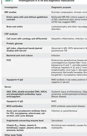
Table 1 Investigations in ATM and diagnostic implications
Investigation Diagnostic purpose
MRI studies Extrinsic compression, intrinsic cord disease
Entire spine with and without gadolinium contrast
McDonald MRI MS criteria support a diagnosis of MS; subclinical optic nerve involvement with LETM may suggest NMO
Brain and orbits Leukodystrophy and other neurodegenerative disorders
CSF analysis
Cell count with cytology and differential Idiopathic inflammation, infection, tumor
Protein, glucose
IgG index, oligoclonal bands (paired studies with serum)
Abnormal in MS; 30% abnormal in NMO and autoimmune TM
Bacterial and viral culture Infection
PCR Enterovirus; parechovirus; herpes simplex virus; cytomegalovirus; Epstein-Barr virus; human herpesvirus 6 and 7; varicella-zoster virus; influenza; hepatitis A, B, and C; human T-lymphotrophic virus type 1;Mycoplasma pneumonia; Bartonella henselae; Borrelia burgdorferi;mycoplasma; tuberculosis
Aquaporin-4 IgG NMO (unlikely to be solely positive in CSF and negative in serum)
Serum
ANA, ENA, double-stranded DNA, ANCA, anti-phospholipid antibodies, lupus anticoagulant
Systemic lupus erythematosus, Sjögren syndrome, antiphospholipid antibody syndrome, Behçet disease
Aquaporin-4 IgG NMO
MOG antibodies MOG antibody–associated disease
Acute and convalescent antibody titers for HIV, mycoplasma, arboviruses, cat-scratch, and Lyme disease
Infection or parainfectious
Angiotensin-converting enzyme level Sarcoidosis
Vitamin B12, folate, vitamin E,
biotidinase, copper, plasma amino acids, ammonia, lactate
Nutritional and metabolic causes for myelopathy
Other body fluids
Immunofluorescence assay for respiratory viruses from nasopharyngeal aspirates/swab
Infection or parainfectious
Throat swab and stool for enterovirus PCR Acute flaccid myelitis
techniques may be challenging in young children because they involve increased imaging time and associated sedation.
PATHOLOGY, PATHOPHYSIOLOGY, AND PATHO-GENESIS OF IDIOPATHIC TM Clinical and patho-logic studies reveal features of inflammation and neu-ronal loss in idiopathic and disease-associated ATM.25
Nevertheless, significant differences in pathogenesis exist, and distinguishing among them is important for understanding disease biology and treatment im-plications. Neurosarcoid, for example, is pathologi-cally associated with noncaseating granulomas within the spinal cord, whereas ATM associated with MS has lymphocytic cell infiltration.25It is important
to note that multiple mechanisms may coexist or occur independently within a disease spectrum, as in lupus-associated ATM in which vasculitis is iden-tified in some and thrombotic infarction in others.26 There is limited information on
immu-nopathogenesis of ATM in children; however, studies in adults are likely to be relevant. In child-hood ATM, as in adult cases, immune-mediated mechanisms are implicated by radiologic findings and CSF reactivity.3Histopathologic adult studies
demonstrate focal infiltration of the spinal cord by
monocytes and CD41 and CD81 T lymphocytes, accompanied by activation of astrocytes and micro-glia.27 Demyelination and axonal loss occur, often
involving the gray matter, a finding that is supported by neuroimaging in adults and children.28,29Necrosis
and cavitation can result in severe disability, especially in NMO.30
The mechanisms of cellular and humoral auto-immune responses contributing to spinal cord inflammation and degeneration remain unclear. Molecular mimicry, a mechanism whereby immune targeting of infection proteins cross-react with neu-ronal proteins that bear molecular similarity, or a less specific super-antigen effect are 2 proposed mechanisms.25
In contrast to the large number of studies charac-terizing the abnormal cellular immune responses in MS, a paucity of similar evaluations are available in ATM. Studies to date interrogate the humoral response, identifying interleukin-6 (IL-6) levels to be markedly elevated in the CSF of adult patients with ATM compared to those with MS and con-trols.31A recent pediatric study confirmed these
re-sults.32 IL-6 is secreted following activation of
astrocytes and microglia, exerting its effect on oligo-dendroglia and axons and mediating cellular injury in spinal cord culture sections.28 There is a correlation
between elevated IL-6 levels and disability in patients with ATM.33Positive results from early-phase
inter-vention studies with monoclonal antibodies to atten-uate IL-6 responses in NMO have direct implications on patients with ATM with elevated IL-6.34
Since the identification of aquaporin-4 (AQP4) antibody in NMO, recent studies have also reported the presence of other autoantibodies in ADS, includ-ing ATM.35 Myelin oligodendrocyte glycoprotein
(MOG) antibodies have been found in childhood-onset ADS and may be an important early predictor of a non-MS course,36 although it remains unclear
whether patients with recurrent demyelination asso-ciated with MOG antibodies warrant the same treat-ment as in AQP4 antibody disease. Because MOG antibody testing is still not universally available (as it is in the United States), the larger implications of this finding remain to be determined.
OUTCOMES, PROGNOSIS, AND MEASUREMENT Since publication of the TMCWG criteria, several case series and cohort studies have been conducted to help understand risk of relapse at first presentation and risk of subsequent disability.2,3,8–10,12 Children
with ATM have a better outcome than adults,8,37with
nearly one-half making a complete recovery by 2 years.8,37 However, in a single-center childhood
ATM study (n547), 43% were unable to walk 30 feet at a median of 3 years follow-up.8Mortality is
Figure MRI spinal images of children with ATM
associated with respiratory failure and a high cervical cord lesion.8,38 The most common sequelae are
sensory disturbances and bladder dysfunction (15%–50%). Approximately one- quarter are nonambulatory or require walking aids, and 10%– 20% never regain mobility or bladder function. The influence of age, time to nadir of symptoms, and time to recovery from nadir in predicting clinical outcome varies between studies.
Studies in pediatric ATM have attempted to define risk factors for relapse and disability at onset of disease. A single-center study of 47 children iden-tified younger age (less than 3 years old), longer time from symptom onset to treatment, higher spinal level, radiologic evidence of longer segmental involvement, presence of T1-hypointense lesions, and lack of white cells in the CSF as predictors of disability.8A
multi-center ATM study (n595) with retrospective ascer-tainment and longitudinal follow-up had a relapse frequency of 17% (n53 NMO and n513 MS). Risk factors for relapse were female sex and abnormal brain MRI, consistent with adult ATM data. Risk factors for disability included severe ASIA scale (A–C) at onset, absence of CSF pleocytosis, spinal lesion with gadolinium enhancement, female sex, and absence of cervicothoracic lesion. ATM may have a relapsing course, which could be categorized as relapsing ATM, a presentation of MS, part of a sys-temic autoimmune disease, or NMOSD in the set-ting of identified antibodies.35,39
Comparison between existing studies regarding disability outcomes presents a challenge because of inconsistent use of core outcomes. The ASIA scale is an internationally accepted scale for the measurement of disability in ATM but has rarely been used in pediatric studies.40 Previous studies
used other measures such as the Expanded Disabil-ity Status Scale, WeeFIM II system, clinician-derived motor recovery ordinal measures, and Paine and Byers scale (poor, fair, and good recov-ery).5,8–10,12The time of CSF sampling (if done at
all) and imaging often varies. Patients with ATM should be followed up longitudinally, irrespective of initial outcome, in part to clarify the diagnosis and also to provide multidisciplinary rehabilitation
interventions (motor disability, urinary/bladder man-agement, psychological and schooling support).
THERAPEUTIC CONSIDERATIONSBecause of lack of controlled clinical trials, there are no US Food and Drug Administration–approved therapies for ATM. Medications are used based on experience and data from open- label studies and retrospective analyses, primarily from studies involving adults. Data suggest that certain conditions have preferential responses to certain therapeutic interventions.41For example, SLE
patients with ATM may respond to cyclophospha-mide whereas patients with NMO benefit from plas-mapheresis (PLEX). In patients without a prior history suggestive of a systemic condition, the treat-ment of TM has to be approached empirically.
In general, no data suggest that first-line therapies (i.e., corticosteroids) worsen the outcome of patients with mimics of TM, including infarcts or infections. Thus, clinicians should empirically treat cases of sus-pected or confirmed ATM. The potential benefit of earlier therapy outweighs the theoretical concerns of treating infectious or vascular etiologies with an anti-inflammatory therapy.
The standard empiric therapy for ATM consists of high-dose corticosteroids. Pediatric patients are usually treated with a 30 mg/kg/dose (maximum 1,000 mg) of methylprednisolone intravenously once a day for 3–5 days.38 Multiple studies have
docu-mented the efficacy and safety of corticosteroids in CNS inflammatory disorders, including ATM. The benefit to patients with ATM was observed in a ret-rospective study, suggesting better short- and long-term outcomes in patients treated with corticosteroids vs patients who did not receive steroids.38
PLEX has been used to treat ATM. Some centers have used this intervention if patients do not respond to corticosteroids, whereas other centers have used the therapy concurrent with corticosteroids if a patient had significant motor or respiratory deficits. Several studies support the use of PLEX in patients with ATM (table 2). PLEX protocols typically involve 5–7 treatment sessions, with each session exchanging 1.1–1.5 plasma volumes. Of note, American Acad-emy of Neurology guidelines published in 2011 recognized the potential benefit of PLEX in patients with adult ATM. Anecdotal reports of IV immuno-globulin (IVIg) 2 gm/kg divided over 2–5 days have not provided conclusive evidence of benefit, but IVIg is often incorporated into the treatment regimen in fulminant disease.16 A UK randomized controlled
trial to determine the benefit of additional treatment with IVIg in adults and children with TM is currently open to recruitment.44
Although there is no consensus on how to handle patients with ATM-plus syndromes therapeutically,
Table 2 Publications reporting therapeutic data for pediatric patients with TM
Author Year
No. of patients (TM patients)
Effective therapy
Patient population
Bigi et al.42 2014 12 (6) PLEX Pediatric
Llufriu et al.43 2009 41 (1) PLEX Pediatric and adult
Defresne et al.38 2001 12 (12) Corticosteroids Pediatric
there is no evidence to suggest a detriment to using the therapies traditionally used for patients with ATM.
CONCLUSIONS AND FUTURE DIRECTIONS ATM is a potentially devastating condition with variable outcome and presents significant cumulative de-mands on health and social care resources. There are no robust controlled trials in children or adults to inform optimal treatment of ATM, with one study in the UK currently open to recruitment.44The
fol-lowing are proposed future directions in ATM: 1. Standardization of clinical assessment and
investi-gation protocols will enable international collabo-rative studies to help define early therapy considerations and delineate prognostic factors for disability and relapse.
2. Defining a basic protocol of core MRI sequences for the evaluation of ATM. New MRI sequences should be investigated as surrogate markers for disability to facilitate early-phase clinical trials. 3. Establishing common outcome measures for use
in ATM. The ASIA scale is an internationally accepted scale for the measurement of disability in ATM but requires adaptation and validation for young children.
4. The role of antibodies such as MOG in mono-phasic and relapsing ATM and their significance in therapeutic approaches remain unclear and require international collaboration.
5. Future clinical trials need to consider adaptive de-signs to include pediatric and adult populations with appropriate statistical considerations.44
AUTHOR CONTRIBUTIONS
All authors contributed extensively to the writing, editing, and consensus-finding process of all sections of the manuscript.
STUDY FUNDING
This supplement is made possible by funding from the MS Cure Fund, Danish MS Society, German MS Society, Italian MS Association, MS International Federation, MS Research Foundation (Netherlands), National MS Society (USA) and Swiss MS Society.
DISCLOSURE
M. Absoud received research grants from Action Medical Research, MS Society, and the NIHR; serves on the data safety monitoring board for a study sponsored by Neurim Pharmaceuticals; and is on the editorial advisory board for theInternational Journal of Language &
Communica-tion Disorders. B. Greenberg has received consulting fees from Novartis,
EMD Serono, and Medimmune and has received grants from Biogen, Chugai, Medimmune, NIH, PCORI, and Acorda. M. Lim received research grants from Action Medical Research, MS Society, and the NIHR; has received consultation fees from CSL Behring; has received travel grants from Merck Serono; and has been awarded educational grants to organize meetings by Novartis, Biogen Idec, Merck Serono, and Bayer. T. Lotze and T. Thomas report no disclosures relevant to the manuscript. K. Deiva received funds as national PI for studies from Merck Serono and travel subsidies and speaker fees from Biogen Idec. Go to Neurology.org for full disclosures.
Received August 19, 2015. Accepted in final form January 4, 2016.
REFERENCES
1. Transverse Myelitis Consortium Working Group. Pro-posed diagnostic criteria and nosology of acute transverse myelitis. Neurology 2002;59:499–505.
2. Krupp LB, Tardieu M, Amato MP, et al. International Pediatric Multiple Sclerosis Study Group criteria for pedi-atric multiple sclerosis and immune-mediated central ner-vous system demyelinating disorders: revisions to the 2007 definitions. Mult Scler 2013;19:1261–1267.
3. Deiva K, Absoud M, Hemingway C, et al. Acute idio-pathic transverse myelitis in children: early predictors of relapse and disability. Neurology 2015;84:341–349. 4. Wolf VL, Lupo PJ, Lotze TE. Pediatric acute transverse
myelitis overview and differential diagnosis. J Child Neu-rol 2012;27:1426–1436.
5. De Goede CG, Holmes EM, Pike MG. Acquired trans-verse myelopathy in children in the United Kingdom–a 2 year prospective study. Eur J Paediat Neurol 2010;14: 479–487.
6. Banwell B, Kennedy J, Sadovnick D, et al. Incidence of acquired demyelination of the CNS in Canadian children. Neurology 2009;72:232–239.
7. Absoud M, Lim MJ, Chong WK, et al. Paediatric acquired demyelinating syndromes: incidence, clinical and magnetic resonance imaging features. Mult Scler 2013;19:76–86. 8. Pidcock FS, Krishnan C, Crawford TO, Salorio CF,
Trovato M, Kerr DA. Acute transverse myelitis in child-hood: center-based analysis of 47 cases. Neurology 2007; 68:1474–1480.
9. Alper G, Petropoulou KA, Fitz CR, Kim Y. Idiopathic acute transverse myelitis in children: an analysis and dis-cussion of MRI findings. Mult Scler 2011;17:74–80. 10. DaJusta DG, Wosnitzer MS, Barone JG. Persistent motor
deficits predict long-term bladder dysfunction in children following acute transverse myelitis. J Urol 2008;180: 1774–1777.
11. Miyazawa R, Ikeuchi Y, Tomomasa T, Ushiku H, Ogawa T, Morikawa A. Determinants of prognosis of acute transverse myelitis in children. Pediatr Int 2003; 45:512–516.
12. Thomas T, Branson HM, Verhey LH, et al. The demo-graphic, clinical, and magnetic resonance imaging (MRI) features of transverse myelitis in children. J Child Neurol 2012;27:11–21.
13. Meyer P, Leboucq N, Molinari N, et al. Partial acute transverse myelitis is a predictor of multiple sclerosis in children. Mult Scler 2014;20:1485–1493.
14. DeSena A, Graves D, Morriss MC, Greenberg BM. Trans-verse myelitis plus syndrome and acute disseminated encephalomyelitis plus syndrome: a case series of 5 chil-dren. JAMA Neurol 2014;71:624–629.
15. Maynard FM Jr, Bracken MB, Creasey G, et al. Interna-tional standards for neurological and funcInterna-tional classifica-tion of spinal cord injury. Spinal Cord 1997;35:266–274. 16. Scott TF, Frohman EM, De Seze J, et al. Evidence-based guideline: clinical evaluation and treatment of transverse myelitis: report of the therapeutics and Technology assess-ment Subcommittee of the American Academy of neurol-ogy. Neurology 2011;77:2128–2134.
17. Marchioni E, Ravaglia S, Montomoli C, et al. Postinfec-tious neurologic syndromes: a prospective cohort study. Neurology 2013;80:882–889.
temporally associated with an outbreak of enterovirus D68 in children in Colorado, USA. Lancet 2015;385: 1662–1671.
19. Rengarajan B, Venkateswaran S, McMillan HJ. Acute asymmetrical spinal infarct secondary to fibrocartilaginous embolism. Childs Nerv Syst 2015;31:487–491. 20. Absoud M, Lim MJ, Appleton R, et al. Paediatric
neuro-myelitis optica: clinical, MRI of the brain and prognos-tic features. J Neurol Neurosurg Psychiatry 2015;86: 470–472.
21. Dhaunchak AS, Becker C, Schulman H, et al. Implication of perturbed axoglial apparatus in early pediatric multiple sclerosis. Ann Neurol 2012;71:601–613.
22. Flanagan EP, Weinshenker BG, Krecke KN, et al. Short myelitis lesions in aquaporin-4-IgG-positive neuromyelitis optica spectrum disorders. JAMA Neurol 2015;72:81–87. 23. Sethi V, Yousry TA, Muhlert N, et al. Improved detec-tion of cortical MS lesions with phase-sensitive inversion recovery MRI. J Neurol Neurosurg Psychiatry 2012;83: 877–882.
24. Kearney H, Miller DH, Ciccarelli O. Spinal cord MRI in multiple sclerosis–diagnostic, prognostic and clinical value. Nat Rev Neurol 2015;11:327–338.
25. Kerr DA, Ayetey H. Immunopathogenesis of acute trans-verse myelitis. Curr Opin Neurol 2002;15:339–347. 26. Nakano I, Mannen T, Mizutani T, Yokohari R. Peripheral
white matter lesions of the spinal cord with changes in small arachnoid arteries in systemic lupus erythematosus. Clin Neuropathol 1989;8:102–108.
27. Krishnan C, Kaplin AI, Deshpande DM, Pardo CA, Kerr DA. Transverse Myelitis: pathogenesis, diagnosis and treatment. Front Biosci 2004;9:1483–1499. 28. Awad A, Stuve O. Idiopathic transverse myelitis and
neuromyelitis optica: clinical profiles, pathophysiology and therapeutic choices. Curr Neuropharmacol 2011;9: 417–428.
29. Beh SC, Greenberg BM, Frohman T, Frohman EM. Transverse myelitis. Neurol Clin 2013;31:79–138. 30. Tobin WO, Weinshenker BG, Lucchinetti CF.
Longitu-dinally extensive transverse myelitis. Curr Opin Neurol 2014;27:279–289.
31. Krishnan C, Kerr DA. Idiopathic transverse myelitis. Arch Neurol 2005;62:1011–1013.
32. Horellou P, Wang M, Keo V, et al. Increased interleukin-6 correlates with myelin oligodendrocyte glycoprotein anti-bodies in pediatric monophasic demyelinating diseases and multiple sclerosis. J Neuroimmunology 2015;289:1–7.
33. Kaplin AI, Deshpande DM, Scott E, et al. IL-6 induces regionally selective spinal cord injury in patients with the neuroinflammatory disorder transverse myelitis. J Clin Invest 2005;115:2731–2741.
34. Trebst C, Jarius S, Berthele A, et al. Update on the diag-nosis and treatment of neuromyelitis optica: recommenda-tions of the Neuromyelitis Optica Study Group (NEMOS). J Neurol 2014;261:1–16.
35. Hacohen Y, Absoud M, Woodhall M, et al. Autoantibody biomarkers in childhood-acquired demyelinating syn-dromes: results from a national surveillance cohort. J Neu-rol Neurosurg Psychiatry 2014;85:456–461.
36. Hacohen Y, Absoud M, Deiva K, et al. Myelin oligodendro-cyte glycoprotein antibodies are associated with a non-MS course in children. Neurol Neuroimmunol Neuroinflamm 2015;2:e81. doi: 10.1212/NXI.0000000000000081. 37. Defresne P, Hollenberg H, Husson B, et al. Acute
trans-verse myelitis in children: clinical course and prognostic factors. J Child Neurol 2003;18:401–406.
38. Defresne P, Meyer L, Tardieu M, et al. Efficacy of high dose steroid therapy in children with severe acute trans-verse myelitis. J Neurol Neurosurg Psychiatry 2001;71: 272–274.
39. Kitley J, Waters P, Woodhall M, et al. Neuromyelitis optica spectrum disorders with aquaporin-4 and myelin-oligodendrocyte glycoprotein antibodies: a comparative study. JAMA Neurol 2014;71:276–283.
40. Graves DE, Frankiewicz RG, Donovan WH. Construct validity and dimensional structure of the ASIA motor scale. J Spinal Cord Med 2006;29:39–45.
41. Greenberg BM, Thomas KP, Krishnan C, Kaplin AI, Calabresi PA, Kerr DA. Idiopathic transverse myelitis: cor-ticosteroids, plasma exchange, or cyclophosphamide. Neurology 2007;68:1614–1617.
42. Bigi S, Banwell B, Yeh EA. Outcomes after early Admin-istration of plasma exchange in pediatric central nervous system inflammatory demyelination. J Child Neurol 2015; 30:874–880.
DOI 10.1212/WNL.0000000000002820
2016;87;S46-S52
Neurology
Michael Absoud, Benjamin M. Greenberg, Ming Lim, et al.
Pediatric transverse myelitis
This information is current as of August 29, 2016
Services
Updated Information &
http://n.neurology.org/content/87/9_Supplement_2/S46.full
including high resolution figures, can be found at:
References
http://n.neurology.org/content/87/9_Supplement_2/S46.full#ref-list-1
This article cites 44 articles, 5 of which you can access for free at:
Subspecialty Collections
http://n.neurology.org/cgi/collection/transverse_myelitis Transverse myelitis
http://n.neurology.org/cgi/collection/autoimmune_diseases Autoimmune diseases
http://n.neurology.org/cgi/collection/all_pediatric All Pediatric
http://n.neurology.org/cgi/collection/all_demyelinating_disease_cns All Demyelinating disease (CNS)
following collection(s):
This article, along with others on similar topics, appears in the
Permissions & Licensing
http://www.neurology.org/about/about_the_journal#permissions
its entirety can be found online at:
Information about reproducing this article in parts (figures,tables) or in
Reprints
http://n.neurology.org/subscribers/advertise
Information about ordering reprints can be found online:
rights reserved. Print ISSN: 0028-3878. Online ISSN: 1526-632X.
1951, it is now a weekly with 48 issues per year. Copyright © 2016 American Academy of Neurology. All ® is the official journal of the American Academy of Neurology. Published continuously since