
International Journal of Nanomedicine 2016:11 6297–6306
International Journal of Nanomedicine
Dove
press
submit your manuscript | www.dovepress.com 6297
R e v I e w
open access to scientific and medical research
Open Access Full Text Article
The effect of exposure to nanoparticles and
nanomaterials on the mammalian epigenome
MI Sierra1
A valdés2
AF Fernández1
R Torrecillas2
MF Fraga2
1Cancer epigenetics Laboratory,
Institute of Oncology of Asturias (IUOPA), Hospital Universitario Central de Asturias (HUCA), Universidad de Oviedo, Oviedo,
2Nanomaterials and Nanotechnology
Research Center (CINN-CSIC), Universidad de Oviedo-Principado de Asturias, el entrego, Spain
Abstract: Human exposure to nanomaterials and nanoparticles is increasing rapidly, but their effects on human health are still largely unknown. Epigenetic modifications are attracting ever more interest as possible underlying molecular mechanisms of gene–environment interac-tions, highlighting them as potential molecular targets following exposure to nanomaterials and nanoparticles. Interestingly, recent research has identified changes in DNA methylation, histone post-translational modifications, and noncoding RNAs in mammalian cells exposed to nanomaterials and nanoparticles. However, the challenge for the future will be to determine the molecular pathways driving these epigenetic alterations, the possible functional consequences, and the potential effects on health.
Keywords: noncoding RNAs, ncRNAs, DNA methylation, histone modification, epigenetics
Introduction
During the last decade, we have witnessed a tremendous increase in the use of nanoparticles and nanomaterials in several areas of human life, chemical industry,
food technology, cosmetics and biomedicine, among others (Table 1).1,2 This rise
in use is the consequence of developments in nanotechnology, defined according to the US Government’s National Nanotechnology Initiative as “the understanding and control of matter at dimensions of roughly 1 to 100 nm, where unique phenomena enable novel applications.”
Particle sizes up to 100 nm determine changes in the physical and chemical properties of materials compared to those observed at larger scales; for instance, chemical reactivity, electrical conductivity, melting point, fluorescence, and magnetism are all affected by size. This is mainly due to the increase in surface area of nanomateri-als compared with larger-scale particles of similar masses, enabling increased contact with the surrounding materials and therefore changes in their reactivity. For example,
titanium dioxide (TiO2) particles lose their white color and become colorless at sizes
below 50 nm. Other types of particles considered to have electrical insulation proper-ties may become conductive at the nanosize, and substances with low solubility can
become more soluble at sizes below 100 nm.3 Furthermore, nanoparticles can combine
(homogeneously or heterogeneously) as aggregates or agglomerates; aggregates are formed by particles strongly linked by molecular-type bonds, while agglomerates
are formed by particles interlinked by van der Waals-type lower energy bonds.4
Also related to the size of nanoparticles is their coefficient of diffusion, which is inversely proportional to their size and ensures that their main transport mechanism in
the environment is air diffusion.4 As a result, independent of their current use in human
endeavors, the effects of human exposure to nanoparticles have attracted interest in the Correspondence: MF Fraga
ed. FINBA-Hospital Universitario Central de Asturias (HUCA), Avda Roma s/n, 33011 Oviedo, Spain
email [email protected]
Journal name: International Journal of Nanomedicine Article Designation: Review
Year: 2016 Volume: 11
Running head verso: Sierra et al
Running head recto: Nanoparticles and the epigenome DOI: http://dx.doi.org/10.2147/IJN.S120104
International Journal of Nanomedicine downloaded from https://www.dovepress.com/ by 118.70.13.36 on 23-Aug-2020
For personal use only.
Number of times this article has been viewed
This article was published in the following Dove Press journal: International Journal of Nanomedicine
Dovepress
Sierra et al
Table 1
e
pigenetic effects of some nanoparticles and their uses in human activities
Nanoparticle
Frequent uses
Epigenetic effect
NP characteristics (shape, size, charge)
Experiment
setting
Biological model
Silver
Antimicrobial (dental plaque biofilms, treatment of infected wounds) Mould- and bacteria-resistant paints Decreased histone methylation (H3K4me3 and H3K79me1)
39
Spherical, 25–30 nm, Negative
In vitro
M
eL cell line (mouse erythroleukemia)
Antimicrobial coatings on surfaces of medical and dental instruments such as grippers, drills, and scalpels Zac1 and Igf2r hypo- and hypermethylation
30
Spherical, 4–24 nm, nd
In vivo
ICR female mice (imprinting control region)
Ultrasensitive label-free detection of biochemical markers
Altered DNA methylation
32
nd, 3–20 nm, –28 m
v
In vitro
HT22 cell line (mouse hippocampal neuronal cells)
Food packaging (prolonged shelf-life of dairy products due to antimicrobial activity)
mi-RNA expression changes
49
nd,
,
100 nm, nd
In vitro
Jurkat
T
cell line (human
T-cell leukemia)
Anti-caking agents in powder products (salt, soups, etc.) Antibacterial fabrics (socks, towels, etc.)
Altered HDAC activity
52
Spherical,
15,
25,
and
80
nm,
nd
Polysaccharide coated NPs of 10, 25–30, and 80 nm
In vitro
C18-4 cell line (mouse germ line stem cells)
Gold
Delivery vehicles in biomedicine
mi-RNA expression changes
47
Spherical, 40 and 100 nm, nd
In vivo
Swiss albino mice (H strain, female)
Contrast agents in imaging techniques
Chromatin condensation and reorganization, and mi-RNA expression changes
51
nd, 20 nm, nd
In vitro
MRC5 cell line (human fetal fibroblasts)
Food and beverages packagings Toothpaste
Quantum dots
Fluorescence-based imaging
Histone deacetylation
37
nd, nd, nd
In vitro
MCF-7 cell line (human breast adenocarcinoma)
Anticancer purposes by means of photodynamic U
v
or photothermal
therapies
Histone modifications
53
nd, 3.4 nm, negative
In vitro
THP-1 cell line (human acute monocytic leukemia)
Solar cells or batteries for energy storing
mi-RNA expression changes
50
nd, 1–2.5 nm, nd
In vitro
NIH/3T3 cell line (mouse fibroblasts)
Nano- hydroxyapatite
Skeletal repair and dental applications
ALP, BSP, and OSC promoter hypomethylation
13
Dihedral,
100
×
10
nm,
-1.83
m
v
In vitro
Mouse bone marrow stromal cells and
MC3T3-e1 (pre-osteoblasts) and
MLO-Y4 (osteocytes) cell lines
Silica
Lighter but stronger and more durable concrete, steel, wood, or glass Global genomic hypomethylation and decreased methyltransferase machinery
16
nd, 15 nm, nd
In vitro
HaCaT cell line (human epidermal keratinocytes)
Also nano-sensors can be incorporated into building materials to monitor changes induced by external factors PARP-1 promoter hypermethylation and inactivity
17
nd, 15 nm, nd
In vitro
HaCaT cell line (human epidermal keratinocytes)
Plastic bottles with nanoparticle coatings improve possibilities for gas containing drinks
mi-RNA expression changes
46
Spherical, 70 nm,
-52.7 m
v
In vivo
BALB/c mice
International Journal of Nanomedicine downloaded from https://www.dovepress.com/ by 118.70.13.36 on 23-Aug-2020
Dovepress Nanoparticles and the epigenome
Anti-caking agents in powder products (salt, soups, etc.) Highly transparent super-hydrophobic coating Breathable textile materials
Titanium dioxide
Sun blocking lotions (sunscreen lotions) and cosmetics
PARP-1 promoter hypermethylation
27
Spherical, 22 nm,
-29.8 m
v
In vitro
A549 cell line (human lung adenocarcinoma)
w
hitening agents used in soaps,
toothpastes, or wet wipes, among others Changed expression levels of methylation machinery and LIN
e1 and
ALU expression levels
14
Spherical, 21.0 nm, negative
In vitro
RA
w
264.7 (mouse macrophages), SA
eC
(human small airway epithelial cells), and THP-1 (human acute monocytic leukemia) cell lines
Remediation: removal of organic pollutants from soil and water
Dysfunction of methylation cycle
54
nd, 10–100 nm, nd
In vitro
HaCaT cell line (human epidermal keratinocytes)
Glass changing color upon light exposition
DNA hypomethylation
55
nd, nd, nd
In vitro
MRC5 cell line (human lung fibroblasts)
mi-RNA expression changes
45
nd, 20 nm, nd
In vivo
C57BL/6BomTac mice
Carbon nanotubes Remediation: removal of organic pollutants from soil and water Global DNA hypomethylation; IFN-gamma and
TNF-α
promoter
hypomethylation; Thy-1 promoter hypermethylation
12
24–45 nm diameter, 1–15 nm length From
-7.69 to
-18.7 m
v
In vivo
C57BL/6 mice
Sea water desalination for drinking purposes
mi-RNA expression changes
50
100–200 nm, nd
In vitro
NIH/3T3 cell line (mouse fibroblasts)
Non-scratchable paints
Increased DNA methylation but decreased DNMTs transcription
18
Several sizes, nd
In vitro
A549 cell line (human lung adenocarcinoma)
Stronger and lighter sport equipments Lighter and stronger vehicles
Copper oxide
Doping materials in semiconductors
Global DNA and transposable elements methylation changes
22
Spherical, 58.7 nm, negative
In vivo
BALB/c male mice
Chemical sensors
Alu hypermethylation
14
Spherical, 58.7 nm, negative
In vitro
RA
w
264.7 (mouse macrophages), SA
eC
(human small airway epithelial cells), and THP-1 (human acute monocytic leukemia) cell lines
Antimicrobial agent Biomedical applications
Abbreviations:
nd, not defined; DNMTs, DNA methyltransferases; NPs, nanoparticles; IFN, interferon, TNF
α
, tumor necrosis factor alpha.
International Journal of Nanomedicine downloaded from https://www.dovepress.com/ by 118.70.13.36 on 23-Aug-2020
Dovepress
Sierra et al
last few years from a safety/health point of view. Since it has recently became obvious that the misregulation of cellular epigenetic mechanisms may be detrimental to health, this has further fueled interest in the study of the effects of
nanopar-ticle exposure on epigenetic regulation.5 While epigenetic
modifications can be very stable and, in some cases, passed on to multiple generations, they can also change dynamically in response to specific cellular conditions or environmental stimuli. The effect of the environment on the epigenome has
been extensively reviewed.6,7 Some authors have already
emphasized that the potential epigenetic properties of nano-materials could not only affect the exposed individuals but
also their offspring.8
Here, we will focus on the potential effect of exposure to nanoparticles and nanomaterials on the epigenome, considering epigenetic regulation as the biological mecha-nism whereby DNA, RNA, and proteins are chemically or structurally modified without changing their primary sequence. These epigenetic modifications play critical roles in the regulation of numerous cellular processes, including gene expression and DNA replication and recombination. Epigenetic regulatory mechanisms include, among others, DNA methylation and hydroxymethylation, histone modifi-cation, chromatin remodeling, RNA methylation, and small and long noncoding RNAs (ncRNAs).
There are many different types of nanoparticles and nano-materials that are available with completely different physical and chemical properties; although there is a natural tendency to assess the physiological effects of nanosubstances in gen-eral, in reality, their impact on the organism will be highly dependent on the substance in question. Furthermore, some authors remark that unintentionally emitted nanoparticles in natural systems can be dynamically affected by the environ-ment, complicating even further the assessment of their effect
on exposed populations.9
DNA methylation changes
in response to exposure to
nanoparticles and nanomaterials
DNA methylation consists in the covalent chemical modi-fication of cytosines at the C5 position with a methyl group and occurs at CpG dinucleotides, which are clustered into CpG islands, which affects DNA accessibility to the cel-lular transcriptional machinery and typically turns off gene expression. This methylation process is catalyzed by DNA
methyltransferases (DNMT3a/DNMT3b and DNMT1).10
Demethylation, in contrast, can occur either passively or through active mechanisms in which 10–11 translocator
proteins, 5-hydroxymethylcytosine formation, and thymidine
glycosidases are all implicated.11
A rapid increase in the use of nanocomposites in the last decade has led several research groups to study alterations in DNA methylation patterns in response to these substances. Examples of nanosubstances studied include carbon-based
nanoparticles,12 nano-hydroxyapatite,13 nanoparticles of
TiO2 and copper oxides (CuO),14,15 and SiO
2.16,17 In general,
these studies have identified a number of both locus-specific and global DNA methylation changes in response to nano-particle and nanomaterial exposure. However, these works provide neither genome-wide data nor correlation with gene expression, which makes it difficult to interpret the extent of DNA methylation changes and their possible functional effects. To our knowledge, the first report on the effect of a nanosized substance on DNA methylation was published
only six years ago.16 In this study, the authors incubated the
human epidermal keratinocyte cell line HaCaT with
increas-ing concentrations of 15 nm SiO2 for 24 h and then analyzed
the global DNA methylation changes using high-performance capillary electrophoresis and immunofluorescence staining. Both assays showed a relative decrease of more than 20%
in global DNA methylation. SiO2 was also associated with
a decrease in maintenance and de novo DNMTs, leading the authors to propose that deregulation of DNMTs might play an important role in the process. However, such a strong effect in such short period of time could also imply active demethylation processes. Global DNA hypomethylation in response to nanosized substances was also recently observed
in vivo.12 In this study, the authors detected significant DNA
hypomethylation in the lungs and blood of mice exposed by airways to a type of multiwall carbon nanotubes (MWCN).
In contrast to the findings of Gong et al16 with regard to
very early DNA methylation changes, the effects were
observed only after seven days of exposure in this study.12
Complicating the scenario even further, a recent study using high-performance liquid chromatography-mass spectrometry (HPLC-MS) found an increase in global methylation in a human lung cancer cell line exposed to different concentra-tions of carbon-based nanoparticles, but a reduction in the expression of DNMTs to different extents depending on the
type of carbon nanoparticle.18 Therefore, it is clear that future
research is still needed to determine the effect of nanoparticle size and type and the effect of in vivo conditions on the time of exposure required to induce DNA methylation changes.
Global DNA methylation mainly depends on the methy-lation level of repeated DNA where the majority of the cytosine methylation occurs in the genome, and that is why
International Journal of Nanomedicine downloaded from https://www.dovepress.com/ by 118.70.13.36 on 23-Aug-2020
Dovepress Nanoparticles and the epigenome
the level of DNA methylation at certain repeats is widely
accepted as a surrogate of global DNA methylation.19 This
is of considerable relevance in this context because some recent studies have found a substantial decrease in repeated DNA methylation in response to ambient-air
microparticu-lates exposure.20,21 In contrast, the effect of different types
of nanoparticles on the methylome of cultured mammalian cells is less clear. Indeed, a recent study that analyzed the DNA methylation changes at LINE1 and Alu repeats in mam-malian cells exposed to different concentrations of printer-emitted engineered nanoparticles and nanoparticles of mild
steel welding fumes, CuO, and TiO2 found both hyper- and
hypomethylation, depending on the cell type and the repeated
DNA sequence involved.14,15 Interestingly, although the DNA
methylation changes were small, nanoparticle exposure was clearly associated with DNMT downregulation and repeated DNA reactivation. It is also worth noting that experiments using mouse models have revealed similar methylation
changes in response to nanoparticle exposure.22,23 These
results highlight the fact that the functional consequences of nanoparticle-dependent DNA methylation changes are still largely unknown and that other molecular mechanisms must be involved in the upregulation of repeated DNA.
In addition to the methylation changes at the global and repeated DNA level, some studies have identified the changes in response to nanoparticle exposure at specific loci. It should be noted that, generally, promoter hypermethyla-tion is associated with gene silencing, whereas the effect of intragenic methylation is not so clear, although it might also
have a role in regulating gene expression.24 A recent work
by Ha et al13 reported a 40% increase in DNA methylation
at the promoter region of the osteoblast lineage commitment gene alkaline phosphatase (ALPL) in murine bone marrow
stromal cells, following treatment with nano-hydroxyapatite, a compound being studied as a therapeutic biomaterial for use as a functional scaffold and implant coating for skeletal
repair and dental applications.25,26 An association was found
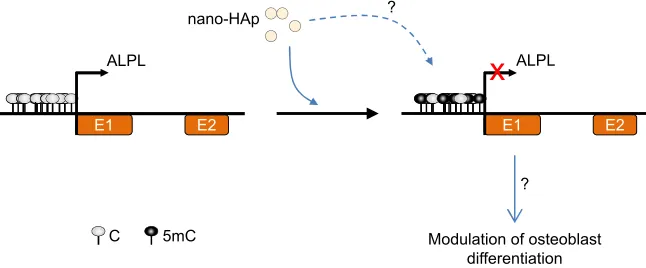
between nano-hydroxyapatite-dependent promoter hyper-methylation and ALPL gene repression and, consequently, osteoblast differentiation, which could have important health implications (Figure 1). Although these results are interest-ing, future research analyzing single CpG methylation at different regions of the ALPL gene is needed to determine the precise role of nanoparticle-dependent DNA methylation changes in gene expression and to determine the molecular mechanism through which nano-hydroxyapatite induces hypermethylation.
In the same vein, promoter-specific hypermethylation of the poly (ADP-ribose) polymerase 1 (PARP-1) was recently reported upon exposure of human lung adenocarcinoma cells
to TiO2 nanoparticles.27 PARP-1 is a DNA-binding protein
involved in DNA repair, proliferation, and chromatin modifi-cation, among other cellular processes. PARP-1 has also been found to form a complex with the DNA methyltransferase DNMT1, the histone H3K9 methyltransferase G9a, and the histone ubiquitin ligase Np95, indicative of a link between
polyADP-ribosylation and the epigenome28 and suggesting
that it contributes to the genome integrity.29 Treatment with
SiO2 nanoparticles has also been found to induce the
aber-rant hypermethylation and inactivation of PARP-1 in human keratinocytes. Interestingly, DNMT1 knockdown restored
PARP-1 promoter methylation levels and its expression,17
further supporting the notion that epigenetic regulations could
be involved in the response to SiO2 nanoparticle exposure.
Nanosized substances do not always induce DNA hyper-methylation at single-copy sequences. For example, using
Figure 1 Possible DNA methylation alteration in response to nano-hydroxyapatite (nano-HAp) exposure in osteoblasts. exposure of early stage differentiating osteoblasts to nano-Hap might induce, through a still unknown molecular mechanism, alkaline phosphatase (ALPL) promoter hypermethylation and gene repression, which in turn could alter lineage commitment and differentiation of bone-forming osteoblasts.
Abbreviations: C, cytosine; mC, methylcytosine; e1, exon 1; e2, exon 2.
P& &
(
0RGXODWLRQRIRVWHREODVW GLIIHUHQWLDWLRQ
$/3/ $/3/
QDQR+$S
( ( (
" "
International Journal of Nanomedicine downloaded from https://www.dovepress.com/ by 118.70.13.36 on 23-Aug-2020
Dovepress
Sierra et al
bisulfite pyrosequencing, Brown et al recently showed a
decrease in the promoter methylation levels of the INF-γ
and TNF-α genes in the lungs of mice exposed by airways
to MWCN.12 In the case of the TNF-α gene, there was a
negative correlation between DNA methylation and gene expression, which suggests that DNA methylation might play a role in the regulation of this gene in response to MWCN and, in consequence, in the regulation of the inflammatory response following MWCNT exposure. In another study, researchers identified hypo- and hypermethylation of the differentially methylated regions of the imprinted genes Zac1 and Igf2r, respectively, in mice embryos exposed to
silver nanoparticles.30 These alterations might have important
implications in disease development, since DNA methylation
plays a critical role in the regulation of gene imprinting,31 a
critical molecular process during embryonic development. Future research is needed to determine the possible functional consequences of these alterations during embryo develop-ment. Silver nanoparticles have also been shown to alter
DNA methylation in mouse hippocampus neuronal cells,32
thus suggesting that this type of nanoparticle is an ontogenic-independent modulator of the epigenome.
Therefore, at this moment, it seems clear that, in general, nanoparticle exposure can induce loss of DNA methylation. But, to our knowledge, there are no reports on DNA methyla-tion levels in human populamethyla-tions exposed to specific types of nanoparticles, although there are reports on methylome changes after exposure to ambient-air pollution, where particulate matter (PM) is included at the nano- and micro-scales. A recent study found that exposure to air pollutants
(in this case, microparticles of NO2, PM10, PM2.5, and O3)
in healthy human individuals led to a small but statistically
significant decrease in global DNA methylation.33 However,
another study found increased or decreased methylation levels of ten different repeated DNA sequences in men
exposed to airborne pollutant.34 The authors attributed this
variability in response to the differences in susceptibility of DNMTs to environmental factors, which could modify their methylation activity on specific DNA repeated sequences. Of note is also a recent paper reporting the lack of
associa-tion between global DNA and ambient PM in dogs.35 The
reason for these differences is not clear and, in consequence, further research is needed to establish the factors determin-ing the effect of microparticulates on methylome in vivo. Moreover, considering that most studies have been carried out on peripheral blood and that DNA methylation patterns are in fact cell-type specific, changes in blood cell composi-tion in response to exposure to airborne pollutants could be
a confounding factor, and should thus be taken into account in future studies.
Effect of nanomaterials and
nanoparticles on histone
post-translational modifications
DNA packaging strength is dependent on other epigenetic marks collectively known as histone modifications. There are a variety of such chemical marks (eg, acetylation, methyla-tion, phosphorylation), all of which modify the terminal amino tails of histones, changing how tightly or loosely the DNA is packaged. In general, when the wrapping is tight, a gene is less accessible to the cellular transcription machinery and is consequently expressed less, and when the wrapping is lost, the gene generally becomes more accessible.
The entry of nanoparticles into the cell nucleus can modulate different cellular functions depending on the chro-matin region affected; while nanoparticle-mediated hetero-chromatin changes cause marked shrinkage of the nucleus,
euchromatin is only marginally modified.36
The effect of nanosized substances on histone post-translational modifications has been studied much less than their effect on DNA methylation. However, some preliminary studies suggest that histone modifications are also important molecular targets for different types of nanoparticles. For example, a recent study found that the nucleus of human breast cancer cells undergoes chromatin condensation and global histone hypoacetylation after quantum dot treatment
with cadmium telluride,37 a nanosubstance currently being
considered as a potential diagnostic, therapeutic, and imag-ing tool. Incubation of MCF-7 cells with cadmium telluride quantum dot for either 4 h or 24 h resulted in global histone H3 hypoacetylation and reduced gene transcription in both cases. Intriguingly, quantum dot treatment increased the expression of some apoptotic genes through the activation of p53. However, the effect of quantum dot exposure on cell death was not investigated in this study. Future research should determine the possible effects of specific nanoparticles on cancer cell death.
Silver nanoparticles have also been recently proposed to affect histone post-translational modifications. These compounds are being utilized in an increasing number of fields and can be found as components of antibacterial coat-ings, antistatic materials, superconductors, and biosensors. The potential of silver nanoparticles to penetrate inside the nucleus is reported to affect various enzymes involved in
chromatin remodeling, such as histone deacetylases.38 In this
regard, a recent study showed that sublethal concentrations
International Journal of Nanomedicine downloaded from https://www.dovepress.com/ by 118.70.13.36 on 23-Aug-2020
Dovepress Nanoparticles and the epigenome
of silver nanoparticles induced a reduction in hemoglobin levels in mouse erythroleukemia cells, possibly through the
diminished methylation of H3K4me3 and H3K79me1.39
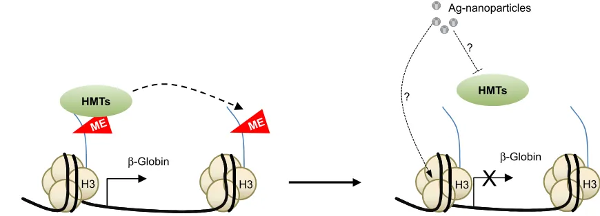
Although the underlying molecular mechanisms are not known, the latter authors presented data suggesting that the effect could be mediated by the inhibition of specific histone methyltransferases or even through the binding of silver nanoparticles to histones H3 and H4 (Figure 2).
The shape, or more precisely the topography, of micro- and nanomaterials used as scaffolds for biological and medical applications seems to affect the cellular epigenome, specifically through histone modification alterations. Further-more, reprogramming somatic cells as pluripotent stem cells
is more efficient when bioengineered substrates are used.40
Supporting this notion, a recent study showed that the topog-raphy of the cell substrate can modulate cell differentiation
and reprogramming.41 Through as yet unknown molecular
mechanisms, microgrooved surfaces lead to increased histone H3 acetylation and methylation which is assumed to play an important role in the regulation of cell differentiation and
reprogramming processes.41
effect of exposure to nanoparticles on
ncRNA expression
The ncRNAs represent another important component of the epigenetic machinery. They can be classified as short ncRNAs (with less than 200 nucleotides) and long ncRNAs (include all larger transcripts). Furthermore, there are sev-eral subtypes of both short (eg, miRNAs, endo-siRNAs, or piRNAs) and long ncRNAs (eg, natural antisense transcripts, long intergenic noncoding RNAs, or sense intronic). Many ncRNAs irrespective of their nature or length are able to
modulate gene expression through their interaction with other epigenetic processes, mainly with the expression and func-tion of proteins involved in histone modificafunc-tions, chromatin
remodeling, and/or DNA methylation.42,43 Therefore, many
ncRNAs are targets of these same processes.43 It is the
plas-ticity of ncRNAs and their capacity for dynamic interaction with DNA, RNA, and proteins which confers on them the ability to mediate in the various epigenetic processes through
which cells respond to diverse external or internal stimuli.42
It is therefore not unexpected that, as with DNA methyla-tion and histone post-translamethyla-tional modificamethyla-tion, the biology of ncRNAs can also be affected by exposure to nanosized
substances.44 The study by Halappanavar et al was one of
the first studies evidencing the effect of nanocompounds
on the expression of ncRNAs.45 Using a mouse model, they
identified significant changes in the expression of 16 miRNAs in lungs of mice exposed to surface-coated nanotitanium
dioxide particles (nanoTiO2), among which mmu-miR-449a
presented the greatest change compared with controls. The
same study also showed that nanoTiO2 also induced lung
inflammation, although the data could not establish a direct link between miRNA alteration and lung injury (Figure 3). Furthermore, a later study from a different group found that
pulmonary instillations of Printex® 90, a type of carbon
black nanoparticle, resulted in a sustained and substantial induction of three miRNAs in mice for up to one month after exposure. Interestingly, the affected miRNAs identified in each study were different, which raises important questions in relation to the possibility of different nanoparticles target-ing specific miRNAs.
In addition to the lungs, nanosized compounds can also affect miRNA expression in other organs in mice. In this
Figure 2 Model explaining the possible molecular mechanisms underlying the effect of Ag-nanoparticles on the regulation of the β-globin gene in mouse erythroleukemia cells. exposure to Ag-nanoparticles induces β-globin repression through still not fully understood molecular mechanisms that might involve inhibition of specific histone
methyl transferases and direct binding of the nanoparticles to histones.
Abbreviations: Me, methylation; H3, histone H3; HMTs, histone methyltransferases; Ag, silver.
+ +
β*ORELQ
+07V
+
β*ORELQ
+07V
0( 0(
$JQDQRSDUWLFOHV
"
"
+
International Journal of Nanomedicine downloaded from https://www.dovepress.com/ by 118.70.13.36 on 23-Aug-2020
Dovepress
Sierra et al
regard, a study by Nagano et al identified changes in the blood levels of three liver-specific miRNAs in adult mice
exposed to70 nm diameter silica nanoparticles.46 They found
that the sensitivity of one of the miRNAs for liver damage was similar to other well-known markers of liver damage. As they can be detected in blood, the authors suggest that altered liver-specific miRNA levels might represent a novel noninvasive biomarker of liver damage in response to nano-particle exposure. In mice, it has even been proposed that nanoparticle exposure has transplacental epigenetic effects. In their study, Balansky et al found that treatment of pregnant mice with gold nanoparticles (average size of 40–100 nm) induced changes in the expression of 28 miRNAs in fetal
tissues.47 As the direct physiological consequences were not
assessed, further research is needed to more fully determine the role of nanoparticles in epigenetic regulation during embryonic development, a time period in which epigenetic regulation plays a critical role in processes such us cell fate
determination and cell differentiation, among others.48
Till date, the effect of nanoparticle exposure on miRNA expression in humans has only been assessed in vitro. A study by Eom et al showed that exposure of Jurkat T cells to silver
nanoparticles altered the expression of 63 miRNAs.49 In line
with this effect in vitro, incubation of NIH/3T murine fibro-blasts with CdTe quantum dots has been shown to induce
significant expression changes in 51 miRNAs.50 In addition,
changes in miRNAs, together with chromatin condensation and reorganization, have been reported upon treatment of
lung fibroblasts with gold nanoparticles.51 Taken together,
the current data suggest that nanomaterial exposure induces alterations in miRNA expression both in vivo and in vitro. However, the underlying molecular mechanisms remain largely unknown.
Conclusion
The rise in the use of nanoparticles in the last decade prob-ably only represents the tip of the iceberg in terms of what the future holds. Given the vast array of fields in which
nanoparticles are being used and studied (Table 1), human exposure can only be expected to grow. The epigenetic effects of this exposure are beginning to be studied, but a great deal of work still needs to be done to reveal the whole picture of the impact of such exposure, not only from the epigenetic point of view, where broader epigenomic and mechanistic data are lacking, but also in terms of concrete data on the properties of nanoparticles, composition, size, charge, and functionalization, and whether or not they are degradable or modifiable by external/ambient conditions (Figure 4). Another important challenge for the future will be to determine the possible effect of the genetic variability on the susceptibility to specific nanoparticle exposure.
Acknowledgments
We thank Ronnie Lendrum for editorial assistance. This work has been financially supported by the Plan Nacional
de I+D+I 2008–2011/2013–2016/FEDER (PI11/01728 to
A.F.F.; PI12/01080 to M.F.F; PI15/00892 to M.F.F.; the ISCIII-Subdirección General de Evaluación y Fomento de la Investigación (Miguel Servet contract: CP11/00131 to A.F.F.); and IUOPA (to M.I.S.). The IUOPA is supported by the Obra Social Cajastur, Spain.
Disclosure
The authors report no conflicts of interest in this work.
Figure 3 Titanium dioxide nanoparticles (nanoTiO2) have been shown to induce
pulmonary inflammation. Although the underlying molecular mechanisms are still
poorly understood, changes in gene and miRNA expression might play an important role in the process.
3XOPRQDU\ LQIODPPDWLRQ QDQR7L2
"
"
0XOWLSOHFKDQJHVLQJHQH DQGPL51$H[SUHVVLRQ
Figure 4 Potential factors affecting nanoparticle exposure-associated epigenetic alterations include, among others, the type and physicochemical characteristics of the nanoparticle, the type of tissue, and interindividual genetic variation. Human exposure to nanoparticles is expected to increase in the coming years. The epigenetic effects of this exposure are beginning to be studied, but a great deal of work still needs to be done to reveal the whole picture of the impact of such exposure, not only from the epigenetic point of view, where broader epigenomic and mechanistic
data are lacking, but also in terms of concrete data on the properties of specific
nanoparticles.
"
7\SHVRIQDQRSDUWLFOHV
,QWHULQGLYLGXDO JHQHWLF YDULDWLRQ 7LVVXHH[SRVHG
International Journal of Nanomedicine downloaded from https://www.dovepress.com/ by 118.70.13.36 on 23-Aug-2020
Dovepress Nanoparticles and the epigenome
References
1. Leung AW, Kalra J, Santos ND, Bally MB, Anglesio MS. Harnessing the potential of lipid-based nanomedicines for type-specific ovarian cancer treatments. Nanomedicine (Lond). 2014;9(3):501–522. 2. Loeve S. Of drug administration, war and oïkos: mediating cancer with
nanomedicines. Nanomedicine (Lond). 2015;10(21):3261–3274. 3. Colvin VL. The potential environmental impact of engineered
nano-materials. Nat Biotechnol. 2003;21(10):1166–1170.
4. Kaluza S, Bladerhaar JK, Orthen B, et al. Work place exposure to nanoparticles. In: Kosk-Bienko J, editor. EU-OSHA Report. European Agency for Safety and Health at Work; 2010.
5. Martínez E, Lagunas A, Mills CA, et al. Stem cell differentiation by functionalized micro- and nanostructured surfaces. Nanomedicine (Lond). 2009;4(1):65–82.
6. Feil R, Fraga MF. Epigenetics and the environment: emerging patterns and implications. Nat Rev Genet. 2012;13(2):97–109.
7. Sierra MI, Fernández AF, Fraga MF. Epigenetics of aging. Curr Genomics. 2015;16:435–440.
8. Stoccoro A, Karlsson HL, Coppedè F, Migliore L. Epigenetic effects of nano-sized materials. Toxicology. 2013;313(1):3–14.
9. Yao Y, Costa M. Genetic and epigenetic effects of nanoparticles. J Mol Genet Med. 2013;7:86.
10. Chen ZX, Riggs AD. DNA methylation and demethylation in mammals. J Biol Chem. 2011;286(21):18347–18353.
11. Kohli RM, Zhang Y. TET enzymes, TDG and the dynamics of DNA demethylation. Nature. 2013;502(7472):472–479.
12. Brown TA, Won Lee J, Holian A, et al. Alterations in DNA methylation corresponding with lung inflammation and as a biomarker for disease devel-opment after MWCNT exposure. Nanotoxicology. 2016;10(4):453–461. 13. Ha SW, Jang HL, Nam KT, Beck GR Jr. Nano-hydroxyapatite modu-lates osteoblast lineage commitment by stimulation of DNA methylation and regulation of gene expression. Biomaterials. 2015;65:32–42. 14. Lu X, Miousse IR, Pirela SV, Melnyk S, Koturbash I, Demokritou P.
Short-term exposure to engineered nanomaterials affects cellular epig-enome. Nanotoxicology. 2016;10(2):140–150.
15. Pirela SP, Miousse IR, Lu X, et al. Effects of laser printer–emitted engi-neered nanoparticles on cytotoxicity, chemokine expression, reactive oxygen species, DNA methylation, and DNA damage: a comprehensive in vitro analysis in human small airway epithelial cells, macrophages, and lymphoblasts. Environ Health Perspect. 2016;124(2):210–219. 16. Gong C, Tao G, Yang L, Liu J, Liu Q, Zhuang Z. SiO(2) nanoparticles
induce global genomic hypomethylation in HaCaT cells. Biochem Biophys Res Commun. 2010;397(3):397–400.
17. Gong C, Tao G, Yang L, Liu J, Liu Q, Zhuang Z. Methylation of PARP-1 promoter involved in the regulation of nano-SiO2-induced decrease of PARP-1 mRNA expression. Toxicol Lett. 2012;209(3):264–269. 18. Li J, Tian M, Cui L, et al. Low-dose carbon-based nanoparticle-induced
effects in A549 lung cells determined by biospectroscopy are associated with increases in genomic methylation. Sci Rep. 2016;6:20207. 19. Yang AS, Estécio MRH, Doshi K, Kondo Y, Tajara EH, Issa JPJ. A
sim-ple method for estimating global DNA methylation using bisulfite PCR of repetitive DNA elements. Nucleic Acids Res. 2004;32(3):e38. 20. Baccarelli A, Tarantini L, Wright RO, et al. Repetitive element DNA
methylation and circulating endothelial and inflammation markers in the VA Normative Aging Study. Epigenetics. 2010;5(3):222–228. 21. Madrigano J, Baccarelli A, Mittleman MA, et al. Prolonged exposure
to particulate pollution, genes associated with glutathione pathways and DNA methylation in a cohort of older men. Environ Health Perspect. 2011;119(7):977–982.
22. Lu X, Miousse IR, Pirela SV, et al. In vivo epigenetic effects induced by engineered nanomaterials: a case study of copper oxide and laser printer-emitted engineered nanoparticles. Nanotoxicology. 2016;10(5): 629–639.
23. Pirela SP, Lu X, Miousse IR, et al. Effects of intratracheally instilled lase printer-emitted engineered nanoparticles in a mouse model: a case study of toxicological implications from nanomaterials released during consumer use. NanoImpact. 2016;1:1–8.
24. Ball MP, Li JB, Gao Y, et al. Targeted and genome-scale strategies reveal gene-body methylation signatures in human cells. Nat Biotechnol. 2009; 27(4):361–368.
25. Zhou H, Lee J. Nanoscale hydroxyapatite particles for bone tissue engineering. Acta Biomater. 2011;7(7):2769–2781.
26. Zhang JZ, Liu GY, Wu QO, Zuo JL, Qin YG, Wang JC. Novel mes-oporous hydroxyapatite/chitosan composite for bone repair. J Bionic Eng. 2012;9(2):243–251.
27. Bai W, Chen Y, Gao A. Cross talk between poly(ADP-ribose) poly-merase 1 methylation and oxidative stress involved in the toxic effect of anatase titanium dioxide nanoparticles. Int J Nanomed. 2015;10: 5561–5569.
28. Sharif J, Muto M, Takebayashi S, et al. The SRA protein Np95 medi-ates epigenetic inheritance by recruiting Dnmt1 to methylated DNA. Nature. 2007;450(7171):908–912.
29. Veith S, Mangerich A. RecQ helicases and PARP1 team up in maintain-ing genome integrity. Agemaintain-ing Res Rev. 2015;23(Pt A):12–28. 30. Zhang XF, Park JH, Choi YJ, Kang MH, Gurunathan S, Kim JH. Silver
nanoparticles cause complications in pregnant mice. Int J Nanomed. 2015;10:7057–7071.
31. Voon HP, Gibbons RJ. Maintaining memory of silencing at imprinted differentially methylated regions. Cell Mol Life Sci. 2016;79(3): 1871–1879.
32. Mytych J, Zebrosky J, Lewinska A, Wnuk M. Prolonged effects of silver nanoparticles on p53/p21 pathway-mediated proliferation, DNA damage response, and methylation parameters in HT22 hippocampal neuronal cells. Mol Neurobiol. Epub 2016 Feb 3.
33. de Prins S, Koppen G, Jacobs G, et al. Influence of ambient air pollution on global DNA methylation in healthy adults: a seasonal follow-up. Environ Int. 2013;59:418–424.
34. Byun H-M, Motta V, Panni T, et al. Evolutionary age of repetitive element subfamilies and sensitivity of DNA methylation to airborne pollutants. Part Fibre Toxicol. 2013;10:28–37.
35. Montrose L, Noonan C, Cho YH, et al. Evaluating the effect of ambi-ent particulate pollution on DNA methylation in Alaskan sled dogs: potential applications for a sentinel model of human health. Sci Total Environ. 2015;512–513:489–494.
36. Mytych J, Wnuk M. Nanoparticle technology as a double-edged sword: cytotoxic, genotoxic and epigenetic effects on living cells. J Biomater Nanobiotechnol. 2013;4:53–63.
37. Choi AO, Brown SE, Szyf M, Maysinger D. Quantum dot-induced epigenetic and genotoxic changes in human breast cancer cells. J Mol Med (Berl). 2008;86(3):291–302.
38. Dubey P, Matai I, Kumar SU, Sachdev A, Bhushana B, Gopinath P. Per-turbation of cellular mechanistic system by silver nanoparticle toxicity: cytotoxic, genotoxic and epigenetic potentials. Adv Colloid Interface Sci. 2015;221:4–21.
39. Qian Y, Zhang J, Hu Q, et al. Silver nanoparticle-induced hemo-globin decrease involves alteration of histone 3 methylation status. Biomaterials. 2015;70:12–22.
40. Tong Z, Solanki A, Hamilos A, et al. Application of biomaterials to advance induced pluripotent stem cell research and therapy. EMBO J. 2015; 34(8):987–1008.
41. Downing TL, Soto J, Morez C, et al. Biophysical regulation of epigenetic state and cell reprogramming. Nat Mater. 2013;12(12):1154–1162. 42. Zhao Y, Sun H, Wang H. Long noncoding RNAs in DNA methylation:
new players stepping into the old game. Cell Biosci. 2016;6:45. 43. Peschansky VJ, Wahlestedt C. Non-coding RNAs as direct and indirect
modulators of epigenetic regulation. Epigenetics. 2014;9(1):3–12. 44. Vrijens K, Bollati V, Nawrot TS. MicroRNAs as potential signatures
of environ-mental exposure or effect: a systematic review. Environ Health Perspect. 2015;123(5):399–411.
45. Halappanavar S, Jackson P, Williams A, et al. Pulmonary response to surface-coated nanotitanium dioxide particles includes induction of acute phase response genes, inflammatory cascades, and changes in microRNAs: a toxicogenomic study. Environ Mol Mutagen. 2011;52(6): 425–439.
International Journal of Nanomedicine downloaded from https://www.dovepress.com/ by 118.70.13.36 on 23-Aug-2020
International Journal of Nanomedicine
Publish your work in this journal
Submit your manuscript here: http://www.dovepress.com/international-journal-of-nanomedicine-journal
The International Journal of Nanomedicine is an international, peer-reviewed journal focusing on the application of nanotechnology in diagnostics, therapeutics, and drug delivery systems throughout the biomedical field. This journal is indexed on PubMed Central, MedLine, CAS, SciSearch®, Current Contents®/Clinical Medicine,
Journal Citation Reports/Science Edition, EMBase, Scopus and the Elsevier Bibliographic databases. The manuscript management system is completely online and includes a very quick and fair peer-review system, which is all easy to use. Visit http://www.dovepress.com/ testimonials.php to read real quotes from published authors.
Dovepress
Dove
press
Sierra et al
46. Nagano T, Higashisaka K, Kunieda A, et al. Liver-specific microRNAs as biomarkers of nanomaterial-induced liver damage. Nanotechnology. 2013;24(40):405102.
47. Balansky R, Longobardi M, Ganchev G, et al. Transplacental clasto-genic and epigenetic effects of gold nanoparticles in mice. Mutat Res. 2013;751–752:42–48.
48. Hochberg Z, Feil R, Constancia M, et al. Child health, developmental plasticity, and epigenetic programming. Endocr Rev. 2011;32(2): 159–224.
49. Eom HJ, Chatterjee N, Lee J, Choi J. Integrated mRNA and micro RNA profiling reveals epigenetic mechnism of differential sensitivity of Jurkat T cells to AGNPs and Ag ions. Toxicol Lett. 2014;229(1):311–318. 50. Li S, Wang H, Qi Y, et al. Assessment of nanomaterial cytotoxicity
with SOLiD sequencing-based microRNA expression profiling. Biomaterials. 2011;32(34):9021–9030.
51. Ng CT, Dheen ST, Yip WCG, Ong CN, Bay BH, Lanry Yung LY. The induction of epigenetic regulation of PROS1 gene in lung fibroblasts by gold nanoparticles and implications for potential lung injury. Biomaterials. 2011;32(30):7609–7615.
52. Braydich-Stolle LK, Lucas B, Schrand A, et al. Silver nanoparticles disrupt GDN/Fyn kinase signaling in spermatogonial stem cells. Toxicol Sci. 2010;116(2):577–589.
53. Conroy J, Byrne SJ, Gun’ko YK, et al. CdTe nanoparticles display tropism to core histones and histone-rich cell organelles. Small. 2008; 4(11):2006–2015.
54. Tucci P, Porta G, Agostini M, et al. Metabolic effects of TiO2 nanopar-ticles, a common component of sunscreens and cosmetics, on human keratinocytes. Cell Death Dis. 2013;4:e549.
55. Patil NA, Gade WN, Deobagkar SS. Epigenetic modulation upon exposure of lung fibroblasts to TiO2 and ZnO nanoparticles: alterations in DNA methylation. Int J Nanomed. 2016;11:4509–4519.
International Journal of Nanomedicine downloaded from https://www.dovepress.com/ by 118.70.13.36 on 23-Aug-2020

![Poly[diaqua(μ2 oxalato κ4O1,O2:O1′,O2′)(μ2 pyrazine 2 carboxylato κ3N1,O:O′)cerium(III)]](data:image/gif;base64,R0lGODlhAQABAIAAAP///wAAACH5BAEAAAAALAAAAAABAAEAAAICRAEAOw==)