International Journal of Nanomedicine
Development of a controlled-release
anti-parkinsonian nanodelivery system
using levodopa as the active agent
Aminu Umar Kura1
Samer Hasan Hussein Al Ali2 Mohd Zobir Hussein3 Sharida Fakurazi1,4 Palanisamy Arulselvan1 1Laboratory of Vaccine and
Immunotherapeutics, Institute of Bioscience, 2Laboratory of
Molecular Biomedicine, Institute of Bioscience, 3Materials Synthesis and
Characterization Laboratory, Institute of Advanced Technology, 4Faculty
of Medicine and Health Science, Pharmacology Unit, Universiti Putra Malaysia, Selangor, Malaysia
Correspondence: Sharida Fakurazi Faculty of Medicine and Health Science Pharmacology Unit, Universiti Putra Malaysia, Selangor 43400, Malaysia Tel +60 3 8947 2117
Fax +60 3 8947 2118
Email [email protected]
Abstract: A new layered organic–inorganic nanocomposite material with an anti-parkinsonian active compound, L-3-(3,4-dihydroxyphenyl) alanine (levodopa), intercalated into the inorganic interlayers of a Zn/Al-layered double hydroxide (LDH) was synthesized using a direct copre-cipitation method. The resulting nanocomposite was composed of the organic moiety, levodopa, sandwiched between Zn/Al-LDH inorganic interlayers. The basal spacing of the resulting nano-composite was 10.9 Å. The estimated loading of levodopa in the nanonano-composite was approxi-mately 16% (w/w). A Fourier transform infrared study showed that the absorption bands of the nanocomposite were characteristic of both levodopa and Zn/Al-LDH, which further confirmed intercalation, and that the intercalated organic moiety in the nanocomposite was more thermally stable than free levodopa. The resulting nanocomposite showed sustained-release properties, so can be used in a controlled-release formulation. Cytotoxicity analysis using an MTT assay also showed increased cell viability of 3T3 cells exposed to the newly synthesized nanocomposite compared with those exposed to pure levodopa after 72 hours of exposure.
Keywords: levodopa, layered double hydroxide, coprecipitation, sustained release
Introduction
Levodopa has remained the gold standard symptomatic replacement therapy for Parkinsonism, a progressive neurodegenerative disease, over the past 40 years. However, long-term treatment is often complicated by dyskinesia, a form of motor dysfunction, which is attributed to the discontinuous and intermittent delivery of levodopa to the brain, resulting in nonphysiological pulsatile stimulation of striatal
dopamine receptors.1,2 Dyskinesia is observed in 40% of patients with Parkinson disease
after 5 years (the honeymoon period) of levodopa use and in over 80% of patients
after 10 years of using levodopa.2
Tailored nanoparticles are good drug delivery agents because of their sustained-release and controlled-sustained-release properties. It is widely believed that reducing the pul-satile stimulation of dopaminergic neurons will reduce the risk of levodopa-induced
dyskinesia.2
Use of nanotechnology in the medical science field and in nanomedicine has reached an advanced stage, where much research has been conducted on the synthesis of nanoparticles that can be used as delivery tools for anticancer agents, central nervous
system tumor agents,3,4 antibiotics,5 and nonsteroidal anti-inflammatory drugs6 in gene
delivery and imaging studies.7,8
Layered double hydroxides (LDHs), also known as hydrotalcite-like com-pounds or anionic clays, are a broad class of inorganic lamellar comcom-pounds with
Dove
press
O r I g I N A L r E S E A r C H
open access to scientific and medical research
Open Access Full Text Article
International Journal of Nanomedicine downloaded from https://www.dovepress.com/ by 118.70.13.36 on 23-Aug-2020
For personal use only.
Number of times this article has been viewed
This article was published in the following Dove Press journal: International Journal of Nanomedicine
a high capacity for anion intercalation. The chemical composition of LDHs is expressed by the general formula
M M (OH)1 x2 (A mH O,
x 3
2
x n
x/n 2
−
+ −
+ +
) ⋅ where M2+ and M3+
are divalent and trivalent metal cations, respectively. These
hydroxides are derived from brucite [Mg(OH)2], and they
can be found in nature or be synthesized in a laboratory via coprecipitation method. The interlayer region contains various amounts of water, which is hydrogen-bonded
to the hydroxide layers and/or to the interlayer anions.9
A characteristic feature of LDH is weak bonding between the interlayer anions and hydroxide sheets that enables exchange
of anions.10
LDHs are less toxic than other conventional drug carriers and are generally biocompatible, making them an
accept-able alternative for drug delivery.11 Anionic drugs, such as
levodopa, which replace the interlayer anions lying between the two positive metal hydroxide sheets, enable a controllable ion-exchange mechanism. pH dependency and the ability for controlled-release are other advantages of using LDHs as drug delivery systems. Further, the positively charged outer layer of the nanohybrid delivery system, which can attract a negatively charged cell membrane, enables easy penetration of LDH into cells. This type of research has not as yet focused on neurodegenerative diseases. Hence, the aim of this study was to intercalate levodopa into LDH for the treatment of Parkinson’s disease.
Materials and methods
Materials
L-3-(3,4-dihydroxyphenyl) alanine (levodopa, 99% purity) was purchased from Sigma-Aldrich (St Louis, MO, USA). Other
chemicals, including Zn(NO3)2 ⋅ 6H2O and Al(NO3)3 ⋅ 9H2O, were
of analytical grade and used without further characterization. Deionized water was used throughout the experiment, unless stated otherwise.
Synthesis
Zn/Al-LDH (the nanolayer) was synthesized in nitrate form by adding a solution of sodium hydroxide (1 molar) dropwise to a solution of zinc and aluminum nitrate, with a molar ratio of 2:1, in deionized water under a nitrogen atmosphere while vigorously stirring until a pH of 7.0 was reached. The mixture was aged in an oil bath for 18 hours
at 70°C. The white precipitate obtained was centrifuged,
washed three times with deionized water, and dried in an
oven overnight at 70°C. The resulting Zn/Al-LDH was used
in a cytotoxicity study and compared with its nanocomposite and levodopa.
Levodopa-Zn/Al-LDH (the nanocomposite) was synthe-sized using a direct coprecipitation method. In brief, a
solu-tion of levodopa (0.08 molar) was added to a Zn(NO3)2 ⋅ 6H2O
and Al(NO3)3 ⋅ 9H2O solutions, at a ratio of 2:1, under
con-stant stirring in the presence of a sustained nitrogen supply at room temperature, and the pH was adjusted to 7.0 using 1.0 molar NaOH. The experiment was protected from direct sunlight exposure because of the sensitivity of levodopa to
light. The mixture was aged at 70°C in an oil bath for 18 hours
and then centrifuged, filtered, washed with deionized water three times, and dried in an oven overnight.
Controlled-release study
Drug release profiles were determined at room temperature using phosphate-buffered saline at pH 4.8 and 7.4. Approximately 300 mg of the nanocomposite was added to 500 mL of the medium. The accumulated amount of levodopa released into the solution was measured at preset time intervals
and at λmax= 280 nm using an ultraviolet-visible
spectropho-tometer (Lambda 35, Perkin-Elmer, Boston, MA, USA).
Cell culture
3T3 cells (fibroblasts) were purchased from the American Type Culture Collection ((ATCC), Manassas, VA, USA), cultured in RPMI 1640 medium supplemented with 10% fetal bovine serum, L-glutamine 15 mmol/L, penicillin 100 U/
mL, and streptomycin 100 µg/mL, and were then incubated
in 5% CO2 at 37°C. Cells at 80%–90% confluence were used
for seeding and treatment.
Seeding and treatment
For the cytotoxicity study, cells were seeded in a 96-well
plate at a density of 0.5 × 10⁵ cells/mL (100 µL/well) and
allowed 24 hours to attach before treatment. A stock solution containing 10 mg/mL of the nanocomposite, the nanohybrid, and levodopa in phosphate-buffered saline was made, diluted in RPMI 1640 complete medium to the desired concentra-tion, and subjected to sonication before being added to the cells. Cell treatment was conducted using a series of dilutions
ranging from 150 µg/mL to 0 µg/mL (the control), and
read-ings were taken 72 hours after treatment using a microplate
reader (EL 800×, Bio-Tek Instruments Inc, Winooski, VT,
USA) after adding MTT (3-(4,5-dimethylthiazol-2-yl)-2,5-diphenyltetrazolium bromide). This assay is dependent upon reduction of the tetrazolium salt by mitochondrial dehydrogenase in viable cells, which forms a blue formazan
product (Mosmann 1983).12 In brief, 20 µL of MTT solution
(5 mg/mL in phosphate-buffered saline) was added to each
Dovepress
Kura et al
International Journal of Nanomedicine downloaded from https://www.dovepress.com/ by 118.70.13.36 on 23-Aug-2020
well, and the plates were kept in an incubator in 5% CO2 at
37°C for 3 hours and centrifuged at 1000 rpm for 5 minutes.
The supernatant was discarded, and 100 µL of dimethyl
sul-foxide was added per well. After keeping this mixture in the dark at room temperature for 30 minutes, absorbance was measured at a wavelength of 570 nm. Experiments were con-ducted in triplicate, and the cytotoxicity was calculated as:
Cell viability (%) = [Average] test/[Average] control × 100.
Characterization
Powder X-ray diffraction patterns were recorded in the 2°–60°
range on a diffractometer (XRD-6000, Shimadzu, Tokyo,
Japan) using CuKα radiation (λ= 1.5418 Å) at 30 kV and
30 mA, with a dwell time of 4 degrees per minute. The Fourier transform infrared spectra of the materials were recorded at
400–4000 cm−1 using a Thermo Nicolet Nexus FTIR (model
Smart Orbit) (International Equipment Trading Ltd. Vernon Hills, IL, USA). The chemical compositions of the samples were analyzed for zinc and aluminum ion content using inductively coupled plasma atomic emission spectrometry (Optima 2000DV, Perkin-Elmer) under standard conditions. An elemental analyzer was used for C, H, N, and S analyses (CHNS-932, LECO Corporation, Saint Joseph, MI, USA). Thermogravimetric and differential thermogravimetric analy-ses (Mettler Toledo, Columbus, OH, USA) were carried out at
a heating rate of 10°C per minute from 20° to 1000°C under
a nitrogen atmosphere (N2 flow rate of 50 mL per minute).
A field emission scanning electron microscope (Nova™ NanoSEM 230, FEI Company, Hillsboro, OR, USA) was used to determine the surface morphology of the samples.
Results and discussion
Powder X-ray diffraction
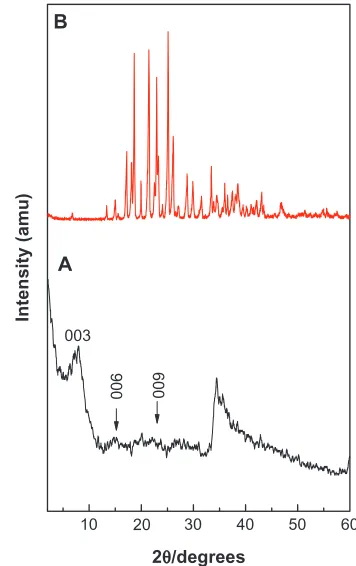
The X-ray diffraction patterns of levodopa intercalated into Zn/Al-LDH and of free levodopa are shown in Fig-ure 1A and B, respectively. As shown in FigFig-ure 1A, the
(003), (006), and (009) reflections were found at 2θ values
of approximately 8.06°, 16.04°, and 24.52°, respectively.
As reported in the literature, the (003) reflection for the
precursor Zn/Al-NO3 LDH appears at 9.98°, indicating a d
spacing of 8.90 Å.13 The shifting of this reflection toward
low 2θ (2θ= 8.06°, corresponding to a d spacing of 10.9 Å)
for the nanocomposite indicates intercalation of levodopa into the interlayer galleries of the LDH. The characteristic reflections of levodopa, shown in Figure 1B, were absent from the X-ray diffraction pattern of the nanocomposite, suggesting that levodopa was intercalated into the interlayer
00
9
00
6
A
003
B
In
te
n
si
ty
(
am
u
)
10 20 30 40 50 60
2θ/degrees
Figure 1 Powder X-ray diffraction patterns prepared using the coprecipitation method for nanocomposite (A) and levodopa (B).
galleries rather than absorbed at the surface. The broader (003) reflection peak in Figure 1A might have been caused by the formation of a secondary phase due to cointercalation
of the counter ion (NO3−).
Spatial orientation of intercalated levodopa
Figure 2A shows the three-dimensional molecular size of levodopa, which was obtained using ChemOffice 2008 soft-ware (Cambridge, MA, USA). The long and short axes (x axis and y axis, respectively) and molecular thickness (z axis) of levodopa were calculated, giving values of 10.4 Å, 7.1 Å, and 6.2 Å, respectively. The X-ray diffraction pattern shows a basal spacing (d) of 10.9 Å for the nanocomposite. The
thick-ness of the Zn/Al-NO3 LDH layer was 4.8 Å.14 Therefore, the
gallery height of LDH after the intercalation processes was 6.1 Å (10.9–4.8 Å). This gallery height of the nanocomposite, 6.1 Å, is similar to the thickness axis of levodopa (6.2 Å). This result indicates that the levodopa anions are accommodated as an alternate monolayer (see Figure 2B).
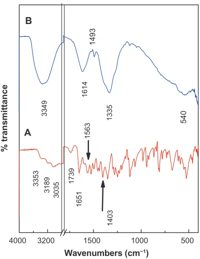
Infrared spectroscopy
The Fourier transform infrared spectra for pristine levodopa and levodopa intercalated into the Zn/Al interlayer are shown in Figure 3. The spectrum for levodopa (Figure 3A) shows many intense, sharp absorption peaks that arise from the
Dovepress Levodopa nanodelivery system for Parkinson’s disease
International Journal of Nanomedicine downloaded from https://www.dovepress.com/ by 118.70.13.36 on 23-Aug-2020
different functional groups present in the molecules, ie, the primary amine, carboxylic acid, benzene ring, and hydroxyl groups.
The carboxylic group for levodopa shows three absorption
peaks: 1245 cm−1 for the C−O stretching, 1739 cm−1 for the
C=O bond, and a broad band at 1400–1440 cm−1for the O−H
bending vibration. The bands at 3353, 3189, and 3035 cm−1
can be attributed to the ν(N−H), ν(O−H), and ν(Ar−H)
vibrations, respectively. The primary amine shows absorption
peaks at 1651 cm−1and 1563 cm−1 that can be attributed to N−H
bending, while the peaks between 1064 cm−1 and 1200 cm−1 are
due to C−N stretching, and the peak at approximately 800 cm−1
indicates N−H (oop) bending. The band at 1496 cm−1 can be
attributed to the C=C bond in the benzene ring.
The Fourier transform infrared spectrum of the levodopa intercalated into Zn/Al-LDH is shown in Figure 3B. The spec-trum of the nanocomposite shows the characteristic bands of pure levodopa, which indicate that the levodopa anions were intercalated into the interlayer galleries of LDH. Some of the bands are slightly shifted. For example, the position of
the band for the C=C bond shifted from 1496 cm−1 for free
levodopa to 1493 cm−1 for the nanocomposite, and the band
at 1118 cm−1 for the C−N bond shifted to 1120 cm−1; these
shifts are due to the interaction between the levodopa anion and the interlayers as a result of the intercalation process.
The appearance of a new broad peak at 1614 cm−1 for the
nanocomposite can be assigned to the asymmetric stretching
vibration of the COO− group.14 The band at 1335 cm−1 is due
to the nitrate anion, which may not be completely removed
from the interlayers during the intercalation process.15
Elemental analysis
Elemental (CHNS) and inductively coupled plasma analy-ses were conducted to determine the organic and inorganic compositions of the nanocomposite. As expected, the levodopa-nanocomposite contained both organic and inor-ganic constituents. This result indicates that the levodopa was incorporated into the Zn/Al-LDH inorganic interlayers.
The percentage loading of levodopa in the nanocom-posite is 16.00%, as shown in Table 1. The C/N ratio for free levodopa is 8.09, and this value is higher than the C/N ratio of the nanocomposite, possibly indicating that the nitrate anion was not completely removed from the interlayers during the intercalation process. This result was
confirmed by the presence of a very broad peak at low 2θ
in the X-ray diffraction pattern. From the elemental chemi-cal analysis and thermogravimetric studies, the empirichemi-cal formula for the nanocomposite can be proposed as: be
Zn0.64Al0.36
( )
OH levodopa NO3 H O.2 2
(
)
0 09( )
⋅0 27 3 03
. . .
−
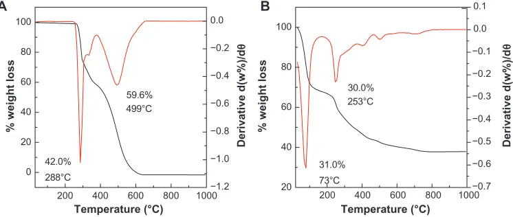
Thermal analysis
The thermal behavior of the compounds before and after intercalation of levodopa into Zn/Al-LDH was examined using thermogravimetric and derivative thermogravimetric analysis. The thermograms of levodopa and the nanocom-posite are shown in Figure 4. The free levodopa (Figure 4A) thermal decomposition exhibited two weight loss events
above 270°C. The first sharp event occurred at 288°C with
a weight loss of 42.0%, which was due to the decomposition
of levodopa. The second weight loss at 381°C–666°C can be
attributed to the combustion of levodopa.
After the intercalation process, the thermal decomposition characteristics of the resulting product were different from
Zn/Al-LDH Zn/Al-LDH
6.2 Å 4.8 Å
Carbon Oxygen Nitrogen Hydrogen
6.2 Å 7.1 Å
10.4 Å
A B
Figure 2 Three-dimensional structure of levodopa (A) and molecular structure model of levodopa intercalated between interlayers of Zn/Al-layered double hydroxide (B).
Abbreviation: LDH,layered double hydroxide.
4000 3200 1500 1000 500
14
03
54
0
13
35
14
93
16
14
33
49
15
63
17
39
16
51
33
53
31
89
B
% transmittance
Wavenumbers (cm−1)
A
30
35
Figure 3 Fourier transform infrared spectra of free levodopa (A) and levodopa-Zn/ Al-layered double hydroxide nanocomposite (B).
Dovepress
Kura et al
International Journal of Nanomedicine downloaded from https://www.dovepress.com/ by 118.70.13.36 on 23-Aug-2020
those of the precursor. The thermal decomposition of the nanocomposite was characterized by two weight loss events,
one at 35°C–162°C with a weight loss of 31.0%, due to the
removal of the external surface-adsorbed and interlayer water
molecules, and a second event at 162°C–960°C with a weight
loss of 30.0%. This second weight loss was due to dehydroxy-lation of the layers and decomposition of levodopa.
Surface properties
In Figure 5, the surface morphologies of the Zn/Al-nanolayer (A and B) and levodopa-nanocomposite (C) are shown. The micrographs were obtained using field emission
scanning electron microscopy at (A) 8000×, (B) 15,000×,
and (C) 30,000× magnifications. Zn/Al-LDH and the
nanocomposite show nonuniform, irregular agglomerates with compact and nonporous plate-like structures, which are commonly observed for LDH and its nanocomposites.
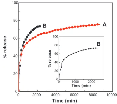
release behavior of levodopa
from the Zn/Al-levodopa nanocomposite
Typical release behavior profiles of levodopa from its nano-composite at different pHs are shown in Figure 6, indicating that the release rate of levodopa from the nanocomposite is pH-dependent. The release rate at pH 7.4 is remarkably lower than that at pH 4.8. The percentage release of levodopa from the nanocomposite reaches approximately 74% within 2400 minutes when exposed to a pH of 4.8. When the pH is changed to 7.4, the release rate of levodopa from the
nanocomposite is obviously lower. The time for 76% of the drug to be released from the nanocomposite is approximately 8600 minutes. This lower release rate of levodopa from the nanocomposite at pH 7.4 indicates that the levodopa-Zn/Al nanocomposite is indeed a potential drug delivery system. The difference in the release rates at pH 4.8 and pH 7.4 may be due to the difference in the release mechanism of levodopa
from the nanocomposite.16 At an acidic/lower pH of 4.8,
Zn/Al-LDH begins to dissolve. At pH 7.4, the Zn/Al-LDH is more stable because of electrostatic interaction between the levodopa anions and the positively charged Zn/Al-LDH layers. Thus, the release mechanism at pH 4.8 should occur through both dissolution of the LDH layers and ion exchange, while the mechanism at pH 7.4 should occur via ion exchange
with the ions in the buffer solution.16
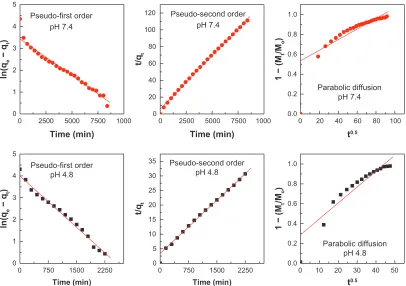
release kinetics of levodopa
from the Zn/Al-levodopa nanocomposite
To investigate the exact mechanism of levodopa release from the Zn/Al-LDH interlayer, the release profile data were fitted using mathematical models that describe various kinetics. The pseudo-first order kinetics mathematical model equation
ln(qe− qt) = lnqe− k1t (qe and qt are the equilibrium release
amount and the release amount at time t, respectively),17 the
pseudo-second order equation t/qt= 1/k2qe2+ t/q
e18 and the
parabolic diffusion equation (1 − Mt /Mo)/t = kt−0.5 + b (M
o and
Mt are the drug content that remained in LDH at the release
times of 0 and t, respectively)19 were used.
Table 1 Elemental chemical compositional for free levodopa and its nanocomposite
Sample Zn%a Al%a C%b N%b C/N Drug%b Zn2+/Al3+ X
Levodopa – – 55.69 6.88 8.09 – – –
Nanocomposite 16.44 3.85 8.56 4.62 1.85 16.00 1.80 0.36
Notes: aCalculated from inductively coupled plasma data; bcalculated from CHNS elemental data.
Abbreviations: Zn, zinc; Al, aluminium; C, carbon; N, nitrogen; X, aluminium mole fraction = mole of Al/(mole of Al + mole of Zn).
200 400 600 800 1000 200 400 600 800 1000
0 20 40 60 80 100
42.0%
59.6%
288°C
499°C
31.0% 30.0%
73°C 253°C
Temperature (°C) Temperature (°C)
% weight loss % weight loss
Derivative d(w%)/d
θ
Derivative d(w%)/d
θ
−
−1.2 −1.0 −0.8 −0.6 −0.4 −0.2 0.0 A
20 40 60 80 100 B
−0.7 −0.6 −0.5 −0.4 −0.3 −0.2 −0.1 0.0 0.1
Figure 4 Thermogravimetric and differential thermogravimetric thermograms of levodopa (A) and levodopa-nanocomposite (B).
Dovepress Levodopa nanodelivery system for Parkinson’s disease
International Journal of Nanomedicine downloaded from https://www.dovepress.com/ by 118.70.13.36 on 23-Aug-2020
alone, cell viability was reduced by more than 40% when
the concentration of the hybrid exceeded 100 µg/mL. We
observed that the levodopa nanocomposite reached a higher cell viability compared with the carrier nanolayer or with the drug alone in a dose-dependent manner after 72 hours of exposure, as shown in Figure 8.
In vitro, cytotoxicity testing is an essential step in analyzing the health hazards of synthesized nanomaterials in terms of the cellular response to a toxicant for a biomedical application. Therefore, in the present study, we investigated the cytotoxic effect of the synthesized nanocomposite, nanohybrid, and pristine levodopa doses using a biological model with 3T3 fibroblast cells and an MTT assay.
It was found that exposure to the nanocomposite at a dose
level of 5–150 µg/mL caused dose-dependent cytotoxicity
in 3T3 cells, as shown in Figure 8, but the same dose of pristine levodopa but more cytotoxicity after 72 hours of exposure compared with the nanocomposite. Findings by other researchers have demonstrated a similar pattern of toxicity in primary rat hepatocytes after exposure to allergic acid and its nanocomposite, ie, the allergic acid caused more toxicity compared with its nanocomposite using the same
dose range after 6 hours of exposure.21
Our results also demonstrated more toxicity in 3T3 cells following exposure to the nanolayer compared with exposure to the nanocomposite at the same doses after 72 hours. In pre-viously reported studies, a decrease in the viability of human umbilical vein endothelial cells was observed following exposure to an LDH nanolayer compared with that following exposure to its nanocomposite, which was functionalized by
poly(sulfobetaine).22 Meanwhile, in another report, Chang
liver cells exposed to an LDH nanolayer also exhibited a slight decrease in viability compared with those exposed to
its nanocomposite containing cetirizine.23 Our findings are
supported by the above-mentioned studies using nanolayers and their corresponding nanocomposites. The results also indicate that our nanocomposite is likely to be a biocompat-ible product because it shows less cytotoxicity than pristine
A B C
Figure 5 Field emission scanning electron micrographs of Zn/Al-layered double hydroxide (A and B), and of the Zn/Al-layered double hydroxide-levodopa nanocomposite (C).
0 2000 4000 6000 8000 10000
0 20 40 60 80 100
0 1000 2000
0 20 40 60 80 100
B
%
r
el
ea
se
Time (min)
A
B
%
r
el
ea
se
Time (min)
Figure 6 Release profiles of levodopa from the nanocomposite at pH 7.4 (A) and pH 4.8 (B).
Note: Inset shows the release profiles of levodopa from the nanocomposite at pH 4.8 from 0 to 2000 minutes.
The fitting results of the drug release profiles at pH 7.4 and 4.8 based on the three different kinetic models are shown
in Figure 7. The correlation coefficient (R2) is tabulated in
Table 2. From Table 2, it can be seen that the release of levodopa from Zn/Al-LDH follows the pseudo-second order equation very well for both pH values, with satisfactory coefficients of 0.9988 (pH 7.4) and 0.9952 (pH 4.8). The rate constant for the
pseudo-second order equation is 2.85 × 10⁻⁵ mg per minute
and 4.29 × 10⁻⁵ mg per minute at pH 7.4 and pH 4.8,
respec-tively (Table 2). The kinetic results obtained during this work are very similar to those from a kinetic study of the release of
camptothecin from Mg/Al-LDH16 and are also similar to those
for perindopril erbumine intercalated into Zn/Al-LDH using
the ion-exchange and coprecipitation methods.20
Cytotoxicity analysis of the
nanocomposite, nanohybrid, and levodopa
Compared with the control, cell viability was found to decrease with increasing concentrations of levodopa, the nanolayer, and the nanocomposite. The 3T3 cell viability was reduced by approximately 40% when the concentration
of levodopa was more than 50 µg/mL. For the nanolayer
Dovepress
Kura et al
International Journal of Nanomedicine downloaded from https://www.dovepress.com/ by 118.70.13.36 on 23-Aug-2020
0 2500 5000 7500 0
1 2 3 4 5
Pseudo-first order pH 7.4
ln(q
e
−
qt
)
t/q
t
t/q
t
ln(q
e
−
qt
)
Time (min)
0 20 40 60 80 100
120 Pseudo-second order pH 7.4
Time (min)
0.0 0.2 0.4 0.6 0.8 1.0
Parabolic diffusion pH 7.4
1 − (M
t
/M
o
)
1 − (M
t
/M
o
)
0 1 2 3 4 5
Pseudo-first order pH 4.8
Time (min)
0 5 10 15 20 25 30
35 Pseudo-second order pH 4.8
Time (min)
0 20 40 60 80 100
0 750 1500 2250 0 750 1500 2250 0.00 10 20 30 40 50
0.2 0.4 0.6 0.8 1.0
Parabolic diffusion pH 4.8
t0.5
t0.5
1000 0 2500 5000 7500 1000
Figure 7 Fits of the levodopa release data for the nanocomposite to the pseudo-first and pseudo-second order kinetics models as well as to the parabolic diffusion model at pH 7.4 and 4.8.
Table 2 Correlation coefficients (R2), and rate constants (k) obtained by fitting the levodopa release data for the nanocomposite in solutions at pH 4.8 and 7.4
pH Saturation
release (%)
R2 Pseudo-second
order
Pseudo-first
order
Pseudo-second order
Parabolic diffusion model
Rate constant k
(mg/min) × 10-5
7.4 76% 0.9839 0.9988 0.8682 2.85
4.8 74% 0.9845 0.9952 0.8838 4.29
0 20 40 60 80 100 120 140
150 100 50 10 5 0.00
% cell viabilit
y
Concentration (µg/mL) Levodopa nanolayer Nanocomposite
Figure 8 In vitro cytotoxicity study of 3T3 cells after 72 hours of exposure to free levodopa, the nanolayer, and the levodopa nanocomposite.
Notes: Using the graph as well as regression analysis, the IC50 (concentration of the drug required to induce 50% cell mortality) for the nanocomposite, nanolayer, and levodopa was 202 ± 3.55 µg/mL, 161 ± 6.42 µg/mL, and 73 ± 6.46 µg/mL, respectively (P 0.005 for all three, showing statistical significance compared with the control).
Dovepress Levodopa nanodelivery system for Parkinson’s disease
International Journal of Nanomedicine downloaded from https://www.dovepress.com/ by 118.70.13.36 on 23-Aug-2020
International Journal of Nanomedicine
Publish your work in this journal
Submit your manuscript here: http://www.dovepress.com/international-journal-of-nanomedicine-journal
The International Journal of Nanomedicine is an international, peer-reviewed journal focusing on the application of nanotechnology in diagnostics, therapeutics, and drug delivery systems throughout the biomedical field. This journal is indexed on PubMed Central, MedLine, CAS, SciSearch®, Current Contents®/Clinical Medicine,
Journal Citation Reports/Science Edition, EMBase, Scopus and the Elsevier Bibliographic databases. The manuscript management system is completely online and includes a very quick and fair peer-review system, which is all easy to use. Visit http://www.dovepress.com/ testimonials.php to read real quotes from published authors.
levodopa, which is very important for the potential use of the nanocomposite in an in vivo study.
Conclusion
Levodopa-Zn/Al-LDH nanocomposites were synthesized using the coprecipitation method with a basal spacing of 10.9 Å and 16.0% (w/w) loading of levodopa. The inter-calation of levodopa into the interlayer spacing of LDH is shown by Fourier transform infrared spectra. The intercalated guest molecules of levodopa were found to be arranged in a monolayer manner between the inorganic interlayers. Release of levodopa from the nanocomposite was found to be gov-erned by pseudo-second order kinetics, and the release time of levodopa from the nanocomposite at pH 7.4 was longer than that at pH 4.8. The cytotoxicity study also demonstrated a decrease in the toxicity potential of levodopa in a normal cell line following its intercalation into Zn/Al-LDH. Hence, successful intercalation of levodopa may improve drug delivery systems for the treatment of parkinsonism as well as decrease its potential toxicity to cells.
Acknowledgment
We would like to thank the Ministry of Science, Technology, and Innovation Malaysia for project funding under nanofund NND/NA/(I) TD11-010.
Disclosure
The authors report no conflicts of interest in this work.
References
1. Poewe W, Antonini A, Zijlmans JCM, Burkhard PR, Vingerhoets F. Levodopa in the treatment of Parkinson’s disease: an old drug still going strong. Clin Interv Aging. 2010;5:229–238.
2. Aviles-Olmos I, Martinez-Fernandez R, Foltynie T. L-dopa-induced dyskinesias in Parkinson’s disease. Eur Neurol J. 2010;2(2):91–100. 3. Choi SJ, Oh JM, Choy JH. Anticancer drug-layered hydroxide
nanohy-brids as potent cancer chemotherapy agents. J Phys Chem Solids. 2008; 69(5–6):1528–1532.
4. Vergoni AV, Tosi G, Tacchi R, Vandelli MA, Bertolini A, Costantino L. Nanoparticles as drug delivery agents specific for CNS: in vivo biodistribution. Nanomedicine. 2009;5(4):369–377.
5. Silion M, Popa MI, Lisa G, Hritcu D. New hybrid compounds containing intercalated ciprofloxacin into layered double hydroxides: synthesis and characterization. Revue Roumaine de Chimie. 2008;53(9):827–831.
6. Del Arco M, Cebadera E, Gutierrez S, et al. Mg, Al layered double hydroxides with intercalated indomethacin: synthesis, characterization, and pharmacological study. J Pharm Sci. 2004;93(6):1649–1658. 7. Wong Y, Markham K, Xu ZP, et al. Efficient delivery of siRNA to
corti-cal neurons using layered double hydroxide nanoparticles. Biomaterials. 2010;31(33):8770–8779.
8. Flesken-Nikitin A, Toshkov I, Naskar J, et al. Toxicity and bio-medical imaging of layered nanohybrids in the mouse. Toxicol Pathol. 2007;35(6):804–810.
9. Xu ZP, Lu GQ. Layered double hydroxide nanomaterials as potential cel-lular drug delivery agents. Pure Appl Chem. 2006;78(9): 1771–1780. 10. Kovanda F, Jindova E, Dousova B, Kolousek D, Plestil J, Sedlakova Z.
Layered double hydroxides intercalated with organic anions and their application in preparation of LDH/polymer nanocomposites. Acta Geodyn Geomater. 2009;6:111–119.
11. Li F, Jin L, Han J, Wei M, Li C. Synthesis and controlled release properties of prednisone intercalated Mg-Al layered double hydroxide composite. Ind Eng Chem Res. 2009;48(12):5590–5597.
12. Mosmann T. Rapid colorimetric assay for cellular growth and survival: Application to proliferation and cytotoxicity assays. J Immunol Methods. 1983;65(1–2):56–83.
13. Xia SJ, Ni ZM, Xu Q, Hu BX, Hu J. Layered double hydroxides as sup-ports for intercalation and sustained release of antihypertensive drugs. J Solid State Chem. 2008;181(10):2610–2619.
14. Al Ali SHH, Al-Qubaisi M, Hussein MZ, Ismail M, Zainal Z, Hakim MN. Controlled release and angiotensin-converting enzyme inhibition properties of an antihypertensive drug based on a perindopril erbumine-layered double hydroxide nanocomposite. Int J Nanomedicine. 2012;7: 2129–2141. 15. Yaghi OM, Li H. T-Shaped molecular building units in the porous
struc-ture of Ag (4, 4′-bpy). NO3. J Am Chem Soc. 1996;118(1):295–296. 16. Tyner KM, Schiffman SR, Giannelis EP. Nanobiohybrids as delivery
vehicles for camptothecin. J Control Release. 2004;95(3):501–514. 17. Dong L, Yan L, Hou W-G, Liu S-J. Synthesis and release behavior of
composites of camptothecin and layered double hydroxide. J Solid State Chem. 2010;183(8):1811–1816.
18. Ho YS, Ofomaja AE. Pseudo-second-order model for lead ion sorption from aqueous solutions onto palm kernel fiber. J Hazard Mater. 2006; 129(1–3):137–142.
19. Kong X, Shi S, Han J, Zhu F, Wei M, Duan X. Preparation of glycy-l-tyrosine intercalated layered double hydroxide film and its in vitro release behavior. Chem Eng J. 2010;157(2–3):598–604.
20. Al Ali SHH, Al-Qubaisi M, Hussein MZ, Zainal Z, Hakim MN. Con-trolled release and angiotensin-converting enzyme inhibitor properties of an antihypertensive drug based on a perindopril erbumine-layered double hydroxide nanocomposite. Int J Nanomedicine. 2012;7:2129–2141. 21. Hussein MZ, Al Ali SH, Zainal Z, Hakim MN. Development of
antiprolif-erative nanohybrid compound with controlled release property using ellagic acid as the active agent. Int J Nanomedicine. 2011;6: 1373–1383. 22. Xu FJ. Preparation and evaluation of well-defined hemocompatible
layered double hydroxide-poly (sulfobetaine) nanohybrids. J Mater Chem. 2012;22(30):15362–15369.
23. Hussein-Al-Ali SH, Al-Qubaisi M, Hussein MZ, Ismail M, Zainal Z, Hakim MN. In vitro inhibition of histamine release behavior of cetirizine intercalated into Zn/Al-and Mg/Al-layered double hydroxides. Int J Mol Sci. 2012;13(5):5899–5916.
Dovepress
Dove
press
Kura et al
International Journal of Nanomedicine downloaded from https://www.dovepress.com/ by 118.70.13.36 on 23-Aug-2020