Functional analysis of
and
in
replicative senescence
Fiona Janet Gregory
Thesis submitted towards the degree of Doctor of Philosophy in University College London
University College London Imperial Cancer Research Fund
Gower Street Lincoln's Inn Fields
ProQuest Number: U643816
All rights reserved
INFORMATION TO ALL USERS
The quality of this reproduction is dependent upon the quality of the copy submitted. In the unlikely event that the author did not send a complete manuscript and there are missing pages, these will be noted. Also, if material had to be removed,
a note will indicate the deletion.
uest.
ProQuest U643816
Published by ProQuest LLC(2016). Copyright of the Dissertation is held by the Author. All rights reserved.
This work is protected against unauthorized copying under Title 17, United States Code. Microform Edition © ProQuest LLC.
ProQuest LLC
789 East Eisenhower Parkway P.O. Box 1346
Abstract
Normal diploid cells have a limited lifespan in culture culminating in a process known as replicative senescence. Since tumour cells have an extended or indefinite lifespan, senescence is viewed as a mechanism for tumour suppression. Senescent cells arrest in the late G1 phase of the cell cycle with the retinoblastoma protein in a predominantly unphosphorylated state. They also accumulate high levels of the cyclin-dependent kinase inhibitors, plb^^'^*^ and p21^^\ both of which are likely to contribute to the arrest.
To dissect the different contributions and p21^“** make to replicative senescence cyclin-CDK-CKI complexes have been examined in senescent human diploid fibroblasts and fibroblasts whose lifespan has been extended by DNA virus oncoproteins. The latter proteins, such as SV40 T-antigen or the E6 and E7 proteins of human papilloma virus, interfere with the functions of p53 and pRb. Abrogating pRb renders cells insensitive to p i w h e r e a s abrogating p53 diminishes the expression of p21^^\ One strain of human diploid fibroblasts, SVts8, have been analysed in detail. These cells express a temperature sensitive form of SV40 T- antigen and are immortal when grown at the permissive temperature but upon inactivation of T-antigen the cells arrest immediately with the characteristics of senescence.
Analysis of the cyclin-CDK complexes by gel filtration has shown that while cyclin D1-CDK4 complexes remain intact in senescent cells, the cyclin D l- CDK6 complexes are completely disrupted, indicating a potential difference between CDK4 and CDK6 in fibroblast senescence. R24P a variant of p i 6^"^“ able to interact with/CDK6 but not ^DK4 was utilised to dissect the effects of CDK4 and CDK6.
For my Grandmother
"Times change, and we change with them."
Acknowledgements
This thesis would not have been written without the help of many people. I wish to thank Gordon Peters for his guidance during the last four years and his encouragement for the future. His editing and relentless striving for correct English have contributed immeasurably to the final outcome of this thesis.
I am grateful to all members of The Molecular Oncology Laboratory, past and present, for their help and advice. The ICRF support services are invaluable, and I am indebted to them. Many thanks, to Thomas Huot for his computing know how, and to Janice Rowe for generously giving her time in tissue culture during my holidays. For the laugher in our chaos, thanks to Shirley Forster.
Special thanks to the volunteers, fundraisers and member of the public who have given their time and money to ICRF.
For her reassurance and friendship during dark days, thanks to Marion James. For their support at all times, thanks to my friends; they know who they are, and they have my gratitude. For my second home these past few months, thanks to James and Helen.
Table of Contents
Page n®
Abstract
2
Dedication
3
Acknowledgements
4
Table of Contents
5
List of Figures and Tables
12
Abbreviations
16
Chapter 1
Introduction
20
1.1. The Cell Cycle 20
1.2. Cycling and cyclin-dependent kinases 23
1.2.1. Regulation of CDK activity 25
1.2.2. Regulation of cyclins 28
1.3. CDK inhibitor families 30
1.3.1. The CIP/KIP family 30
1.3.2. The INK4 family 33
1.3.3. One locus two proteins: and ARF 35
1.4. The G l/S transition 37
1.4.1. The retinoblastoma protein 37
Page n” 1.4.3. Regulation of E2F transcription factors 40 1.4.4. Exogenous signals affecting pRb phosphorylation 43
1.5. The cyclin Dl-pl6^*^'*“-pRb pathway in human cancer 44
1.5.1. Inverse correlation of cychn D 1, p 1 and pRb mutations 44
1.6. The p53 tumour suppressor pathway 46
1.6.1. p53 : structure and function 46 1.6.2. Activation of p53: covalent modifications 48
1.6.3. Activation of p53 by ARF 50
1.6.4. Signals to ARF 51
1.7. Cellular senescence 53
1.7.1. The M1/M2 model of cellular senescence 55
1.7.2. The telomere hypothesis 57
1.7.3. The role of pi6°^K4a p21^^^ during senescence 59
1.8. Aims of this thesis 61
Chapter 2
Materials and Methods
62
2.1. General
62
2.1.1.
Laboratory organisation62
2.1.2.
Solutions62
2.2. Mammalian Cell Culture
67
Page n“
2.2.2. Inducible cell lines 67
2.2.3. Storage and recovery of cells 69
2.2.4. Proliferation assays 69
2.2.5. Flow cytometric analysis for DNA content 70 2.2.6. Flow cytometric analysis for bromodeoxyuridine incorporation 70 2.2.7. Transient transfection of eukaryotic cells 71
(i) Lipofection 11
(ii) Calcium phosphate precipitation 72 2.2.8. Retroviral infection of mammalian cells 72 2.2.9. Anchorage-independent assays 73 2.2.10. Detection of senescence-associated p-galactosidase activity 73 2.2.11. [^^S] labelling of cellular proteins 73
2.3. Protein biochemistry 74
2.3.1. Antibodies 74
2.3.2. Preparation and standardisation of total cell lysates 74
(i) For western blotting 74
(ii) For immunoprécipitations followed by western blotting 11
(Hi) For kinase assays 11
( iv) For gel filtration analysis 11
2.3.3. Immunoprécipitation from cell lysates 77 2.3.4. SDS polyacrylamide gel electrophoresis (SDS-PAGE) 78
2.3.5. Western blotting 79
2.3.6. Bacterial expression and purification of GST-pRb 80 2.3.7. Cyclin-dependent kinase assays 81
(i) Radioactive method 81
(ii) Non-radioactive method 81
2.3.8. Gel filtration chromatography 82
Page n“
2.4. DNA techniques 83
2.4.1. Oligonucleotides 83
2.4.2. Automated DNA sequencing 84
2.4.3. PCR based TRAP assay 85
2.4.4. Restriction enzyme digestion and agarose gel electrophoresis 86 2.4.5. Purification of DNA from agarose 86
2.4.6. Ligation of DNA 86
2.4.7. Preparation of competent bacteria 87 2.4.8. Transformation of bacteria using electroporation 87 2.4.9. Small scale preparation of plasmid DNA (minipreps) 88 2.4.10. Large scale preparation of plasmid DNA (maxipreps) 88
Chapter 3
Collaboration of
and
in senescence-like arrest of
conditionally immortalised human fibroblasts
89
3.1. Telomerase activity in SVtsS cells at permissive and restrictive
temperatures
90
3.2. Rearrangement of cyclin D-CDK complexes in SVtsS cells at
permissive and restrictive temperatures
92
3.3. Analysis of cyclin D-CDK complexes in proliferating and arrested
SVtsS cells ^
96
3.4. Composition of cyclin D-CDK complexes in proliferating and
arrested SVtsS cells
99
3.5. Ectopic expression of CIP/KIP proteins inhibits proliferation of
SVtsS cells at the permissive temperature
105
3.6. Reassembly of cyclin Dl-CDK complexes by ectopic expression
of CIP/KIP proteins 110
Page n'
3.7. pRb phosphorylation in SVtsS cells at 34°C and 38.5°C 114
3.8. Mechanism of G1 arrest in SVtsS cells shifted to the restrictive
temperature 116
3.9. SvtsS cells at the permissive temperature are transformed by Ras 118
Chapter 4
Dynamics of cyclin-CDK-CKI associations during the
implementation and bypass of M l phase senescence
121
4.1. Accumulation of pl6*^*^'*“ and p21‘^”’* as HDFs approach
senescence 121
4.2. Association of CDK4 and CDK6 with cyclin D1 in TIG3
fibroblasts 124
4.3. Analysis of cyclin D-CDK complexes in young and
senescent HDFs 124
4.4. Persistence of cyclin D1-CDK4 complexes in senescent TIG3
fibroblasts 126
4.5. Reduction in cyclin D1-CDK6 complexes in senescent cells 128 4.6. Rearrangement of cyclin D-CDK-CKI complexes in TIG-ER
fibroblasts expressing the HPV-16 E6 or E7 gene products 130
4.7. Analysis of cyclin D-CDK complexes in control and TIG/E6 cells 132 4.8. Composition of cyclin D-CDK complexes in control and TIG/E6
cells 133
4.9. Analysis of cyclin D-CDK complexes in control and TIG/E7 cells 136 4.10 Composition of cyclin D-CDK complexes in control and TIG/E7
cells 139
Page n'
4.12. Accumulation of disrupts cyclin Dl-CDK complexes in
senescing TIG/E6 cells 144
4.13. Cyclin D1-CDK4 complexes persist in the absence of pl6"^^^"
and p21^“’^ 146
Chapter 5
Functional évaluation of the melanoma associated R24P variant
ofpl6^ ^"" 151
5.1. R24P variant of pl6*^'^'‘* inhibits proliferation of immortal cell lines 151 5.2. U20S cell line engineered for the inducible expression of the
pl^iNK4a yariant R24P 154
5.3. pl6*^*^'*“ variant R24P disrupts cyclin D1-CDK6 complexes in
U20S cells 159
5.4. Cyclin Dl-CDK4-pl6"^^^" ternary complexes are not detectable
in vivo 162
Chapter 6
Discussion
1676.1. Reversible senescence and the telomere hypothesis 168
6.2. Cyclin-CDK-CKI dynamics in the bypass of M l 170
6.3. CDK4 and CDK6 complexes contain different inhibitors in
TIG3 fibroblasts 173
Page n°
6.4. Cyclin-CDK-CKI interactions in HDFs expressing HPV-16
E6 or E7 175
6.5. Cyclin D1-CDK4 complexes persist in the absence of pl6*^*^'‘“
andp21^“** 177
6.6. pl6™‘^'‘“ exists in binary complexes or as a monomer 178 6.7. Melanoma associated p l6 “^*^'*“ variant that distinguishes CDK4
from CDK6 179
6.8. Concluding remarks 181
List o f figures and tables
Page n'
Chapter 1
Figure 1.1. Mammalian cell division cycle 22 Figure 1.2. Principles of CDK regulation 26 Figure 1.3. Major cyclin-CDK complexes throughout the cell division cycle 29 Figure 1.4. Genomic organisation of 9p21 and alternative products encoded
by the INK4a locus 36
Figure 1.5. Regulation of the Gl/S transition 42 Figure 1.6. Perturbations in the pRb-cyclin D-pl6°^*^'^“ pathway in human
cancers 45
Figure 1.7. Connection of pRb and p53 pathways through the INK4a locus 52 Figure 1.8. M1/M2 model for immortalisation of HDFs 56
Chapter 2
Table 2.1. Cell lines 68
Table 2.2. Antibodies 75
Table 2.3. Oligonucleotides 84
Chapter 3
Figure 3.1. Arrested SVts8 cells express senescence-associated
p-galactosidase activity 91
Page n°
Figure 3.2. SVts8 cells express telomerase at both the permissive and
non-permissive temperature 93
Figure 3.3. Cyclin-CDK-CKI interactions in SVts8 cells cultured at 34°C
and 38.5°C 95
Figure 3.4. Elution profile of gel filtration protein standards 97
Figure 3.5. Size fractionation of SVts8 cell lysates cultured at the
permissive and non-permissive temperatures 98
Figure 3.6. Size fractionation of cyclin D1 complexes in SVts8 cells cultured
at34°Cor38.5°C 101
Figure 3.7. Size fractionation of CDK4 complexes in SVts8 cells cultured
at 34°C or 38.5°C 102
Figure 3.8. Size fractionation of p21^°’’ complexes in SVts8 cells cultured
at 34°C or 38.5°C 103
Figure 3.9. Size fractionation of p l6 ^ ’^'‘“ complexes in SVts8 cells cultured
at 34°C or 38.5°C 104
Figure 3.10. Subcellular location of cyclin D1 in SVts8 cells cultured at
the permissive and non-permissive temperatures 106
Figure 3.11. Ectopic expression of CIP/KIP proteins in SVts8 cells cultured at
the permissive temperature 108
Figure 3.12. Growth arrest of SVts8 cells at 34°C following ectopic expression
ofp21^°'^ and p 2 7 ^ ' 109
Figure 3.13. Ectopic expression of p21^“** and p27™*' inhibits proliferation
of SVts8 cells at the permissive temperature 111
Figure 3.14. Presence of ectopically expressed p21^°** in large complexes
with reassembled cyclin D1/CDK4 112
Figure 3.15. Presence of ectopically expressed p27™*^ in large complexes
with reassembled cyclin D1/CDK4 113
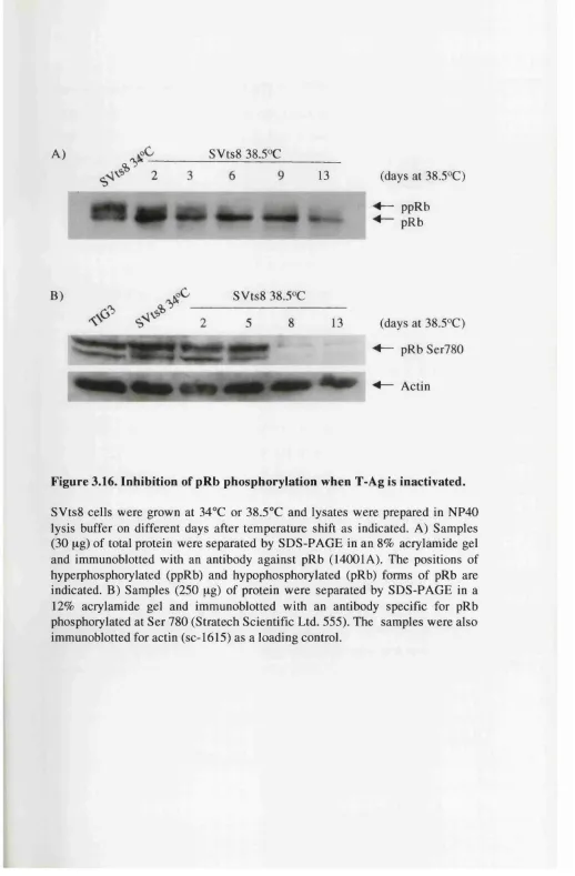
Figure 3.16. Inhibition of pRb phosphorylation when T-Ag is inactivated 115
Figure 3.17. Inhibition of CDK2 kinase activity when T-Ag is inactivated 117
Page n”
Chapter 4
Figure 4.1. Association of CDK4 and CDK6 with cyclin D1 in TIG3
fibroblasts 123
Figure 4.2. Size fractionation of cyclin-CDK-CKI complexes from young,
early senescent and late senescent TIG3 fibroblast cell lysates 125
Figure 4.3. Size fractionation of CDK4 complexes in young, early senescent and late senescent TIG3 fibroblast cell lysates 127
Figure 4.4. Size fractionation of CDK6 complexes in young and late
senescent TIG3 fibroblast cell lysates 129
Figure 4.5. Cyclin-CDK-CKI interactions in TIG/pBabe, TIG/E6 and
TIG/E7 cells 131
Figure 4.6. Size fractionation of cyclin-CDK-CKI complexes in
TIG/pBabe and TIG/E6 cells 134
Figure 4.7. Size fractionation of CDK4 complexes in TIG-ER cells
expressing E6 135
Figure 4.8. Size fractionation of p i c o m p l e x e s in TIG-ER cells
expressing E6 137
Figure 4.9. Size fractionation of cyclin-CDK-CKI complexes in
TIG/pBabe and TIG/E7 cells 138
Figure 4.10. Size fractionation of CDK4 complexes in TIG-ER cells
expressing E7 140
Figure 4.11. Size fractionation of plb^^^'*® complexes in TIG-ER cells
expressing E7 141
Figure 4.12. Lifespan extension in TIG-ER fibroblasts expressing E6 143
Figure 4.13. Upregulation of disrupts cyclin D1-CDK4 complexes
in TIG-ER cells expressing E6 145
Figure 4.14. Consequences of the Leiden deletion in the INK4a locus 147
Page n” Figure 4.15. Cyclin D1-CDK4 interactions in Leiden and Leiden/E6
fibroblasts 149
Chapter 5
Figure 5.1. R24P variant of arrests U20S and MCF7 cells 153 Figure 5.2. Inducible expression of the R24P variant of p i i n different
cell clones 155
Figure 5.3. G1 arrest after induced expression of the R24P variant
ofplti"""""' 156 Figure 5.4. Interaction of the R24P variant of with CDK4
and CDK6 158
Figure 5.5. R24P variant of p i6^^'*'' disrupts cyclin D 1-CDK6 interactions
in IPTG induced R24P2 cells 160
Figure 5.6. R24P variant of p i6®^’^'*“ inhibits the phosphorylation of pRb 161 Figure 5.7. Association of CDK4 and p i6”^"^“ at different times after
p 1 induction 164
Abbreviations
Agm absorbance
ARF alternative reading frame (pl4"^) ATCC American tissue culture collection ATP adenosine 5’-triphosphate
BCA bicinchoninic acid
bmi-1 B cell-specific Mo-MLV integration site 1 bp base pair(s)
BSA bovine serum albumin BrdU 5-bromo-2'-deoxyuridine
°C degrees Centigrade CAK CDK activating kinase cdc/CDC cell division cycle
CDK(s) cyclin dependent kinase(s) cDNA complementary DNA
CIP/KIP CDK interacting protein / kinase inhibitory protein CKI CDK inhibitor
CO2 carbon dioxide
C-terminus carboxy terminus
DMEM Dulbecco’s modified Eagle medium DMSO dimethyl sulphoxide
DNA deoxyribonucleic acid
dNTP deoxynucleoside 5-triphosphate DOPE dioleyl phosphatidylethanolamine
DOSPA 2,2-dioleyloxy-N-[2(sperminecarboxyamido)ethyl]-N, N-dimethyl-1 propanium trifluoroacetate
DTT dithiothreitol
E6 Human papilloma virus type 16 E6 protein E7 Human papilloma virus type 16 E7 protein
ECL enhanced chemiluminescence
E. coli Escherichia coli
EDTA ethylenediaminetetraacetic acid (disodium salt)
EGTA ethylene glycol-bis(p-aminoethyl ether)-N,N,N',N'-tetraacetic acid ER ecotropic receptor
FACS fluorescence activated cell sorting PCS foetal calf serum
FITC fluorescein isothiocyanate
g gravity (centrifugal force) GO quiescence
G1 gap phase 1 of the cell division cycle G2 gap phase 2 of the cell division cycle GST glutathione S transferase
h hour
HBS HEPES buffered saline HDF(s) human diploid fibroblast(s)
HEPES N-(2-hydroxyethyl)piperazine-N’-(2-ethanesulfonic acid) HFK human foreskin kératinocytes
HiSg hexahistidine
HMEC human mammary epithelial cells HPV16 human papilloma vims type 16 HRP horseradish peroxidase
hTERT human telomerase reverse transcriptase hTR human telomerase RNA
ICRF Imperial Cancer Research Fund Ig immunoglobulin
INK4 inhibitor of CDK4 BP immunoprécipitation
IPTG isopropyl P-D-thiogalactopyranoside
kb kilobase
M moles per litre MO mortality stage 0 M l mortality stage 1 M2 mortality stage 2
MAPK mitogen-activated protein kinase MDM2 murine double minute 2
MEFs mouse embryo fibroblasts MEK MAPK/ERK kinase
min minute
MPF maturation/M-phase promoting factor N-terminus amino terminus
NP40 nonidet P40 (octylphenoxy polyetoxy ethanol) PAGE polyacrylamide gel electrophoresis
PBSA phosphate buffered saline PCNA proliferating cell nuclear antigen PCR polymerase chain reaction PD(s) population doubling(s)
PMSF phenylmethylsulphonyl fluoride pRb retinoblastoma protein
PVDF polyvinylidene difluoride RNA ribonucleic acid
rpm revolutions per minute R point restriction point
SA-p-gal senescence-associated P-galactosidase
S. cerevisiae Saccharomyces cerevisiae
SDS sodium dodecyl sulphate
sec second
Sf9 Spodoptera frugiperda
S phase DNA synthesis phase of the cell division cycle
S. pombe Schizosaccharomyces pombe
SV40 Simian Virus 40
T-Ag SV40 large T antigen
TEMED N,N,N',N'-tetramethylethylenedianiine TGF-p transforming growth factor beta
TRAP Telomeric Repeat Amplification Protocol Tris tris(hydroxymethyl) aminomethane Tween 20 polyoxyethylenesorbitan monolaurate UV ultra violet
v/v volume for volume w/v weight for volume
g m
n P
Chapter 1
Introduction
Cell division is fundamental to all living organisms and is subject to tight regulatory controls. Accurate cell division is dependent on the correct order of events, mechanisms to guard against errors and the ability to respond to the changing environment. In multicellular organisms, it is necessary to control cell numbers and limit the proliferative capacity of cells (i.e. senescence). Failure to control these processes can result in aberrant division, aneuploidy, and unrestrained proliferation, characteristics associated with cancer cells. This thesis describes experiments relating to cell cycle control and investigates the mechanism of senescence.
1.1. The Cell Cycle
The process of eukaryotic cell division, the cell cycle, is divided into four phases. DNA replication (S phase) and mitosis (M phase; i.e. the division of the nucleus into daughter nuclei) are the two key events and are separated by so-called gap phases (G1 and G2). G1 occurs prior to S phase, and G2 prior to M phase. DNA synthesis and mitosis are strictly ordered events, and their respective onsets and completions are carefully orchestrated, indicating that specific control mechanisms dictate the strict sequential order of events. These control mechanisms or checkpoints are generally feedback mechanisms ensuring that the next phase of the cell cycle is not initiated before the preceding step is completed (Hartwell, 1989). Checkpoint mechanisms also ensure the accurate transmission of genetic information, which is essential for the long-term survival of the organism. At the Gl/S checkpoint, the cell integrates positive and negative environmental signals, as well as internal signals, to determine whether or not it should become committed to enter a round of DNA replication. The second major checkpoint, the G2/M
checkpoint ensures the cell only divides if DNA synthesis is complete. At this point, the cell also integrates signals resulting from DNA repair, so that it only divides once the errors in the DNA have been repaired. If the cellular DNA is damaged by external agents, entry into S phase can be delayed or the cell may undergo programmed cell death (apoptosis). There are also mechanisms that limit the proliferative capacity of somatic cells protecting against the unrestrained proliferation of individual cells, as well as potentially contributing to the ageing of the organism.
Normal animal cells possess a unique regulatory mechanism that enables them to switch between proliferative and quiescent states. When conditions are sub- optimal, for example, if cell density is high or serum or nutrients are insufficient, cells will cease proliferating and enter a state termed quiescence or GO. When conditions become more favourable and cells are supplemented with complete medium, they can re-enter the cycle and proliferate, first synthesising their DNA before dividing. The ability to switch between quiescence and proliferation is determined at a stage in G l, the restriction point, after which the cell no longer requires serum and protein synthesis to enter S phase (Pardee, 1974). Once the cell has traversed the restriction point, it can complete the remainder of the cell cycle in the absence of further exposure to mitogens. Alternatively, if the signals received during this period have not been propitious for growth, the cell may arrest and re enter GO, or may commit itself to enter into a postmitotic, differentiated state. The manunalian cell division cycle is summarised in Figure 1.1. Most animal cells in vivo exist in a non-proliferating state in which they remain viable and metabolically active. These cells arise from proliferating cells whose metabolic patterns are switched to quiescence at some time during differentiation.
GO
Restriction point G2/M
checkpoint
0 2
Figure 1.1. Mammalian cell division cycle
constitutive or aberrant activation. Tumour suppressor genes, as their name implies, are negative regulators of cellular proliferation and their inactivation by mutation results in the loss of a crucial brake on tumour growth. One hallmark of a tumour suppressor is that recessive mutant alleles occur in the germ line of cancer prone families. As originally proposed by Knudson, the disease develops if the remaining wild type allele is inactivated by somatic mutation or chromosome loss (Knudson, 1971). The same genes can also be involved in the formation of sporadic tumours if both alleles are disrupted by somatic mutations.
1.2. Cyclins and cyclin-dependent kinases
Our present knowledge of cell cycle regulation stems largely from genetic analyses in fission yeast {Schizosaccharomyces pombe) and budding yeast
{Saccharomyces cerevisiae), and a biochemical analysis using various invertebrate and vertebrate oocytes and eggs. In both types of yeast, a large collection of cell division cycle (cdc) mutants were isolated that arrested the cell cycle at specific points, including START, a point in G l analogous to the restriction point in mammalian cells. The analysis of these mutants led to the identification of a kinase, designated in budding yeast and p34‘^‘‘‘"^ in fission yeast, essential for passage through START and mitosis (Murray and Kirschner, 1989). The human homologue designated CDC2 was cloned by complementation of a fission yeast
cdc2^' mutant strain (Lee and Nurse, 1987). As with the yeast proteins, human p3 4CDC2 found to be phosphorylated and to have protein kinase activity (Draetta
etal., 1987).
identified in sea urchin eggs whose levels cycle because they are abruptly degraded at mitosis (Evans et al., 1983).
Since the cloning of a number of closely related mammalian kinases have been identified. These kinases are named cyclin-dependent kinases (CDKs) because their activity requires association with a regulatory subunit, or cyclin, and function as serine/threonine protein kinases that target critical substrates required for cell cycle progression. The major transitions in the cycle are regulated by the sequential activation of CDKs, which themselves are tightly controlled by complex mechanisms.
Although yeast cell cycle transitions are governed by a single kinase, higher eukaryotes have evolved multiple CDKs to regulate different stages of their cell cycle (Meyerson and Harlow, 1994). These different CDKs are highly conserved, being closely related in size (35-40 kDa), and sequence (>40% identity) (Morgan, 1995). The typical CDK catalytic subunit contains a 300 amino acid catalytic core that is completely inactive when monomeric and unphosphorylated. The crystal structure of human CDK2, like other protein kinases, reveals a smaller N-terminal lobe, dominated by a beta sheet and the large PSTAIRE (single letter amino acid code) helix, and C-terminal lobe that is primarily helical (De Bondt et al., 1993; Morgan, 1996). Monomeric CDK2 is inactive because of two structural constraints, the substrate binding site is blocked by an extended flexible loop (the T-loop) and a number of key residues involved in ATP-phosphate binding are unfavourably positioned for efficient phosphate transfer (De Bondt et ah, 1993; Morgan, 1996). The structure of CDK2 is altered upon cyclin binding and phosphorylation.
The cyclins are a remarkably diverse family of proteins, ranging in size from about 35 to 90 kDa. The sequence homology within the family is concentrated in a 100-residue region known as the cyclin box, which is necessary for CDK binding and activation (Kobayashi et ah, 1992; Lees and Harlow, 1993). The structure of cyclin A has been determined as a monomer (Brown et ah, 1995) and in a complex with CDK2 (Jeffrey et ah, 1995b). The two structures are essentially the same indicating that CDK2 binding does not alter the structure of cyclin A. The structure of a cyclin consists of two characteristic folds, each represented by five a-helical
bundles connected to one another by a short linker peptide (Brown et al., 1995). The cyclin box is composed of the first five-helix core. The second five-helix bundle does not show sequence similarities despite having the same structure. In mammalian cells nine cyclins have been identified to date, designated A-I, and have been divided into a number of classes based on sequence similarity and physiological function.
1.2.1. Regulation of CDK activity
The levels of CDKs remain relatively constant throughout the cell cycle but their activity is regulated by complex mechanisms (Figure 1.2.). The most significant is the binding of the regulatory cyclin (Connell-Crowley et al., 1993). For example, cyclin A binding to CDK2 increases kinase activity by several orders of magnitude (Connell-Crowley et a l, 1993). The reason for this becomes apparent when the cyclin A-CDK2 crystal structure is compared to monomeric CDK2 (Jeffrey et al., 1995b). Cyclin A binding imposes major structural changes within the PSTAIRE region of CDK2. The T-loop no longer blocks the substrate binding site, and the critical Thr 160 residue within the loop is further exposed making it more accessible for phosphorylation by the CDK activating kinase, CAK (see below). Full CDK activation requires phosphorylation at this conserved threonine (Thr 161 in human CDC2, Thr 160 in CDK2, Thr 172 in CDK4) (Desai et a l, 1995; Gould et a l, 1991; Krek and Nigg, 1991; Solomon et a l, 1992). The effect of phosphorylation is to stabilise the cyclin-CDK interaction, and possibly improve binding to the protein substrate (Connell-Crowley et a l, 1993; Russo et a l, 1996).
CAK activity is attributed to a complex between CDK7 (Fesquet et a l,
1993; Poon et a l, 1993; Solomon et a l, 1993) and cyclin H (Fisher and Morgan, 1994; Makela et a l, 1995). The association of CDK7 with cyclin H is enhanced by an assembly factor MATl (Devault et a l, 1995; Fisher et a l, 1995; Tassan et a l,
Ubiquitin-mediated proteolysis cyclin
synthesis
CKI binding
complex
phosphorylation T t r n f ' assembly
CAK ---^ T'GO'
T14YI5 dephosphorylation
KAPl
phosphorylation dephosphorylation WEEl/MYTl CDC25
Figure 1.2. Principles of CDK regulation
transcription initiation and in nucleotide excision repair (Feaver et a l, 1994; Roy et al., 1994; Serizawa et al., 1995; Shiekhattar et al., 1995; Svejstrup et al., 1996). In line with this more general role, CAK is a nuclear enzyme with constitutive activity during the cell cycle (Poon et al., 1993; Tassan et al., 1995). Thr 160 is dephosphorylated by KAPl phosphatase (Poon and Hunter, 1995).
As well as being activated by Thr phosphorylation CDKs are negatively regulated by phosphorylation at a conserved tyrosine, (Tyr 15 in CDC2 Gould et al., 1991) and in some cases at the adjacent threonine, Thr 14, (Krek and Nigg,
1991; Norbury et al., 1991). These residues are hidden beneath the T-loop in monomeric CDK2, but when cyclin A is bound to CDK2 they are relatively accessible. It is likely that the phosphorylation of these residues hinders substrate access to the catalytic site of the enzyme (Morgan, 1997). This inhibitory phosphorylation contributes to the timing of mitosis in many organisms (Dunphy, 1994; Lew and Kombluth, 1996). Prior to mitosis, CDC2-cyclin B complexes are held in an inactive state by phosphorylation at these sites. At the end of 02, rapid dephosphorylation prompts CDC2 activation and mitosis. Phosphorylation of Thr 14 and Tyr 15 is carried out by the WEEl and MYTl phosphatases (Liu et al.,
1997; Mueller et a l, 1995). Déphosphorylation of both sites is carried out by dual specificity phosphatases of the CDC25 family (Draetta and Eckstein, 1997).
Most cyclin-CDK complexes interact with high affinity without the need for assembly factors (Connell-Crowley e ta l, 1993; Desai et a l, 1995; Morgan, 1995). The CAK complex, CDK7-cyclin H, is an obvious exception since it requires the presence of MATl (see above). The assembly of cyclin D-CDK4 complexes appears to be mediated by a molecule(s) whose activity is regulated by growth factors, because quiescent fibroblasts engineered to constitutively express cyclin D do not assemble cyclin D-CDK4 complexes (Matsushime et a l, 1994). A possible candidate is the human homologue of CDC37 which interacts with CDK4 in vivo
family of CDK inhibitors are also likely to be involved in cyclin D-CDK4 assembly (discussed in section 1.3.).
1.2.2. Regulation of cyclins
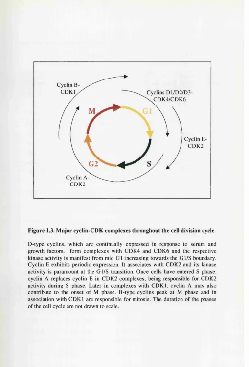
Each cyclin has a unique expression pattern and the timing of expression is a key determinant of the phase of the cell cycle at which their associated CDK is active (Figure 1.3.). A variety of cyclin-CDK complexes are formed during the cell cycle, and each is presumably dedicated to the phosphorylation of a precise set of target proteins. D-type cyclins are induced during early G l, and they bind and activate CDK4 and CDK6. Cyclin E-CDK2 and cyclin A-CDK2 complexes form later in G l as cells approach S phase. Cyclin B is synthesised during S phase and binds to CDC2 (CDKl), to activate it during G2 and M.
Expression of the D-type cyclins depends on the continuous presence of growth factors and growth factor withdrawal leads to rapid cyclin D destruction, regardless of the position of the cell in the cell cycle (Lukas et aL, 1996; Matsushime et al 1991). However, after entering S phase the cell can complete cell division without cyclin D expression (Ajchenbaum et at., 1993; Matsushime et al.,
1991). Growth factor signalling via the Ras-Raf-Mek pathway leads to increased expression of cyclin D1 (Filmus et al., 1994; Liu et a l, 1995; Lloyd et al., 1997; Sewing et al., 1997; Winston et a l, 1996). Although the level of D-type cyclins fluctuate little during the remainder of the cell cycle, the expression of cyclin A (Pines and Hunter, 1990), cyclin E (Dulic et al., 1992; Koff et al., 1992; Ohtsubo et al., 1995) and cyclin B (Pines and Hunter, 1990) is highly periodic.
In addition to transcriptional mechanisms, cyclin levels are controlled significantly by regulated proteolysis. Degradation of mitotic cyclins, the key event governing the exit from mitosis, is mediated through a conserved amino-terminal ‘destruction box’ that targets the proteins for degradation via the ubiquitin pathway (Glotzer et al., 1991). In contrast, yeast G l and human D-type cyclins lack destruction boxes but instead contain PEST sequences, rich in proline, serine, threonine and acidic amino acids. PEST motifs are found in a number of unstable
Cyclin B-
C D K l / Cyclins D1/D2/D3-
\C D K 4/C D K 6
Cyclin E- CDK2
G2
Cyclin A- CDK2
Figure 1.3. Major cyclin-CDK complexes throughout the cell division cycle
proteins and have been proposed to target these proteins for rapid turnover (Rogers
et a l, 1986). Cyclin E lacks both a destruction box and a strong PEST sequence. It is targeted for degradation via autophosphorylation of cyclin E-CDK2 on residue Thr 380 of cyclin E (Clurman et a l, 1996; Won and Reed, 1996). Cyclin D1 is targeted for degradation by phosphorylation on Thr 286 (Diehl et a l, 1997) by glycogen synthase kinase-313 (GSK-3p) (Diehl e ta l, 1998).
The third mechanism to influence cyclin levels is subcellular localisation. During G l, cyclin D1 progressively accumulates in the nucleus, but during S phase cyclin D1 relocalises to the cytoplasm (Baldin et a l, 1993). In direct contrast to cyclin D l, GSK-3p is predominantly cytoplasmic during G l phase, but a significant proportion enters the nucleus during S phase. A highly stable cyclin D l mutant, with an alanine substituted for threonine at position 286 (T286A), is unable to be phosphorylated by GSK-3p, and remains in the nucleus throughout the cell cycle (Diehl e ta l, 1998).
1.3. CDK inhibitor families
An additional level of cell cycle regulation is achieved through expression of CDK inhibitor (CKI) proteins. CKIs are negative regulatory proteins that bind to cyclin-CDK complexes or directly to CDKs and inhibit their catalytic activity (Sherr and Roberts, 1995). There are two distinct families of CKI based on primary sequence comparisons and probable modes of action.
1.3.1. The CIP/KIP family
The CIP/KIP family includes p21^^^ (El-Deiry et a l, 1993; Gu et a l, 1993; Harper et a l, 1993; Noda et a l, 1994), p27^^^ (Hengst et a l, 1994; Polyak et a l,
1994a; Polyak et a l, 1994b; Toyoshima and Hunter, 1994) and p57*^“’^ (Lee et a l,
1995; Matsuoka et a l, 1995). p21^^^^ was cloned via a number of approaches, implicating it in a wide range of cellular activities, such as checkpoint control (Gu
et a l, 1993; Harper et a l, 1993; Xiong et a l, 1993), differentiation (Halevy et a l.
1995; Jiang and Fisher, 1993; Steinman et al., 1994), senescence (Noda et al. 1994; Tahara et a l, 1995; Zhang et a l, 1994) and apoptosis (El-Deiry et a l, 1994; El- Deiry et a l, 1993). p27™*^ appears to be primarily responsible for regulating CDK activity in response to extracellular growth inhibitory signals (Polyak et a l, 1994b; Slingerland et a l, 1994; Toyoshima and Hunter, 1994) and restricting cellular proliferation during the course of development (Fero et a l, 1996; Kiyokawa et a l,
1996; Nakayama et a l, 1996). p21^^^ and p27^* are widely expressed in tissues, unlike p57^^^ which is highly tissue specific (Lee et a l, 1995; Matsuoka et a l,
1995). There is a good correlation between p57™^ expression and the differentiated state of cells, suggesting that p57^^^ plays a role in cell cycle exit associated with terminal differentiation during mouse development (Matsuoka et a l, 1995).
The proteins are grouped together largely because they share a 60 amino acid domain of similarity (39% to 47%) in the amino-terminal half of each protein which is necessary for cyclin binding and CDK inhibitory functions. They also have nuclear localisation signals near the carboxy terminus, but outside these regions, the three proteins have no resemblance except for a short segment of similarity near the carboxy-terminus of p27^^^^ and p57^^^^ (Chen et a l, 1995; Luo et a l, 1995; Toyoshima and Hunter, 1994).
The CIP/KIP proteins exhibit a broad specificity for cyclin-CDK complexes
in vitro, binding efficiently to cyclin A-CDK2, cyclin E-CDK2, cyclin D-CDK4 and less efficiently to cyclin B-cdc2 (El-Deiry et a l, 1993; Harper et a l, 1993; Lee
et a l, 1995; Matsuoka et a l, 1995; Polyak et a l, 1994b; Toyoshima and Hunter, 1994). p21^“’* and p27^^^^ bind to cyclin-CDK complexes, making contacts with both subunits but do not associate with kinase subunits unless a cyclin is present (Chen et a l, 1995; Hall et a l, 1995; Harper et a l, 1995). CKI association with cyclin-CDK complexes in vitro is correlated with a reduction in CDK kinase activity (Harper et a l, 1993; Lee et a l, 1995; Matsuoka et a l, 1995; Polyak et a l,
role as inhibitors of G l cyclin-CDK complexes. However, cell lines engineered for inducible expression of p21^“’* elicit both a G l and a G2/M arrest (Bates et al.,
1998b; Cayrol et al., 1998; Medema et al., 1998), and recent reports have indicated other functional activities for the CIP/KIP proteins apart from CDK inhibition (see below).
Surprisingly for a protein which blocks cell cycle progression, p21^^^^ expression was also shown to be induced when quiescent cells were stimulated to proliferate (Li et al., 1994a; Noda et al., 1994), and cyclin-CDK complexes in proliferating cells have been reported to include p21^°*^ (Harper et al., 1995; Zhang
et al., 1994). This apparent paradox led to the suggestion that p21^*^ can exist in active and inactive cyclin complexes depending on the concentration of p21^^^^ (Zhang et al., 1994). It was proposed that cyclin-CDK complexes are active when associated with one molecule of p21^^^ and inactive when they contain multiple p21^^^ molecules, at least for cyclin D-CDK4 complexes (Harper et a l, 1995; Zhang et al., 1994). These results run contrary to the crystallographic data on cyclin A-CDK-p27*^^^* ternary complexes (Russo et al., 1996). The structure reveals that p2 7 Kipi separate binding sites on the cyclin and CDK subunits consistent with
cooperative binding, but indicates that complexes containing multiple p27^* molecules would be thermodynamically unstable (Russo et al., 1996). Subsequent
in vivo data confirmed the structural indications, that cyclin A-CDK2 complexes are fully inhibited by one molecule of p21 (Hengst et al., 1998).
However, it may not be appropriate to extrapolate data for cyclin A-CDK2 complexes to cyclin D-CDK4 complexes. The association of cyclin D with CDK4 may require an assembly factor (Kato et a l, 1994; Matsushime et al., 1994) and there is evidence that the CIP/KIP proteins may perform such a function promoting kinase activity at low concentrations and inhibiting kinase activity at higher concentrations (LaBaer et al., 1997). p21^^' and p27™’* appear to target cyclin D l- CDK4 to the nucleus (Cheng et a l, 1999; LaBaer et a l, 1997; Reynisdôttir and Massagué, 1997) and in the absence of p 2 l‘^°’* and p27^^’’\ the majority of the cyclin D l remains in the cytoplasm during the cell cycle (Cheng et a l, 1999). The
current data indicate that although CIP/KIP proteins can exist in both CDK4 and CDK2 complexes the latter are the prime targets for inhibition.
Although there have been reports that p21^^^^ expression during the cell cycle is controlled by the E2F family of transcription factors (Hiyama et al., 1998; and see later in section 1.4.3.) the major regulator of p21^“** is believed to be the p53 tumour suppressor (see section 1.6.). Thus, p53 binding sites are present in the p21^^^ promoter and it was cloned in a screen for p53 responsive genes (El-Deiry et al. 1993; El-Deiry gr a/. 1995).
In contrast, p27’“ ’* transcription is constant throughout the cell cycle but the protein is more stable in quiescent cells. p27™'^ is regulated in part by proteolysis through the ubiquitin/proteasome pathway, and it has been suggested that quiescent cells contain lower amounts of ubiquitinating activity compared to proliferating cells (Pagano etal., 1995).
1.3.2. The INK4 family
Members of the INK4 family of inhibitors are structurally unrelated to the CIP/KIP family, p lb ”^*^"*® is the prototype and the other family members (Serrano et a l, 1993), include p l 5 ™ (Hannon and Beach, 1994), plB'^*'^ (Hirai et a l, 1995) and pi9°^K4d (Quan et a l, 1996; Hirai et a l, 1995). p i6^^^"^“ was first identified through its association with CDK4 in a two-hybrid assay system (Serrano et a l,
1993). It specifically binds CDK4 and CDK6, and because of its specificity as an inhibitor of CDK4 (and CDK6), the protein was designated INK4.
presumably required to provide stabilising interactions between the helices and P- strands of adjacent modules. The genes encoding pl6°^*^'^“ and pl5^^"^^ are tandemly linked on human chromosome 9p21 and the proteins share 80% similarity at the primary sequence levels (Hannon and Beach, 1994). It is therefore clear that pl6iNK4a pj^iNK4b have arisen from a relatively recent gene duplication. pjgiNK4c p2çiNK4d^ Qn thc othcr hand, are only 40-50% identical to p lb ”^*^"^® and to each other, and are found on different chromosomes.
However, the four proteins are indistinguishable biochemically, all being specific inhibitors of CDK4 and CDK6 and able to impose a G l arrest when overexpressed in various cell hues (Guan et al., 1994; Lukas et al., 1995; Serrano et al., 1995; Sherr and Roberts, 1995). They bind directly to CDK4 and CDK6 in vitro, and there is no evidence that they associate with other CDKs (Guan et al.,
1994; Guan et al., 1996; Hara et al., 1996; Hirai et al., 1995; Parry et a i, 1995; Serrano et al., 1993). The INK4 proteins are thought to induce a G l arrest by forming binary associations with CDK4 and CDK6, preventing the D cyclins from interacting with CDK4/6. Support for this hypothesis comes from observations that in cells with high levels of endogenous p l 6 ^ ^ \ CDK4 is only associated with pl6iNK4a not with cyclin D (Hirai et al., 1995; Li et al., 1994b; McConnell et al.,
1999; Parry et al., 1995; Parry et al., 1999; Xiong et al., 1993) and in vitro,
prevents the binding of CDK4/6 to cyclin D (Guan et al., 1996; Lukas et al., 1995; Parry et al., 1995). However, it has also been reported that p i 6^^"^“ can bind to and inhibit preassembled cyclin D-CDK4/6 complexes in vitro, without dissociating cyclin D (Hirai et a l, 1995; Koh et al., 1995; Serrano et a l, 1993). In addition, after overexpression of pl5”^’^'‘‘’ or pl9^'*^, it is apparently possible to detect ternary complexes between cyclin D, CDK4 and INK4 proteins (Adachi et a l, 1997b; Hirai
et a l, 1995; Reynisdôttir and Massagué, 1997). This topic is discussed in more detail in Chapter 5. Although the INK4 proteins are specific inhibitors of CDK4 and CDK6, they can indirectly inhibit CDK2 activity (McConnell et a l, 1999; Reynisdôttir and Massagué, 1997) by influencing the redistribution of p27™"^ from cyclin D-CDK4 complexes to CDK2 complexes.
The structure of the INK4 proteins does not change significantly upon binding to CDK6 (Brotherton et a l, 1998; Russo et a l, 1998). plô"^'*® and p l9 ”^*^'“ bind to one side of the catalytic cleft, opposite where the cyclin would bind, interacting with both the N- and C-lobes of CDK6. However, the structure of CDK6 is distorted upon INK4 binding, principally affecting the ATP-binding site and indirectly blocking cyclin binding (Brotherton et al., 1998; Russo et al., 1998). The binding of an INK4 protein to CDK4/6 would effectively preclude a CIP/KIP protein binding to the same complex.
Despite the biochemical and structural similarities of the INK4 family members, only pl6^^^"^^ has the attributes of a tumour suppressor. This may be related to the observation that p i l e v e l s increase significantly as primary cells reach the end of their finite lifespan in culture (Alcorta et al., 1996; Hara et al.,
1996; Loughran et al., 1996; Reznikoff et al., 1996; Wong and Riabowol, 1996). This would be consistent with a role for pl6^^’^'‘‘* in establishing the G l arrest associated with senescence, and a need to escape senescence would provide a strong selection for the inactivation of plb^^^'^'* during the emergence of an immortal clone. The final confirmation that p i 6”^*^'^ was indeed a tumour suppressor came from the generation of plb^^^'^-null mice, p i 6’^’^'^“ proved non-essential for viability and proper development (Serrano et al., 1996).
1.3.3. One locus two proteins: and ARF
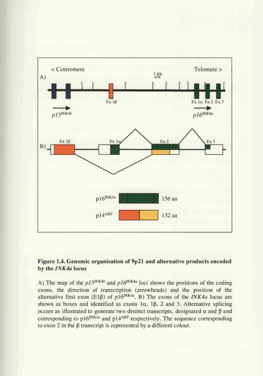
The plh^^*^"^® locus has the unusual capacity to give rise to two distinct transcripts from two different promoters (Figure 1.4.). p i6^^"^“ is encoded by exons la , 2 and 3, and an entirely unrelated protein p l4 "^, is encoded by exons ip, 2 and 3 with the shared exon 2 being encoded by an alternative reading frame (ARF) from that of p lb ^ ’^'^^ (Duro et al, 1995; Quelle et al., 1995a; Quelle et al., 1995b; Stone
< Centromere
A)
J _ L
E x ip
p l 5 l N K 4 b
Telomere >
W
Ex l a Ex 2 Ex 3►
B) J
Ex Ip Ex 3
p l6 lN K 4 a
p j ^ A R F
156 aa
132 aa
Figure 1.4. Genomic organisation of 9p21 and alternative products encoded by the INK4a locus
1.4. The Gl/S transition
The transition from a serum dependent state to a serum independent state coincides with the cell traversing the restriction point. The controls governing the restriction point have been the focus of attention for a number of years, leading to the identification of the retinoblastoma tumour suppressor protein as a key player in the Gl/S transition.
1.4.1. The retinoblastoma protein
The retinoblastoma susceptibility gene, RB-1 on chromosome 13ql4, encodes a ubiquitously expressed 928 amino acid (110 kDa) nuclear phosphoprotein designated pRb (Friend et al., 1987; Lee et al., 1987a; Lee et al.,
1987b). There is a wealth of genetic evidence implicating pRb as a tumour suppressor, with the RB-1 gene being mutated in 30% of human tumours (Weinberg, 1995). Individuals that inherit a mutant copy of the gene are predisposed to the development of childhood retinoblastoma and, in later life sarcoma. Furthermore, when wild-type pRb is re-introduced into retinoblastoma or osteosarcoma cell lines that do not express pRb, cellular proliferation is inhibited (Huang etal., 1988).
pRb is the prototype of a family of proteins that include p i07 (Ewen et al.,
1.4.2. Control of pRb phosphorylation
The levels of pRb do not vary substantially during the cell cycle, although levels decline in quiescent or arrested cells. The major regulation of pRb function is by cell cycle dependent phosphorylation (Buchkovich et al., 1989; Chen et al.,
1989; DeCaprio et al., 1992; DeCaprio et al., 1989; Mihara et al., 1989). These phosphorylation events change the mobility of pRb in denaturing acrylamide gels. Hyperphosphorylated forms of pRb migrate considerably slower than hypophosphorylated pRb. Analysis of pRb in serum stimulated cells revealed that it exists in a hypophosphorylated state in GO and early Gl phase but undergoes rapid phosphorylation in late Gl and remains in a hyperphosphorylated state until the end of mitosis where it is dephosphorylated by protein phosphatase type 1 (Durfee et al., 1993). Suppression of cellular proliferation by serum starvation is associated with the dephosphorylation of pRb (Stein et al. 1990) and DNA viral oncoproteins bind preferentially to the hypophosphorylated form of pRb (DeCaprio et al., 1988; Dyson et al., 1989; Ludlow et al., 1989; Whyte et al., 1988), suggesting that this form is normally active in growth suppression. The hyperphosphorylated form is conversely the inactive form. DNA viral oncoproteins bind to pRb through a specific motif, LxCxE (in the single letter code for amino acids, where x is any amino acid) and this motif is necessary for cellular transformation (Dowdy et al.,
1993). The LxCxE motif is conserved in a number of viral oncoproteins, as well as in several cellular pRb-binding proteins (Dowdy et a l, 1993).
There are sixteen consensus phosphorylation sites for CDKs in the primary sequence of human pRb (Connell-Crowley et al., 1997; Lees et al., 1991; Zarkowska and Mittnacht, 1997). It is not clear whether pRb is phosphorylated on all these sites in vivo and differentially phosphorylated forms of pRb are observed (DeCaprio et a i, 1992; Mittnacht et al., 1994). Furthermore, the contribution of individual phosphorylation sites to the inactivation of pRb remains controversial. Both Ser 608 and Ser 780 have been identified as among the sites that are initially phosphorylated (Kitagawa et al., 1996; Zarkowska and Mittnacht, 1997), and the latter site is believed to be specifically targeted by cyclin D-CDK4 complexes
(Kitagawa et al., 1996). The phosphorylation of different sites on pRb affects its ability to interact with various partner proteins. For example, Ser 780 phosphorylation prevents pRb from binding E2F (Kitagawa et al., 1996); Ser 780/811 phosphorylation prevents pRb binding to the transcription factor c-abl (Knudsen and Wang, 1996); while phosphorylation of Thr 821/826 abolishes pRb binding to LxCxE-containing proteins such as SV40 T-antigen (Knudsen and Wang, 1996; Zarkowska and Mittnacht, 1997).
Purified cyclin D1-CDK4/6, cyclin E-CDK2 and cyclin A-CDK2 complexes are all capable of phosphorylating pRb in vitro. Cyclin E and D l regulate different rate-limiting events in the Gl/S transition. When overexpressed concurrently they accelerate the G l/S transition (Resnitzky et al., 1994), but their individual expression has different outcomes (Resnitzky and Reed, 1995). The overexpression of cyclin D l leads to the immediate appearance of hyperphosphorylated pRb, while cyclin E overexpression does not (Resnitzky and Reed, 1995). Furthermore, only overexpression of cyclin E, and not cyclin D l, is capable of overcoming a Gl arrest imposed by p i 6^’^’^'^“ or a non-phosphorylatable form of pRb (Lukas et al., 1997). Cyclin E remains both rate-limiting and essential for the Gl/S transition in pRb- positive and pRb-negative cells (Ohtsubo et al., 1995; Wimmel et al., 1994). These results implicate pRb as the exclusive target of cyclin D-associated kinases, while cyclin E-associated kinases probably target additional factors necessary for cell cycle progression.
1992; Meyerson and Harlow, 1994; Moberg et a l, 1996). Inhibition of CDK2 kinase activity, through use of a dominant negative CDK2, results in partially phosphorylated pRb (Lundberg and Weinberg, 1998). This result not only suggests that cyclin D-CDK4/6 complexes are unable to fully inactivate pRb in vivo but they also indicate that D-type cyclin-directed phosphorylation of pRb is restricted to a subset of sites on pRb (Lundberg and Weinberg, 1998). Furthermore, cyclin E- CDK2 is unable to phosphorylate pRb in the absence of prior phosphorylation by cyclin D-CDK4/6 complexes (Lundberg and Weinberg, 1998).
1.4.3. Regulation of E2F transcription factors
The transcription factor E2F was originally identified as a cellular protein whose DNA-binding activity was responsible for the activation of the adenovirus E2A promoter (Kovesdi et a/., 1987; La Thangue and Rigby, 1987; Yee et al.,
1987). E2F turns out to be a series of heterodimers (Helin et at., 1993b) composed of any one of six E2F subunits (E2F 1-6), in combination with either of two DP polypeptides, DP-1 or DP-2 (Dyson, 1998). E2F 1-5 act predominantly as transcriptional activators, having both a DNA-binding domain and transcriptional activation domain (Dyson, 1998; Helin, 1998). E2F-6, on the other hand, only has a DNA-binding domain and is therefore likely to repress transcription by binding to E2F sites in promoters, preventing access to other members of the family (Morkel
et at., 1997; Trimarchi et at., 1998). When overexpressed, E2F 1-5 induce the expression of different patterns of E2F target genes to differing degrees (DeGregori
etal., 1997).
Although "free" E2F dimers can be detected in cells, a major proportion of the E2F occurs in complexes with pocket proteins (Bagchi et al., 1991; Bandara and La Thangue, 1991; Cao et al., 1992; Chellappan et al., 1991; Chittenden et al.,
1991; Cobrinik et al., 1993; Devoto et al., 1992; Shirodkar et al., 1992). pRb, pl07 and p i 30 bind to different E2F molecules at various times during the cell cycle, antagonising the transcriptional activity of the otherwise free E2F (Sardet et al.,
1997). pRb binds to E2F-1 through E2F-4, while p i07 and p i 30 preferentially bind
E2F-4 and E2F-5. pRb-E2F complexes are found mostly during Gl phase, while pl07-E2F-4 complexes persist throughout the cell cycle and contain cyclin E or cyclin A at different cell cycle phases. Complexes of E2F-4 and E2F-5 with p i 30, predominate in quiescent cells.
E2F binding sites are found in the promoters of several genes that encode proteins necessary for DNA synthesis, such as dihydrofolate reductase (DHFR), thymidine kinase and DNA polymerase a , and E2F binding is required to activate transcription of these genes (DeGregori et a l, 1995a). The current model of Gl progression suggests that, in early G l, pRb binds the transactivation domain of E2F, directly inhibiting transactivation by E2F (Flemington et al., 1993; Helin et a l,
1993a; Hiebert et a l, 1992). This is achieved because pRb blocks the formation of the preinitiation complex, a key step in commencing transcription (Ross et a l,
1999). The phosphorylation of pRb relieves E2F-1, resulting in E2F-1 dependent activity and the transcription of genes required for S phase (DeGregori et a l,
1995b; Hollingsworth Jr et a l, 1993; Weinberg, 1995). In support of this model, ectopic expression of E2F-1 blocks cells from entering quiescence, and can induce already quiescent cells to enter S phase (Johnson et a l, 1993; Qin et a l, 1994). The control of the Gl/S transition is summarised in Figure 1.5.
Mitogenic signals
i
cyclin D cyclin D
CDK4/6) < CDK4/6
cyclin E
pl5,pl6, 18,pl9
t
Anti-mitogenic signals
Figure 1.5. Regulation of the Gl/S transition
Surprisingly, E2F-1 can function as a tumour suppressor or an oncogene. E2F-1 knockout mice exhibit a broad range of tumours (Field et al., 1996; Yamasaki et al., 1996), yet overexpression of E2F-1 in transgenic mice promotes tumorigenesis (Pierce et al., 1998). Deregulated expression of E2F-1 can lead to the transformation of an established rat embryo fibroblast cell line (Singh et al., 1994), or, in cooperation with an activated Ras oncogene, can lead to transformation of primary rat embryo cells (Johnson et al., 1994). It is likely that the tumour suppressive qualities of E2F-1 are due, at least in part, to its ability to repress gene expression in conjunction with pRb (Dyson, 1998; Yamasaki et al., 1998). Additionally, when overexpressed in normal cells, E2F-1 causes high levels of apoptosis (Kowalik et al., 1995; Qin et al., 1994; Shan and Lee, 1994; Wu and Levine, 1994). Exactly how E2F-1 induces apoptosis is unclear", but this will be discussed in greater detail in section 1.6.4. Direct interaction of pRb with E2F does prevent E2F-induced apoptosis. Since the transactivation and apoptotic functions of E2F-1 are separable, the ability of pRb to inhibit E2F-induced apoptosis is not due to suppression of the E2F-1 transactivation function (Hsieh et al., 1997).
1.4.4. Exogenous signals affecting pRb phosphorylation
1.5. The cyclin D l-pl6 -pRb pathway in human cancer
Cancer cells frequently show some degree of dysregulation of restriction point control. Indeed, since a high proportion of tumours have alterations in at least one of the main regulators of this transition, particularly pRb, CDK4, cyclin D l and pl6iNK4a (pigure 1.6.) (Hall and Peters, 1996; Ruas and Peters, 1998; Sherr, 1996), that it has been extrapolated that all cancers may have defects in this regulatory mechanism.
1.5.1. Inverse correlation of cyclin D l, pl6*'^’^‘*“ and pRb mutations
The functional links between cyclin D l, CDK4, plb^^'^'^ and pRb make it possible to build a simple rationale for these observations. Thus, loss of pRb function would be expected to result in unrestrained proliferation. This is not entirely true as evidenced by cells from RbV^' nullizygous mouse embryos which show normal development up to day 4 and then die due to a failure in erythropoiesis (Jacks et al, 1992). The overexpression of cyclin Dl and CDK4, which occurs as a consequence of DNA amplification and other mechanisms, would promote the phosphorylation and hence inactivation of pRb, again leading to uninhibited growth. However, ectopic expression of cyclin D l has quite varied outcomes, depending on the host cell and levels of expression achieved. Finally, loss of pl6iNK4a fujiQtion would permit unrestrained phosphorylation of pRb by CDK4.
Despite its naivety, this simple model can in part account for the fact that alterations in components of the pathway tend to be mutually exclusive. This is most readily apparent with p lb ^ ’^'^® and pRb, where there is an inverse correlation in their expression/mutation (lung cancers) (Okamato et al., 1995; Otterson et al.,
1994; Shapiro et al., 1995). Since the ability of to arrest cells is dependent on pRb (Fâhraeus et al., 1996; Guan et a l, 1994; Koh et al., 1995; Lukas et al.,
1995; Medema et al., 1995; Serrano et al., 1995), there is no additional selection against pl6^’^’^'^^ in pRb-negative cells. Indeed pRb-negative cells generally have abnormally high levels of plb*^*^'^^ (Hara et al., 1996; Li et al., 1994b). This is partly
Amplification Translocation Retroviral insertion
Cyclin Dl
CDK4
pl6
pRb
G2
Mutation Deletion Tumour viruses
Amplification Mutation
Mutation Deletion Méthylation
Figure 1.6. Perturbations in the pRb-cyclin D-pl6'^*^** pathway in human cancers
explained by a feedback loop through which pRb negatively regulates to a fairly modest degree (Hara et a l, 1996), and partly by the accumulation of p i 6^"^“ that accompanies replicative senescence (see later) (Alcorta et a l, 1996; Hara et a l,
1996; Wong and Riabowol, 1996). In such settings, there can be enough to sequester all the available CDK4 and CDK6 so that cyclin D-CDK4 complexes are no longer detectable (Bates et a l, 1994; Parry et a l, 1995; Tam et a l, 1994; Xiong
e ta l, 1993).
1.6. The p53 tumour suppressor pathway
p53 is a well characterised tumour suppressor gene that is mutated or lost in over 50% of human cancers (Hollstein et a l, 1994). In addition to the frequent somatic mutations of p53 in sporadic cancers, germline mutations of one allele of the human gene are associated with an inborn predisposition to cancer, called Li- Fraumeni syndrome (Malkin et a l, 1990). Individuals with Li-Fraumeni syndrome are highly prone to the development of sarcomas and a variety of other tumour types, including carcinoma of the breast and brain. p53 null mice also develop tumours at an early age (Donehower et a l, 1992; Jacks et a l, 1994). p53 null mouse embryo fibroblasts fail to arrest in Gl following DNA damage, consistent with other indications that p53 is as a critical regulator of this checkpoint (Donehower et a l, 1992; Jacks et a l, 1994).
1.6.1. p53: structure and function
p53 functions as a transcription factor, capable of regulating the expression of a number of downstream genes. It is a modular protein consisting of five domains with distinct but inter-dependent functions. The core of p53 is a sequence specific DNA-binding domain that is resistant to proteases and contains a Zn^^ ion that is necessary for DNA binding activity (El-Deiry et a l, 1992). Greater than 90% of the documented missense mutations in p53 occur in the DNA binding domain (Hollstein et a l, 1994). The native protein binds DNA as a tetramer, a consequence
of intermolecular interaction of the tetramerisation domain in the carboxy-terminal half of the protein (Jeffrey et al.y 1995a). The carboxy-terminal domain, which is protease sensitive, and highly basic, is also subject to post-translational modifications, but the significance of these modifications remains uncertain. The DNA binding domain is separated from the activation domain by a series of repeated proline residues forming an SH3 binding domain. This region can interact with signal transduction molecules containing SH3 domains such as the c-abl oncogene (Yuan et al., 1996). The acidic transcriptional activation domain near the amino terminus enables p53 to interact with the basal transcription machinery, positively activating gene expression. This region is also involved in regulating the stability and activity of p53 via interactions with MDM2 (see later) (Haupt et al.,
1997; Kubbutat etal., 1997).
Under normal circumstances, p53 is expressed at low levels and has a short half life. In response to cellular stress, such as DNA damage (Hartwell, 1992), hypoxia (Graeber et al., 1996), nucleotide deprivation (Linke et al., 1996), loss of adhesion and oncogene imbalance (Serrano et al., 1997), the p53 protein is stabilised, resulting in either cell cycle arrest or apoptosis depending on the context and target genes activated (Giaccia and Kastan, 1998; Gottleib and Oren, 1995; Ko and Prives, 1996; Levine, 1997). The cell cycle arrest occurs in both the G l and G2 phases. The major executor of the Gl arrest is p21^^^' which is directly controlled by p53 through two specific response elements in the p21^^' promoter (El-Deiry et al., 1993; El-Deiry et al., 1995). p21^^^ also binds to PCNA and may contribute to the arrest by inhibiting the processivity of DNA replication. Inducible expression of p2 icipi recently been demonstrated to elicit a G l and G2/M arrest, probably