Abnormalities of somite development in the
absence of retinoic acid
MALCOLM MADEN
1*, ANTHONY GRAHAM
1, MAIJA ZILE
2and EMILY GALE
1 1MRC Centre for Developmental Neurobiology, King’s College London, London, United Kingdom and 3Department of Food Science and Human Nutrition, Michigan State University, East Lansing, Michigan, USAABSTRACT We describe the effects of an absence of retinoic acid (RA) on the development of somites in the quail embryo. RA was removed by generating vitamin A deficient quail embryos whereupon the resulting defects in the embryos can be analysed. The effect on the somites is threefold. Firstly, they are half the size of normal, but the total number of somites is the same as normal. There has therefore been some global regulation event. Secondly, by TUNEL staining and TEM we show that the lateral halves of all of the somites undergo apoptosis between stages 11 and 14. This effect is confined to the sclerotome of the somites. Thirdly, some of the genes involved in somite differentiation are down-regulated such as fgf-4, fgf-8, engrailed and myogenin whereas others we examined such as cek-8, Delta, follistatin and myf5 are not affected. These studies reveal remarkably specific effects of RA on developmental gene pathways in the embryo.
KEY WORDS: somites, quail, retinoic acid, cell death, myogenesis
0214-6282/2000/$20.00
© UBC Press Printed in Spain www.ehu.es/ijdb
*Address for reprints: The Randall Institute, King’s College London, 26-29 Drury Lane, London WC2B 5RL, United Kingdom. FAX: 0171-497-9078. e-mail: [email protected]
Abbreviations used in this paper: RA, retinoic acid; HPLC, high pressure liquid chromatography; TEM, transmission electron microscopy; TUNEL, termi-nal transferase mediated dUTP-biotin nick end labelling; FGF, fibroblast growth factor; TGF, transforming growth factor; PBS, phosphate buffered saline; HRP, horseradish peroxidase.
Introduction
Understanding the multiple roles that retinoic acid (RA) plays in the developing embryo has been enormously facilitated by the vitamin A-deficient embryo from which RA is absent because its metabolic precursor, vitamin A, has been removed from the mater-nal diet. Such a system was first developed by Thompson (Thomp-son et al., 1969) using the chick embryo, and subsequent work in avians has utilised the quail embryo (Heine et al., 1985; Dersch and Zile, 1993). More recently, mice and rats have also been used to the same effect (Morriss-Kay and Sokolova, 1996; Dickman et al., 1997; White et al., 1998). Analyses of these vitamin A-deficient embryos have revealed which systems of the embryo develop abnormally in the absence of RA. These include the nervous system, the cardiovascular system, the craniofacial region, and the limb.
Quail embryos produced in this way are, by all measurable criteria, devoid of RA. HPLC of 2100 embryos has failed to detect any RA (Dong and Zile, 1995), an F9 reporter cell line fails to be activated (Chen et al., 1996) and the retinoic acid receptor β (RARβ) is not expressed in A- embryos (Kostetskii et al., 1996, 1998). In the nervous system of such vitamin A-deficient quail embryos, we have shown that the defects include a missing posterior hindbrain, an absence of neurite outgrowth into the periphery from the spinal cord and extensive neural crest cell death (Maden et al., 1996, 1997, 1998a; Gale et al., 1999). In the cardiovascular system, the heart does not loop correctly and the
vitelline veins fail to form (Heine et al., 1985; Dersch and Zile, 1993). Examining the limbs of these vitamin A-deficient quails has demonstrated how precise the information can be about which gene pathways RA is involved in. The expression of some genes such as 8 was unaffected, whereas the expression of shh, fgf-4, bmp-2 and engrailed were all down-regulated, revealing precise information about the control which RA exerts on the anteroposte-rior and dorsoventral axes of the developing limb (Stratford et al., 1999).
sclerotome inducer, neural tube expression of Wnts and noggin involved in myogenesis, dorsal neural tube and lateral mesoderm expressing bmp-4 involved in dermomyotome and myotome differ-entiation and in proportioning tissue between the somite and lateral mesoderm (Gossler and Hrabe de Angelis, 1998; Jiang et al., 1998; Tajbakhsh and Sporle, 1998).
We examine here representatives of genes from some of these pathways and describe the morphological features of somites which develop in the absence of RA. Such somites are much smaller than normal, suggesting a defect in the cellular counting mechanism, several of the genes referred to above are down-regulated in expression and the lateral halves of the somites undergo apoptosis. This reveals previously unsuspected roles for RA in somite development.
Results
By observing the relative sizes of normal quail embryos and those that had developed in the absence of detectable vitamin A (A-embryos) at comparable stages, it was immediately apparent that the A- embryos were about half the size of normal in the rostrocaudal axis. Although the posterior half of the hindbrain was missing in A-embryos (Maden et al., 1996) which would have an effect on body length, the most obvious feature was that the trunk of A- embryos was dramatically reduced in size. This was not due to a reduction in the number of somites because the somite number and embryo morphology of both normal and A- embryos was consistent with that predicted by incubation time. It was clear that the reduction in rostrocaudal dimension was due to a reduction in the size of each somite. We investigated this phenomenon further by examining in detail the development of the somites to determine whether they are smaller in A- embryos when they first form or whether they become smaller during their differentiation. To do this, we mea-sured the rostrocaudal dimensions of each somite in a group of normal and A- embryos once they had reached 15-19 somite stages.
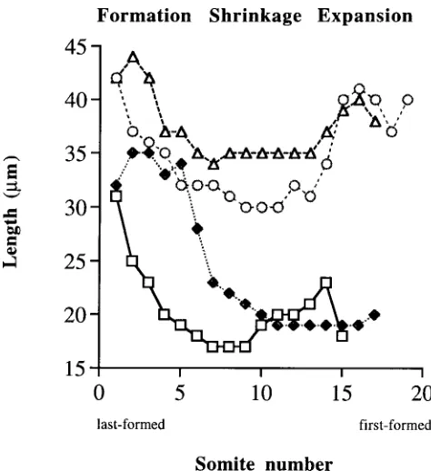
Somite sizes during their formation
In Figure 1, the data from 4 representative embryos are pre-sented (2 normal and 2 A-) at 15,17 and 19 somite stages. If we assume that the size distribution of posterior (last formed) to anterior (first formed) somites at any one stage is representative of the development of an individual somite, then in normal embryos there seemed to be three phases of somite development. The last-formed somites had a size of 42 µm and moving rostrally they then seemed to shrink as the somites in the middle of the trunk had a size of only 30-35 µm. The somites at the head end of the embryo had presumably expanded as they had recovered in size and were 40
µm. In Figure 1 these three phases are referred to as formation, shrinkage and expansion and they may equate to the three morphological steps in somite development referred to above, namely segmentation, epithelialization and differentiation (Keynes and Stern, 1988).
Of course, estimating the size of somites in this static way is a very crude measure and one which ignores any difference in somite size that may be due to rostro-caudal position along the body axis. Neither do we propose any mechanistic explanation for this change in size –it could be due to differences in cell matrix composition or even be due to a fixation artefact, but what is
important is that it is intended to be a comparative measuring tool between normal and A- embryos.
The rostrocaudal length of the last-formed somite in A- embryos is about 25% smaller than that of normal embryos being 31-32 µm (Fig. 1). This is also visible in many of the in situs shown in Figure 3, but particularly the cek-8 and Delta in situs in Figure 3A-D. After their initial formation, the data in Figure 1 show that the somites in the middle of the body of an A- embryo undergo a shrinkage which is somewhat more severe than normal, reducing to about 20 µm and then they do not enter the expansion phase to recover their size. Instead, they stay reduced in size (about 20 µm) by which time they are 50% smaller than normal in rostrocaudal dimension. This vast discrepancy in size can be seen in sections through the anteriormost somites of such embryos at the same magnification (Fig. 2A,B) and it remains throughout the development of these A-embryos.
Cell death in the lateral halves of somites
We next considered whether there were any abnormal regions of cell death in the somites. To examine this, we studied the patterns of cell death in normal and A- quail embryos by the whole-mount TUNEL technique which we have previously used to identify a highly localised region of cell death in the presumptive myelen-cephalon of the neuroepithelium (Maden et al., 1997), and also by a TUNEL technique using sectioned material.
The patterns of cell death in the normal quail embryo have been described previously and will not be repeated here, apart from a brief summary. From early somite stages apoptosis appeared in several discrete regions of the embryo: the anterior neuropore; the cells of the neural tube which had just completed closure and located as a mid-line group at a constant distance from the last formed somite (see Fig. 2C); a crescent at the very posterior end of the embryo; neural crest cells derived from the midbrain, rhombomere 3, rhombomere 5 and rhombomeres 7 and 8. In later stage embryos (stage 15 onwards), apoptosis is seen at a low level in developing organs such as the eye, otic vesicle and heart, and Homma et al. (1994) have described apoptosis in the dorsal and ventral regions of the chick neural tube which peak at stages 17 and 18. Apoptosis of neural crest in the chick hindbrain has been previously documented (Jeffs et al., 1992; Graham et al., 1993),
but despite a previous report of cell death in the normal sclerotome (Sanders, 1997) we could not detect apoptosis in the normally developing somites or sclerotome of the quail embryo (Fig. 2C).
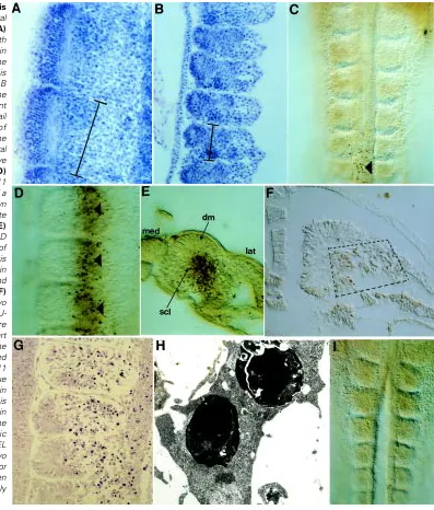
In the A- quails, however, in addition to the presumptive myelen-cephalic cell death, there was a striking pattern of TUNEL stained cells specifically in the lateral halves of each somite (Fig. 2D). This phenomenon started at the 13/14 somite stage (stage 11) and continued through stage 14. It seemed to occur as a global phenomenon rather than in a rostrocaudal wave. Furthermore, there was a remarkably sharp mediolateral border apparently through the middle of each somite which divided healthy somite cells in the medial half from dead and dying somite cells in the lateral half (Fig. 2D). However, in sections of this material it was clear that the apoptotic cells were almost exclusively within the sclerotome and not in the dermamyotome (Fig. 2E). In the dorsal Fig. 2. Sizes and patterns of apoptosis
in normal and A-somites. Horizontal sections through a stage 19 normal (A)
and A- (B) quail embryo stained with haematoxylin to show the difference in size of the somites. In both cases the rostrocaudal dimension of one somite is marked with a bar and is half the size in B as in A. The sections are through the rostralmost somites. (C) Whole-mount TUNEL stained normal stage 11 quail embryo showing the absence of apoptosis, apart from a few cells in the midline (arrowhead) which are in the neural tube and are some of those that have recently completed neurulation. (D)
Whole-mount TUNEL stained A- stage 11 quail embryo showing the presence of a large number of apoptotic cells (dark brown dots) in the lateral halves of each somite (arrowheads). Medial to the left. (E)
Section through a whole-mount from D showing more precisely the location of the apoptotic cells. The mediolateral axis is marked and the apoptotic cells are in the lateral part of the sclerotome (scl) and not in the dermamyotome (dm). (F)
view of the embryo in Figure 2D, therefore, we are looking through the overlying dermomyotome at the sclerotome. As further confir-mation of this unusual phenomenon, another batch of A- embryos were sectioned in paraffin wax and the sections subjected to a different TUNEL technique. Again, apoptotic cells were seen in the lateral part of the sclerotome and perhaps also straying into the intermediate or lateral mesoderm, but not in the dermamyotome (Fig. 2F).
In order to confirm that this phenomenon showed all the hall-marks of apoptosis, another group of A- embryos were prepared for TEM. Thin sections of these embryos confirmed the presence of apoptotic cells only in the lateral halves of each somite (Fig. 2G). In the EM dark heterochromatin condensations and degenerating nuclear and cytoplasmic structures were seen in the majority of the cells in the lateral somite (Fig. 2H).
As proof that this localised cell death is due to the absence of retinoids in the embryo, a group of A- embryos were rescued by the injection of retinol (2 mg) into the egg prior to gastrulation. This
single procedure results in the complete rescue of A- embryos so that they are indistinguishable from normal embryos and the somitic cell death was completely prevented (Fig. 2I –compare with Fig. 2D).
Gene expression patterns in the somites
Retinoids have not previously been considered as having a role in somite development and so we had no clues as to which gene pathways might be perturbed in the somites by the absence of RA. In order to begin such an analysis we performed an initial series of in situ hybridisations with a variety of genes representative of each of the different phases of somite development.
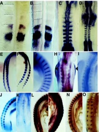
The first genes we examined were two that are expressed in the segmentation phase of somitogenesis, namely cek-8 and Delta. Cek-8 (EphA4) is expressed in the forming somites of mouse, chick and zebrafish embryos (Irving et al., 1996; Durbin et al., 1998) and is responsible for the generation of the somite boundary. In normal quail embryos cek-8 was expressed in the currently forming somite Fig. 3. Gene expression patterns in normal and A-somites. Cek-8 in situ hybridisation of a normal (A) and A- (B) stage 10 quail embryo. The patterns are the same –in both cases the currently forming somite expresses cek-8 as well as the anterior part of the most recently formed somite. The difference in somite size can be seen (somite borders are marked with dashed lines) as both are at the same magnification. (C,D) Delta in situ hybridisation of a normal (C) and A- (D) stage 10 quail embryo. The patterns are the same –in both cases the posterior halves of each somite express Delta. The difference in somite size can clearly be seen as both images are at the same magnification. (E,F) Follistatin in situ hybridisation of a normal (E) and A- (F) stage 19 quail embryo. The patterns are the same –in both cases follistatin is expressed throughout the dermamyotome of each somite. (G) Normal expression pattern of fgf-8 in a stage 19 normal quail embryo in the centre of the myotome. The expression of fgf-4 is exactly the same, though not shown here. (H) Expression of fgf-8 in a stage 19 A- quail embryo showing the complete absence of expression in the somites, but unaffected expression in the apical ectodermal ridge of the limb bud (aer). (I)
Down-regulation of fgf-4 expression in the somites of a stage 19 A- quail embryo (compare with G). (J,K)
Engrailed expression in a stage 19 normal (J) and A- (K)
quail embryo. The normal expression in the dermamyotome of the normal embryo has been almost completely down-regulated in the A- embryo. (L,M)
and at a lower level in the most recently formed somite (Fig. 3A). In A- quails the pattern of expression of cek-8 was identical (Fig. 3B), except that the somites were clearly smaller. Delta is a component of the Delta-Notch signalling pathway and is also involved in the act of generating uniformly sized groups of cells which segregate from each other (review, Jiang et al., 1998). Delta is expressed in the posterior half of each somite as it forms (Bettenhausen et al., 1995). The same pattern was observed in both normal and A- quails (Fig. 3C,D), although the smaller somites were clearly apparent in the A- embryos.
As there seemed to be no effect on segmentation genes, we then turned to genes involved in somite differentiation and exam-ined 4 of them, follistatin, fgf-4, fgf-8 and engrailed. Follistatin is expressed in the dermamyotome of the chick embryo (Connolly et al., 1995) and the same is true of the quail embryo (Fig. 3E). In A-embryos the expression of follistatin is unaltered (Fig. 3F). Fgf-4 and fgf-8 are both expressed in the myotome of the somite (Niswander and Martin, 1992; Vogel et al., 1996) and are thought to be involved in muscle differentiation. They are similarly ex-pressed in normal quail embryos (Fig. 3G). In A- quails, however, the expression of both of these genes was either completely (Fig. 3H) or partially (Fig. 3I) down-regulated. The down-regulation of fgf-8 was a phenomenon specific to the somites because the expression in the apical ectodermal ridge of the limb bud was completely unaffected (Fig. 3H and Stratford et al., 1999) as was its expression at the isthmus –the midbrain/hindbrain border in the developing central nervous system (Gale et al., 1999). Engrailed is a gene which, like follistatin, is expressed in the dermamyotome of the somite (Davis et al., 1991) and the same is true in normal quail embryos (Fig. 3J). In the A- embryo, expression of engrailed in the somite was strongly or completely down-regulated (Fig. 3K).
Because of the down-regulation of fgf-4 and fgf-8 we expected to see some disturbance in muscle differentiation. Consequently, we examined two genes involved in myogenesis, namely myf5 and myogenin and surprisingly noted that there was a differential effect between these genes. Whereas myf5 was expressed at a similar level in both A- embryos and normal (Fig. 3L,M), myogenin was down-regulated in A- embryos (Fig. 3N,O).
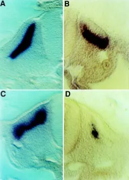
To ensure that these differences in expression levels were not due to the size difference between the normal and A- quails we cut vibratome sections of all of these in situs to examine them in more detail. The differences were confirmed in all cases and, as an example, sections of myf5 in situs which show no difference between normal and A- are shown (Fig. 4A,B) in comparison with sections of myogenin in situs (Fig. 4C,D), which show a strong down-regulation in the A- embryos.
Discussion
Previous studies using the A- quail model system have provided valuable insights into the gene pathways and embryonic systems in which RA is involved. For example, in the developing limb we have shown that RA is concerned with the control of anteroposte-rior polarity and outgrowth because the shh, fgf-4 and bmp-2 genes were down-regulated in A- quails (Stratford et al., 1999). Similarly, in the control of dorsoventral polarity, the engrailed gene was down-regulated and the limb bud resembled a double dorsal structure. In the developing hindbrain, genes normally expressed in the posterior hindbrain such as fgf-3, krox-20 and Hoxb-1 were
down-regulated (Maden et al., 1996), and genes in the apoptotic pathway such as Msx-2 were up-regulated (Maden et al., 1997). In the developing heart of the A- quail, looping does not occur correctly, the heart tube is closed at the sinus venosus and the genes RARα, RARβ and GATA-4 are down-regulated (Kostetskii et al., 1998, 1999). These studies reveal the precise nature of the altered developmental responses in embryos which develop in the absence of RA.
Here we describe another embryonic system which is grossly abnormal in the A- quail embryo, namely the somites. The most obvious defect we have observed is that the somites are about half the size of normal, although they segment at approximately the same rate as normal and the embryos have the same number of somites as normal at an equivalent morphological stage. Not only are the A- somites smaller at their initial segmentation, but they fail to expand as normal somites do (Fig. 1). Subsequently, the lateral halves of these somites, specifically the lateral halves of the sclerotome, die by apoptosis and some of the genes involved in the somitic differentiation pathways are down-regulated. To confirm that this was a specific effect of the lack of retinoids, embryos could Fig. 4. Sections through myf5 and myogenin whole-mount in situ hybridisations of stage 19 embryos. (A,B) Expression of myf-5 in the myotome of a normal (A) and A- (B) quail embryo showing an identical pattern in the myotome. (C,D). Expression of myogenin in the normal (C)
be rescued from these defects by the injection of a single dose of retinol into the egg prior to gastrulation (Gale et al., 1999). Follow-ing this treatment, all the defects seen in the A- embryos were prevented.
Unfortunately, because of the abnormal formation of the sinus venosus of the heart the vitelline veins fail to form and these A- quail embryos die from stage 20 onwards, after 3 days of incubation. It is therefore impossible to determine what would happen to the verte-brae or musculature of the A- quail following this lateral sclerotomal cell death. Neither can we examine whether other later events such as interdigital cell death are disturbed in the A- embryo.
Size regulation of somites
The small, but perfectly formed somites we have seen here are a fascinating example of global regulation –achieving constant num-bers or proportions of a developmental unit despite large variations in total body size. The only other situation in which this has been seen in somites is by experimentally decreasing the amount of embryonic tissue available in the early gastrula stage embryo. This has been done both in Xenopus (Cooke, 1975) and chick (Bellairs and Veini, 1984) by removing large pieces of tissue, for example the posterior third of the area pellucida or the whole of the primitive streak at a defined stage of development. Since somite number is conserved in each of these situations and the effect is to make smaller somites at the same rate as normal, we would not expect any change in the cyclical expression of the oscillatory genes such as hairy1 (Palmeirim et al., 1997) or lunatic fringe (Forsberg et al., 1998; McGrew et al., 1998) and indeed we did not detect any change in cek-8 or Delta, two genes linked to the segmentation process. Single gene knockouts have not generated such a global phenotype either. A disruption of segment border formation in the case of Delta (Hrabe de Angelis et al., 1997), lunatic fringe (Evrard et al., 1998; Zhang and Gridley, 1998), Notch (Conlon et al., 1995) or presenilin (Wong et al., 1997) knockouts, smaller but irregularly shaped and less cohesive somites in the case of N-cadherin knockouts (Radice et al., 1997), or a failure to form somites at all in the case of paraxis knockouts (Burgess et al., 1996).
The models which have been formulated to explain the consis-tency of somite number within a species and following experimental perturbation have either involved a wavefront travelling along the length of the rostro-caudal axis of the embryo, the “clock and wavefront” model (Cooke and Zeeman, 1975) or been of a more local nature involving prepatterns of somite clusters (Bellairs and Veini, 1984) or groups of cells in the segmental plate displaying cell division synchrony (Keynes and Stern, 1988; Primmett et al., 1989). It is difficult to relate the results described here for the A- quail to the clock and wavefront model where the rate of travel of the wavefront across a field of cells determines somite size because in amniotes, somite formation begins while mesoderm is still being generated and the length of the rostrocaudal axis is not known. It is much easier to explain the small somites in A- embryos in terms of the cell cycle
model. Since the rate of generation of somites seemed the same in both normal and A- embryos we would not predict a change in cell cycle time of segmental plate cells in A- quails, rather a decrease in the number of cells cycling. This is a clear and testable hypothesis which we intend to explore in the future.
Apoptosis in the lateral halves of the somites
It is striking that only the lateral halves of the sclerotome showed apoptotic cells both in TUNEL whole-mounts, TUNEL sections and in the TEM. This very precise localisation of cell death is reminis-cent of the equally discrete apoptotic area that we have previously observed in the presumptive hindbrain of the A- quail embryos (Maden et al., 1997). Therefore, the lack of RA in a developing embryo does not result in widespread cell death in all of the tissues in an uncoordinated fashion, but it is reflected in very specific areas suffering this fate, namely a stripe of mesenchyme and then neuroepithelium in the region of the presumptive hindbrain, the lateral halves of the sclerotomes and we have also observed that the ventral part of the developing eye is another region where localised apoptosis occurs.
The fact that only the lateral halves of the somites undergo apoptosis was a surprising observation. It does not seem to be related to a difference in endogenous retinoid distribution within the somite, as studies using the F9 reporter cell system (e.g. Maden et al., 1998b) failed to detect any mediolateral differences. The only other differences that have been observed in the response of the somite to RA concern the stimulation of proliferation and the stimulation of glycosaminoglycan synthesis. But in this case the differential response was between sclerotome (which responded strongly to RA) and dermamyotome (which responded poorly) (Vasan, 1993). However, this stimulation of GAG synthesis in the sclerotome by RA may reveal the cause of the failure of the A-somites to expand (Fig. 1) –in the absence of RA the sclerotomal matrix fails to be laid down. An additional reason for the failure of expansion would also be the cell death itself.
Cell fate studies in the chick embryo have revealed that a particular region of the primitive streak, some 200 µm behind Hensen’s node, gives rise to the lateral halves of the somites (Selleck and Stern, 1991; Psychoyos and Stern, 1996). The same also seems to be the case in the mouse embryo (Wilson and Beddington, 1996). Thus it may be that this population of cells in this specific region of the primitive streak expresses a particular gene whose domain of expression is established by the presence of RA at an early developmental stage and in the absence of RA the gene fails to be activated. It would be interesting to attempt to identify such a gene using this A- system.
somitic mesoderm such that at a high concentration the presomite is converted into lateral plate, but at a lower concentration the medial component of the somite was converted into the lateral component. It is possible that in the A- quail lateral plate the expression of BMP-4 is disturbed, resulting in an abnormal expres-sion of genes such as sim-1 (Pourquie et al., 1996) and this would be an interesting hypothesis to test. Other genes such as pax1 and pax9 may also be involved and further studies will examine these possibilities as well.
Of course, the major question to be resolved is precisely why and how these discrete regions of apoptosis come about. RA is known to have an effect upon the apoptotic programme, but in the majority of cases it is the presence of excess RA which induces apoptosis in a wide range of cell types both in vivo and in vitro rather than the lack of it (Zhang and Jetten, 1997). Indeed, this is the basis of the use of retinoids in cancer treatment. In a small number of cases, retinoids can inhibit apoptosis, for example in thymocytes, but the relevance of this to apoptosis in the A- embryo is not obvious. The downstream genes induced during apoptosis are rapidly becoming characterised, but the uniqueness of the A-embryo is that we can begin to ask about the upstream genes which induce the known apoptotic pathway in such discrete localisations.
Somite differentiation
We also examined the expression of some of the genes involved in somite differentiation. These were follistatin, fgf-4, fgf-8, engrailed, myf5 and myogenin. Some of these were expressed perfectly normally, that is, follistatin and myf5, but the others were either strongly or completely down-regulated. The role of engrailed, a gene expressed in the dermamyotome (Davis et al., 1991), is not clear, but it was strongly down-regulated demonstrating altered differentiation of this compartment of the somite. This gene is also strongly down-regulated in the A- limb bud (Stratford et al., 1999), unlike fgf-8 whose expression was only altered in the somite and not in the limb bud (Fig. 3H and Stratford et al., 1999) or in the isthmus (Gale et al., 1999).
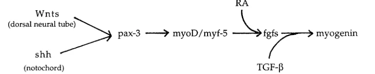
The role of the fgfs, being expressed in the myotomal compart-ment of the somite is thought to be involved in muscle differentia-tion. FGF induces muscle and mesoderm in Xenopus animal caps (Slack et al., 1987), bFGF and TGF-β promote myogenesis in chick somites (Stern et al., 1997) and fgf-4 fails to be expressed in myf5 mutant mice (Grass et al., 1996). These observations therefore place fgf-4 (in combination with TGF-β) downstream of myf5 in somitic muscle differentiation with RA playing a role on the pathway between these two genes because myf5 was expressed normally in our A- quails (Fig. 5).
The differential response of the two myogenic genes was surprising and reveals an unsuspected role for RA in muscle differentiation. Myf5 and myoD are responsible for myoblast deter-mination whereas myogenin is involved in the later event of turning the determined myoblast into a differentiated myotube (Weintraub, 1993). This is reflected in their sequential activation (Pownall and Emerson, 1992). Myf5 is activated by a combination of signals from the neural tube and notochord, namely shh from the notochord and Wnts from the dorsal neural tube (Munsterberg et al., 1995) and this pathway also involves the Pax-3 gene (Rawls and Olson, 1997) (Fig. 5). Since myf5 was unaffected by a lack of RA then we would not expect an effect on the Wnts, shh or Pax-3 in this region of the embryo and indeed this seems to be the case at least for shh and Pax-3 as we have previously shown that these genes are
expressed normally in A- embryos (Maden et al., 1996). Thus, RA may feed into the pathway between myf5/myoD and myogenin, upstream of the fgfs/TGF-β (Grass et al., 1996; Stern et al., 1997) as a hitherto unsuspected regulator of myogenin expression (Fig. 5).
Materials and Methods
Eggs from Japanese quail (Coturnix coturnix japonica) were obtained from the Poultry Research Farm at Michigan State University. Birds were fed a normal or a vitamin A-deficient diet as described previously (Dersch and Zile, 1993). Eggs were collected daily, incubated until the desired stage and fixed in 4% paraformaldehyde. Routine histology was performed on them and sections stained with haematoxylin. In situ hybridisation was performed according to established protocols. Whole-mounts were cleared in 80% glycerol for photography and then sectioned on a vibratome at a thickness of 80 µm.
The terminal transferase mediated dUTP-biotin nick end labelling (TUNEL) technique was a modification of a method developed in Droso-phila (White et al., 1994). Embryos were fixed in 4% paraformaldehyde, washed in PBS/1% Triton-X 100 (PBX) and the endogenous peroxidase inactivated by incubation in 0.1% hydrogen peroxide overnight at 4°C. The embryos were again washed in PBX followed by a wash in 1X terminal transferase buffer/2.5 mM CoCl2 in PBX. They were incubated at 37°C in the same buffer but including 0.5 units of terminal transferase per ml and 10 mM dUTP (2:1 dUTP:d-UTP-biotin) for 3 h. The embryos were washed in PBX and incubated overnight with rocking with streptavidin-HRP diluted 1:300 in PBX. Finally, they were washed in PBX and then stained with diaminobenzidine. To examine cell death in sectioned material, paraform-aldehyde fixed embryos were embedded in paraffin wax, sectioned at 10 µm and the sections used in conjunction with an Oncor apoptosis kit.
To perform rescues on A- embryos, retinol was dissolved in ethanol at its maximum solubility and then mixed with a solution of Tyrode’s solution: A- egg extract 20:3. Each A- egg was injected prior to gastrulation with a volume of this mixture into the albumen so that 2 mg retinol was delivered. For electron microscopy, embryos were fixed in 2.5% glutaraldehyde in 0.1M sodium cacodylate buffer with 5% sucrose. After two rinses in this buffer, the embryos were fixed for 1 h in 1% osmium tetroxide, rinsed in distilled water three times and stained en bloc with 2% aqueous uranyl acetate. They were dehydrated in an ascending series of ethanols ending with propylene oxide and embedded in Agar 100 (Epon 812 substitute). Semithin sections were cut and stained with toluidine blue. Ultrathin sections were then cut from the selected area, mounted on grids and double stained with aqueous uranyl acetate (5 min) and Reynold’s lead citrate (3 min). The tissue was examined using a Joel 200CX transmission electron microscope.
Acknowledgements
This work was supported by The Wellcome Trust (MM and EG) and the USDA (MZ). We are very grateful to the following for gifts of probes: Drs. Elizabeth Pownall (myf5, myogenin), Fernando Giraldez (cek-8), Ivor Mason (fgf-4, fgf-8), Cairine Logan (en), David Ish-Horowicz (Delta) and to Chris Rawlinson for the electron microscopy.
References
BELLAIRS, R. and VEINI, M. (1984). Experimental analysis of control mechanisms in somite segmentation in avian embryos. II. Reduction of material in the gastrula stages of the chick. J. Embryol. Exp. Morphol. 79: 183-200.
BETTENHAUSEN, B., DE ANGELIS, M.H., SIMON, D., GUENET, J-L. and GOSSLER, A. (1995). Transient and restricted expression during mouse embryogenesis of Dll1, a murine gene closely related to Drosophila Delta. Development 121: 2407-2418.
CHEN, Y., DONG, D., KOSTETSKII, I. and ZILE, M.H. (1996). Hensen’s node from vitamin A-deficient quail embyro induces chick limb bud duplication and retains its normal asymmetric expression of Sonic hedgehog (Shh). Dev. Biol. 173: 256-264.
CONLON, R.A., REAUME, A.G. and ROSSANT, J. (1995). Notch1 is required for the coordinate segmentation of somites. Development 121: 1533-1545.
CONNOLLY, D.J., PATEL, K., SELEIRO, E.A.P., WILKINSON, D.G. and COOKE, J. (1995). Cloning, sequencing and expressional analysis of the chick homologue of Follistatin. Dev. Genet. 17: 65-77
COOKE, J. (1975). Control of somite number during morphogenesis of a Vertebrate, Xenopus laevis. Nature 254: 196-199.
COOKE, J and ZEEMAN, E.C. (1975). A clock and wavefront model for control of the number of repeated structures during animal morphogenesis. J. Theor. Biol. 58: 455-476.
DAVIS, C.A., HOLMYARD, D.P., MILLEN, K.J. and JOYNER, A.L. (1991). Examining pattern formation in mouse, chicken and frog embryos with an En-specific antise-rum. Development 111: 287-298.
DERSCH, H. and ZILE, M.H. (1993). Induction of normal cardiovascular development in the vitamin A-deproved quail embryo by natural retinoids. Dev. Biol. 160: 424-433.
DICKMAN, E.D., THALLER, C. and SMITH, S.M. (1997). Temporally-regulated retinoic acid depletion produces specific neural crest, ocular and nervous system defects. Development 124: 3111-3121.
DONG, D. and ZILE, M.H. (1995). Endogenous retinoids in the early avian embryo. Biochem. Biophys. Res. Commun. 217: 1026-1031.
DURBIN, L., BRENNAN, C., SHIOMI, K., COOKE, J., BARRIOS, A., SHANMUGALINGAM, S., GUTHRIE, B., LINDBERG, R. and HOLDER, N. (1998). Eph signalling is required for segmentation and differentiation of the somites. Genes Dev. 12: 3096-3109.
EVRARD, Y.A., LUN, Y., AUEHLA, A., GAN, L. and JOHNSON, R.L. (1998). lunatic fringe is an essential mediator of somite segmentation and patterning. Nature 394: 377-381.
FORSBERG, H., CROZET, F. and BROWN, N.A. (1998). Waves of mouse lunatic fringe expression, in four-hour cycles at two-hour intervals, precede somite bound-ary formation. Curr. Biol. 8: 1027-1030.
GALE, E., ZILE, M. and MADEN, M. (1999). Hindbrain respecification in the retinoid-deficient quail. Mech. Dev. 89: 43-54.
GOSSLER, A. and HRABE DE ANGELIS, A. (1998). Somitogenesis. Curr.Top. Dev. Biol. 38: 225-287.
GRAHAM, A., HEYMAN, I. and LUMSDEN, A. (1993). Even-numbered rhombomeres control the apoptotic elimination of neural crest cells from odd-numbered rhombomeres in the chick hindbrain. Development 119: 233-245.
GRASS, S., ARNOLD, H-H. and BRAUN, T. (1996). Alterations in somite patterning of Myf-5-deficient mice: a possible role for FGF-4 and FGF-6. Development 122: 141-150.
HEINE, U.I., ROBERTS, A.B., MUNOZ, E.F., ROCHE, N.S. and SPORN, M.B. (1985). Effects of retinoid deficiency on the development of the heart and vascular system of the quail embryo. Virchow’s Arch. B [Cell Pathol.] 50: 135-152.
HOMMA, S., YAGINUMA, H. and OPPENHEIM, R.W. (1994). Programmed cell death during the earliest stages of spinal cord development in the chick embryo: a possible means of early phenotypic selection. J. Comp. Neurol. 345: 377-395.
HRABE DE ANGELIS, M.H., McINTYREE, J. and GOSSLER, A. (1997). Maintenance of somite borders in mice requires the Delta homologue Dll1. Nature 386: 717-721.
IRVING, C., NIETO, M.A., DASGUPTA, R., CHARNAY, P. and WILKINSON, D.G. (1996). Progressive spatial restriction of Sek-1 and Krox-20 gene expression during hindbrain segmentation. Dev. Biol. 173: 26-38.
JEFFS, P., JAQUES, K. and OSMOND, M. (1992). Cell death in cranial neural crest development. Anat. Embryol. 185: 583-588.
JIANG, Y-J., SMITHERS, L. and LEWIS, J. (1998). Vertebrate segmentation: The clock is linked to Notch signalling. Curr. Biol. 8: R868-R871.
KEYNES, R.J. and STERN, C.D. (1988). Mechanisms of vertebrate segmentation. Development 103: 413-429.
KOSTETSKII, I., JIANG, Y., KOSTETSKAIA, E., YUAN, S., EVANS, T. and ZILE, M. (1999). Retinoid signalling required for normal heart development regulates GATA-4 in a pathway distinct from cardiomyocyte differentiation. Dev. Biol. 206: 206-218.
KOSTETSKII, I., LINASK, K.K. and ZILE, M.H. (1996). Vitamin A deficiency and the
expression of retinoic acid receptors during early cardiogenesis in quail embryo. Roux’s Arch. Dev. Biol. 205: 260-271.
KOSTETSKII, I., YUAN, S.-Y., KOSTETSKAIA, E., LINASK, K. K., BLANCHET, S., SELEIRO, E., MICHAILLE, J.-J., BRICKELL, P. and ZILE, M. (1998). Initial retinoid requirement for early avian development coincides with retinoid receptor coexpression in the precardiac fields and induction of normal cardiovascular development. Dev. Dyn. 213: 188-198.
MADEN, M., GALE, E., KOSTETSKII, I. and ZILE, M. (1996). Vitamin A-deficient quail embryos have half a hindbrain and other neural defects. Curr. Biol. 6: 417-426.
MADEN, M., GALE, E. and ZILE, M. (1998a). The role of vitamin A in the development of the central nervous system. J. Nutr. 128: 471S-475S.
MADEN, M., GRAHAM, A., GALE, E., ROLLINSON, C. and ZILE, M. (1997). Positional apoptosis during vertebrate CNS development in the absence of endogenous retinoids. Development 124: 2799-2805.
MADEN, M., SONNEVELD, E., VAN DER SAAG, P.T. and GALE, E. (1998b). The distribution of endogenous retinoic acid in the chick embryo: implications for developmental mechanisms. Development 125: 4133-4144.
MCGREW, M.J., DALE, J.K., FRABOULET, S. and POURQUIE, O. (1998). The lunatic fringe gene is a target of the molecular clock linked to somite segmentation in avian embryos. Curr. Biol. 8: 979-982.
MORRISS-KAY, G.M. and SOKOLOVA, N. (1996). Embryonic development and pattern formation. FASEB J. 10: 961-968.
MUNSTERBERG, A.E., KITAJEWSKI, J., BUMCROT, D.A., MCMAHON, A.P. and LASSAR, A.B. (1995). Combinatorial signalling by Sonic hedgehog and Wnt family members induces myogenis bHLH gene expression in the somite. Genes Dev. 9: 2911-2922.
NISWANDER, L. and MARTIN, G.R. (1992). Fgf-4 expression during gastrulation, myogenesis, limb and tooth development in the mouse. Development 114: 755-768.
PALMEIRIM, I., HENRIQUE, D., ISH-HOROWICZ, D. and POURQUIE, O. (1997). Avian hairy gene expression identifies a molecular clock linked to vertebrate segmentation and somitogenesis. Cell 91: 639-648.
POURQUIE, O., FAN, C-M., COLTREY, M., HIRSINGER, E., WATANABE, Y., BREANT, C., FRANCIS-WEST, P., BRICKELL, P., TESSIER-LAVIGNE, M. and LE DOUARIN, N.M. (1996). Lateral and axial signals involved in avian somite patterning: a role for BMP4. Cell 84: 461-471.
POWNALL, M.E. and EMERSON, C.P. (1992). Sequential activation of three myogenic regulatory genes during somite morphogenesis in quail embryos. Dev. Biol. 151: 67-79.
PRIMMETT, D.R.N, NORRIS, W.E., CARLSON, G.J., KEYNES, R.J. and STERN, C.D. (1989). Periodic segmental anomalies induced by heat shock in the chick embryo are associated with the cell cycle. Development 105: 119-130.
PSYCHOYOS, D. and STERN, C.D. (1996). Fates and migratory routes of primitive streak cells in the chick embryo. Development 122: 1523-1534.
RADICE, G.L., RAYBURN, H., MATSUNAMI, H., KNUDSEN, K.A., TAKEICHI, M. and HYNES, R.O. (1997). Developmental defects in mouse embryos lacking N-cadherin. Dev. Biol. 181: 64-78.
RAWLS, A. and OLSON, E.N. (1997). MyoD meets its maker. Cell 89: 5-8.
SANDERS, E.J. (1997). Cell death in the avian sclerotome. Dev. Biol. 192: 551-563.
SELLECK, M.A.J. and STERN, C.D. (1991). Fate mapping and cell lineage analysis of Hensen’s node in the chick embryo. Development 112: 615-626.
SLACK, J.M.W., DARLINGTON, B.G., HEATH, J.K. and GODSAVE, S.F. (1987). Mesoderm induction in early Xenopus embryos by heparin-binding growth factors. Nature 326: 197-200.
STERN, H.M., LIN-JONES, J. and HAUSCHKA, S.D. (1997). Synergistic interac-tions between bFGF and a TGF-β family member may mediate myogenic signals from the neural tube. Development 124: 3511-3523.
STRATFORD, T., LOGAN, C., ZILE, M. and MADEN, M. (1999). Abnormal antero-posterior and dorsoventral patterning of the limb bud in the absence of retinoids. Mech. Dev. 81: 115-125.
TAJBAKHSH, S. and SPORLE, R. (1998). Somite development: constructing the vertebrate body. Cell 92: 9-16.
TONEGAWA, A., FUNAYAMA, N., UENO, N. and TAKAHASHI, Y. (1997). Mesoder-mal subdivision along the mediolateral axis in chicken controlled by different concentrations of BMP-4. Development 124: 1975-1984.
VASAN, N.S. (1993). Nonunifomity within embryonic somites: differential responssse to retinoic acid in vitro. Dev. Growth Differ. 35: 385-393.
VOGEL, A., RODRIGUEZ, C. and IZPISUA-BELMONTE, J-C. (1996). Involvement of FGF-8 in initiation, outgrowth and patterning of the vertebrate limb. Development 122: 1737-1750.
WEINTRAUB, H. (1993). The MyoD family and myogenesis: redundancy, networks and thresholds. Cell 75: 1241-1244.
WHITE, J.C., SHANKAR, V.N., HIGHLAND, M., EPSTEIN, M.L., DELUCA, H.F. and CLAGETT-DAME, M. (1998). Defects in embryonic hindbrain development and fetal resorption resulting from vitamin A deficiency in the rat are prevented by feeding pharmacological levels of all-trans-retinoic acid. Proc. Natl. Acad. Sci. USA 95: 13459-13464.
WHITE, K., GRETHER, M.E., ABRAMS, J.M., YOUNG, L., FARELL, K. and STELLER, H. (1994). Genetic control of programmed cell death in Drosophila. Science 264: 677-683.
WILSON, V. and BEDDINGTON, R.S.P. (1996). Cell fate and morphogenetic move-ment in the late mouse primitive streak. Mech. Dev. 55: 79-89.
WONG, P.C., ZHENG, H., CHEN, H., BECHER, M.W., SIRINATHSINGHJI, D.J.S., TRUMBAUER, M.E., CHEN, H.Y., PRICE, D.L., VAN DER PLOEG, L.H.T. and SISODIA, S.S. (1997). Presenlini 1 is required for Notch1 and Dll1 expressison in the paraxial mesoderm. Nature 387: 288-292.
ZHANG, H. and GRIDLEY, T. (1998). Defects in somite formation in lunatic fringe-deficient mice. Nature 394: 374-377