Development of axon pathways in the zebrafish central
nervous system
JENSEN HJORTH and BRIAN KEY*
Department of Anatomy and Developmental Biology, School of Biomedical Sciences and Centre for Functional and Applied Genomics, University of Queensland, Brisbane, Australia
ABSTRACT The zebrafish has a number of distinct advantages as an experimental model in developmental biology. For example, large numbers of embryos can be generated in each lay, development proceeds rapidly through a very precise temporal staging which exhibits minimal batch-to-batch variability, embryos are transparent and imaging of wholemounts negates the need for tedious histological preparation while preserving three-dimensional spatial relationships. The zebrafish nervous system is proving a convenient model for studies of axon guidance because of its small size and highly stereotypical trajectory of axons. Moreover, a simple scaffold of axon tracts and nerves is established early and provides a template for subsequent development. The ease with which this template can be visualized as well as the ability to spatially resolve individual pioneer axons enables the role of specific cell-cell and molecular interactions to be clearly deciphered. We describe here the morphology and development of the earliest axon pathways in the embryonic zebrafish central nervous system and highlight the major questions that remain to be addressed with regard to axon guidance.
KEY WORDS:
brain, tracts, growth cone, spinal cord, guidance, navigation
0214-6282/2002/$25.00
© UBC Press Printed in Spain www.ijdb.ehu.es
*Address correspondence to: Dr. Brian Key. Department of Anatomy and Developmental Biology, School of Biomedical Sciences and Centre for Functional and Applied Genomics, University of Queensland, Brisbane 4072, Australia. Fax: +61-7-3365-2955. e-mail: [email protected]
Abbreviations used in this paper: AC, anterior commissure; CaP, caudal primary; CNS, central nervous system; CoPA, primary commissural ascending neuron; CoSA, secondary ascending commissural neuron; cyc, Cyclops; DLF, dorsal longitudinal fascicle; DoLA, dorsal longitudinal ascending interneuron; drc dorsorostral cluster; DVDT, dorsoventral diencephalic tract; hpf hours post-fertilization; isl-1 islet-1; MiP, middle primary; MLF, medial longitudinal fasciculus; noi, no-isthmus; nTPC, nucleus of the tract of the posterior commissure; POC, post-optic commissure; RoP, rostral primary; SOT, supraoptic tract; TPC, tract of the posterior commissure; TPOC, tract of the posterior longitudinal commissure; VaP, variable primary; vcc ventrocaudal cluster; VeLD, ventral longitudinal descending interneuron; VLT, ventral longitudinal tract; vrc ventrorostral cluster; zdcc, zebrafish deleted in colon cancer.
Introduction
Embryonic development of the zebrafish occurs in a transpar-ent, extra-embryonic membrane known as the chorion which is approximately 1 mm in diameter. Following fertilization of the egg, zebrafish development proceeds through a number of morphologi-cally distinct stages including: zygote (0-0.75 hpf) (hours postfertilization); cleavage (0.75-2.25 hpf); blastula (2.25-5.25 hpf); gastrula (5.25-10 hpf); and segmentation (10-24 hpf) (Kimmel et al., 1995). At approximately 35 hpf the larva hatches from the chorion and becomes free swimming.
During segmentation the central nervous system (CNS) trans-forms from a flat sheet of ectodermal cells, the neural plate, to a solid three dimensional structure morphologically subdivided into developmental compartments. These subdivisions are easily iden-tified by visible morphological landmarks. In many vertebrates, the neural tube forms by a process known as primary neurulation which involves the evagination of the neural plate until the lateral edges or crest meet and fuse at the dorsal midline, forming a hollow tube. In contrast, cell movements in the neural plate of zebrafish lead to the formation of a solid rod-like structure known as the neural keel at approximately 12 hpf. Shortly after, through a process of “secondary neurulation”, this transient tissue is
hol-lowed to form the lumen of the neural tube (Papan and Campos-Ortega, 1994).
the major subdivisions of the brain. A prominent feature is the ventral flexure which folds the ventral surface of the neural tube back on itself (Ross et al., 1992; Kimmel et al., 1995).
The forebrain can be subdivided into telencephalon, located in a dorso-rostral position above the optic stalk, and diencephalon, located below the optic stalk but extending dorso-caudally at the level of the ventral flexure (Ross et al., 1992). The ventral flexure marks the level of the future mid-diencephalic boundary and dorsal positioned epiphysis (see Fig. 3 in Lauderdale et al., 1997). The presumptive boundary between the forebrain and midbrain is caudal to the ventral flexure. Its precise location has not been defined, although the borders of several gene expression patterns are thought to demar-cate this boundary (Krauss et al., 1991a,b). At 24hpf a small forebrain ventricle is apparent as well as a rudimentary evagination in the roof of the forebrain which will form the epiphysis (Ross et al., 1992; Wilson and Easter, 1991). The midbrain has a prominent ventricle at this age which separates the tectum dorsally and the tegmentum ventrally (Wilson et al., 1990; Kimmel et al., 1995). The cerebellum is also now clearly evident as two abutting folds in the neuroepithe-lium lying in the region of the midbrain/hindbrain boundary. Rhombomere 1 is located directly ventral to the cerebellum (Kimmel et al., 1995). The hindbrain at this age consists of seven discrete and functionally distinct rhombomeres (Trevarrow et al., 1990; Kimmel et al., 1995) around a large ventricle which tapers caudally to the spinal cord. At 24 hpf the spinal cord is essentially a continuous structure at this age with no obvious morphological distinctions along its rostro-caudal axis (Kimmel et al., 1995).
Development of a Template of Neurons
An attractive attribute of the zebrafish as a model for vertebrate developmental neurobiology is the clear and distinct formation of a small neuronal cluster in each major subdivision of the brain. During the first stages of segmentation, primary neurogenesis begins in the midbrain and is followed shortly after in other brain subdivisions. By 24 hpf the first neuronal clusters have extended axons which form a simple scaffold of axon tracts and commis-sures connecting adjacent subdivisions. These first neurons are termed “primary neurons” and can be distinguished from later developing neurons (Kimmel, 1993). Primary neurons typically have larger soma which extend axons long distances in the brain often to pioneer pathways, rather than making small local connec-tions (Metcalfe et al., 1990; Kimmel, 1993). Primary neurons in all regions of the developing CNS express islet-1 (isl-1, a LIM homeobox gene), indicating that they may share a common early regulatory program (Korzh et al., 1993). In fact, overexpression of the zebrafish homologue of delta causes a reduction in isl-1 positive cells throughout the neural tube (Dornseifer et al., 1997). In addition, the ned-1 zebrafish mutant exhibits a neural degeneration phenotype which does not affect primary neurons (Grunwald et al., 1988). In the spinal cord, primary and secondary motor neurons can be distinguished based on their temporal requirements for shh (sonic hedgehog) signalling. shh expression in either the floor plate or the notochord is required for specification of secondary motor neu-rons. However, Shh signals emanating from the gastrula axial mesoderm are required earlier in development to specify primary motor neurons (Beattie et al., 2000).
Although the appearance of post-mitotic neurons occurs in the spinal cord at 12 hpf, they are not evident until 16 hpf in the developing
brain (Fig. 1A). An early marker for post-mitotic neurons is the presence of the intracellular enzyme acetylcholine esterase. En-zyme histochemistry reveals that the first brain neurons begin to develop in discrete, bisymmetrical, stereotypical clusters at 16 hpf (Ross et al., 1992). These clusters have been confirmed also by examining the expression of the post-mitotic neuronal markers HNK-1 and α-acetylated tubulin (Chitnis and Kuwada, 1990; Metcalfe et al., 1990; Wilson et al., 1990; Ross et al., 1992).
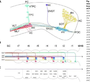
The first post-mitotic neurons in the rostral brain develop at 16 hpf in the ventral midbrain and are referred to as either the ventrocaudal cluster (vcc) (Ross et al., 1992) or the nucleus of the medial longitudinal tract (Chitnis and Kuwada, 1990). By 24 hpf this cluster consists of approximately 40 neurons. In the ventral diencephalon, below the optic recess, approximately 11 cells emerge as the first neurons of the ventrorostral cluster (vrc) at approximately 16 hpf (Ross et al., 1992). By 24 hpf the number of neurons in this cluster has increased to 125 (Ross et al., 1992). The first neurons of the telencephalon form shortly after those of the vcc within the dorsorostral cluster (drc), which lies dorsal to the optic recess (Ross et al., 1992). At this age an average of five cells can be detected by acetylcholine esterase histochemistry (Wilson et al., 1990; Ross et al., 1992). By 24 hpf the average number of cells present in the drc has increased to 165 (Ross et al., 1992). At 18 hpf a third small cluster of 1-2 neurons develops in the dorsal forebrain as the epiphyseal cluster, the anlagen of the epiphysis (Ross et al., 1992). By 24 hpf approximately 18 neurons contrib-ute to this cluster. Additional small clusters of neurons have developed in the midbrain by 24 hpf. These neurons emerge along the dorsoventral axis extending from the vcc to the roof of the midbrain. Collectively, these groups of neurons have been named the nucleus of the posterior commissure (nTPC) (Chitnis and Kuwada, 1990).
Axonogenesis in the Rostral Brain
The result of primary neurogenesis is the formation of discrete neuronal clusters in the zebrafish brain whose axons pioneer the early axon scaffold (Fig. 1A). By 24 hpf a bilaterally symmetrical, stereotypical set of five axon tracts and four commissures have formed. The first axons to navigate the neuroepithelium of the zebrafish brain emerge from the vcc at approximately 16 hpf (Chitnis and Kuwada, 1990; Ross et al., 1992). These axons grow caudally to pioneer the medial longitudinal fasciculus (MLF), which is part of the larger ventral longitudinal tract (VLT) (Wilson et al., 1990; Chitnis and Kuwada, 1990; Ross et al., 1992), the major longitudinal tract which connects the midbrain with the hindbrain. The dorsal portion of the VLT contains axons from the TPOC and dorsal midbrain which go on to contribute to the dorsal longitudinal fascicle of the hindbrain. The ventral portion of the VLT contains the MLF which continues into the hindbrain and spinal cord (Kimmel et al., 1982). Interestingly, the pioneer axons of the vcc never grow rostrally (Ross et al., 1992). Only later, do neurons from this cluster project axons rostrally into the tract of the post-optic commissure (TPOC) (Ross et al., 1992).
TPOC. The second tract pioneered by axons of the drc is the anterior commissure (AC). Similar to axons of the POC in the diencephalon, axons of the AC cross the rostral surface of the telencephalon in a thick, tight fascicle. By 24 hpf approximately 27 axons contribute to this commissure (Wilson et al., 1990). Whether these axons make local connections with neurons of the contralat-eral drc, or grow ventrally into the contralatcontralat-eral SOT is unknown. The AC and POC are clearly distinct commissures which lie above and below the level of the optic recess. These commissures border the site of the future optic chiasm which contains special-ized glial cells expressing noi (no-isthmus, a member of the zebrafish Pax family) (Macdonald et al., 1997; Shanmugalingam et al., 2000).
At approximately 20 hpf the dorsoventral diencephalic tract (DVDT) is pioneered by a single axon growing ventrally from neurons of the epiphyseal cluster (Chitnis and Kuwada, 1990; Wilson and Easter, 1991; Ross et al., 1992). This axon courses ventrally until it encoun-ters axons of the TPOC growing caudally. At this point it turns rostrally and grows within the TPOC but in the opposite direction to the other axons in this tract (Wilson et al., 1990). In the midbrain, at approxi-mately 20 hpf, neurons of the nTPC project axons ventrally to pioneer
Fig. 1. Schematic representation of primary neurogenesis and axonogenesis in the em-bryonic zebrafish brain (A) and hindbrain (B,C) at 24 hpf. Rostral is to the right and dorsal is to the top in all panels. (A) At 24 hpf a simple scaffold of axon tracts and commissures are present in the anterior brain. In the telencephalon, neurons of the dorso-rostral cluster (drc) extend axons to form the supra-optic tract (SOT) and the anterior commissure (AC). Neurons of the ventro-rostral cluster (vrc) in the diencephalon project axons which grow in the tract of the post-optic commis-sure (TPOC) and the post-optic commiscommis-sure (POC). In the ventral midbrain, neurons of the ventro-caudal cluster (vcc) extend axons caudally in the medial longitudinal fasciculus (MLF) which extends into the hindbrain. Axons from the TPOC and MLF continue towards the hindbrain as the ventral longitudinal tract (VLT). Neurons of the vcc also grow axons ventrally in the ventral com-missure (VC). Neurons located along the dorso-ventral axis of the midbrain are collectively known as the nucleus of the tract of the posterior com-missure (nTPC). These neurons project axons ventrally to form the tract of the posterior com-missure (TPC) and dorsally to form the posterior commissure (PC). Neurons of the epiphyseal cluster (ec), located in the dorsal diencephalon, grow axons ventrally to form the dorso-ventral diencephalic tract (DVDT). (B) In the hindbrain at 24 hpf, specific, identifiable neurons are present in each rhombomere (r). These neurons project axons which extend caudally in either the ipsilat-eral ventral longitudinal fasciculus (VLF) or the ipsilateral dorsal longitudinal fasciculus (DLF). Alternatively, their axon may cross the floor plate (fp) and extend in the contralateral VLF or the contralateral DLF. Not shown here is the MLF which continues into the spinal cord. Neurons represented by the same colour are thought to be segmental homologues. (C) Names of neurons represented in (B) based on their relative positions and axon trajectories in each rhombomere. c, contralateral; Ca or C, caudal; D, dorsal; l, VLF; i, ipsilateral; L, lateral; M, medial; m, MLF; M, Mauthner; MHB, midbrain/hindbrain boundary; Mi, middle; R, rostral; Ro, rostral; SC, spinal cord; V, ventral.
reach the vcc and continue to grow beyond these neurons into more caudal regions of the brain (Chitnis and Kuwada, 1990). This tract exhibits different degrees of fasciculation along its rostrocaudal axis. Initially, axons are tightly bundled as they emerge from the vrc. In the region of the posterior diencephalon these axons sort out into 4-6 fascicles which remain segregated as they grow into the midbrain and encounter neurons of the vcc. The most ventral of these fascicles appears to remain tightly fasciculated as it merges into the VLT (Chitnis and Kuwada, 1990). Some neurons of the vrc project axons rostrally, pioneering the post-optic com-missure (POC). Axons in this comcom-missure cross the rostral surface of the diencephalon and course into the contralateral TPOC. The POC is tightly fasciculated and by 24 hpf contains approximately 41 axons (Wilson et al., 1990).
Neurons of the drc begin to extend axons in two directions to pioneer separate tracts by 18 hpf. The supra-optic tract (SOT) is formed by axons of the drc which project ventrally from the telencephalon, pass caudal to the optic stalk, and then grow into the region of the vrc and TPOC (Chitnis and Kuwada, 1990; Ross et al., 1992). At this point axons of the SOT turn either caudally or rostrally among axons of the already established POC and
A
B
the tract of the posterior commissure (TPC). Cells of the nTPC also extend axons dorsally to establish the posterior commissure (PC) and contribute to the contralateral TPC (Chitnis and Kuwada, 1990; Wilson et al., 1990). TPC axons course ventrally to a position just dorsal of the vcc. Neuroanatomical labelling of axons in the TPC has revealed that at this point they turn caudally where they contribute to dorsal fascicles of the VLT (Wilson et al., 1990). At 20 hpf the ventral commissure of the midbrain is formed by ventral growing axons from the vcc which cross the midline. Unlike other commissures in the brain at this time, the ventral commissure is not a tightly fasciculated bundle of axons (Chitnis and Kuwada, 1990; Wilson et al., 1990).
Factors Guiding Axon Growth in the Rostral Brain
In zebrafish, axons of both the TPOC and the MLF grow for considerable distances caudally without deviating from their longitu-dinal trajectory. Do these axons respond to an innate rostrocaudal polarity in the neuroepithelium or to long distance soluble tropic factors guiding them caudalward? This seems unlikely since some axons joining these tracts course rostrally rather than caudally. Perhaps these axons grow in tight fascicles and hence prefer to grow on their own surface with total disregard for external cues? However this seems unlikely since the TPOC does not course as a single large bundle but instead splits into numerous small fascicles at a specific points along its trajectory. What has become apparent recently is that axons in these tracts probably respond to multiple cues as they navigate to their targets. This is certainly true for the different dorsoventrally oriented tracts that join the TPOC. When axons of the TPOC are lesioned, preventing them from growing into the midbrain, approximately half of the TPC axons still make their correct caudalward turn and continue to project to the hindbrain, while the remainder take aberrant pathways (Chitnis and Kuwada, 1991). Similar aberrant growth of the TPC axons was observed in the cyclops mutant (cyc; this gene encodes a TGF-β like protein related to mouse NODAL; Sampath et al., 1998) which lacks the TPOC (Patel et al., 1994). These observations suggest that multiple factors are most likely acting at the TPC-TPOC choice point to ensure correct pathfinding. Indeed, a similar conclusion can be made from results of experimen-tal manipulation of epiphyseal neurons which pioneer the DVDT. The pioneer axon in this tract initially grows ventrally until it encounters caudal growing axons of the TPOC. The DVDT axon is not respon-sive to the cue enabling caudal growth of TPOC axons, since it makes a near 90o rostral turn and grows among TPOC axons coursing in the
opposite direction (Wilson and Easter, 1991). When epiphyseal neurons are transplanted to ectopic locations in the brain, 93% continue to extend axons ventrally (Kanki and Kuwada, 2000). It appears as though the brain neuroepithelium has intrinsic dorsoven-tral polarity throughout its longitudinal axis which can be read by DVDT axons. When ectopic epiphyseal axons encounter the TPOC they continue to turn, but now do so randomly in either rostral or caudal directions. Thus, local cues at the TPOC-DVDT junction are probably responsible for initiating rostral growth of the epiphyseal axons. Collectively, these in vivo studies on the formation of the zebrafish axon scaffold indicate that both widespread ventrodorsal polarity and as well as local discrete cues act in concert to guide these axons.
Early studies favoured the existence of distinct pathways that were etched out in the neuroepithelium and defined by unknown chemical factors (Singer et al., 1979; Katz et al., 1980). Although
the idea arose that these pathways were continuous and homoge-neous, it is equally likely that they are established through a quilt-like distribution of cues arising from distinct patches or domains of neuroepithelium. The developing anterior vertebrate brain is pat-terned by a complex array of regulatory genes expressed in a spatially restricted manner (Simeone et al., 1992). Subpopulations of cells expressing different combinations of transcription factors develop and establish distinct cellular domains within the brain. The brain is consequently subdivided into a number of distinct transverse and longitudinal domains (Hauptmann and Gerster, 2000). These domains were viewed as possible guidance cues for growing axons; axons were postulated to grow along the borders of these domains or between apposing domains (Wilson et al., 1993; Macdonald et al., 1994).
The first evidence to indicate that regulatory gene expression domains may guide axons in vivo came from the observation that pax6 expression domains appeared to define some axon tracts in zebrafish anterior brain (Krauss et al., 1991a). Subsequent analy-ses revealed that the expression domains of pax2, shh, noi, axial and wnt1 also seemed to demarcate axon tracts (Macdonald et al., 1994, 1997). Indirect evidence for this hypothesis emerged from studies of the zebrafish cyc mutant. The altered expression bor-ders of some of these genes in the cyc mutant correlated with the altered positions of axon tracts (Macdonald et al., 1994). Similarly, in wild type animals which had been treated with lithium chloride to perturb normal brain development, the altered expression patterns of these genes also coincided with the altered location of axon tracts (Macdonald et al., 1994). This indirect functional data indi-cated that a possible role for combinations of transcription factors in vivo was to create molecular boundaries along which growing axons extended. Further evidence implicating these regulatory genes in border formation came from analysis of the noi zebrafish mutant. In wild type animals, axons of the POC cross the rostral forebrain in a tight fascicle. The transcription factor noi is ex-pressed in cells which are located at the dorsal border of axons in the POC (Macdonald et al., 1997). In noi mutant animals some axons in the POC grow dorsally into regions of the brain where noi is normally expressed, indicating a potential role for this gene in limiting the spread of axons in the POC (Macdonald et al., 1997). However, this defect is relatively minor and certainly does not affect the subsequent separation and expansion of the AC and POC.
The present of a chemorepulsive activity in cells lying between the POC and AC is more strongly supported by recent observations in ace/fgf8 zebrafish mutants (Shanmugalingam et al., 2000). The patterning and morphology of this region of the neuroepithelium is distorted in ace mutants which leads to more severe aberrant mixing of axons between the AC and POC. While it could be argued that this region is not actively repelling axons but is merely a non-permissive barrier for axon growth, and its reduction in ace mutants allows intermixing to occur, the end result is effectively the same. Like ace, semaz2 is also expressed by cells dorsal to the POC and potentially could be providing guidance cues to axons in this commissure (Halloran et al., 1999). Interestingly, in Xenopus embryos AC and POC axons normally intermix (Anderson and Key, 1999) which suggests that there are fundamental differences in the axon guidance role of this inter-commissural region between these vertebrates.
ventral diencepahlon (Patel et al., 1994). Similar aber-rant growth is observed in whitetail mutants (Halloran et al., 1999). Interestingly, in both embryos there is a dramatic reduction in the expression of semaz2 by a patch of neuroepithelial cells in the ventral diencepha-lon where DVDT axons normally turn (Halloran et al., 1999). At present the expression pattern of the Sema receptor Neuropilin-1 has not been examined in zebrafish brain and the role of Semaz2 remains to be directly tested. At least in noi mutants there are physi-cal abnormalities that could also account for the aber-rant turning behaviour of the epiphyseal axons.
Data emerging from studies in rodents has revealed that in some brain regions TPOC axons coursed directly through domains of neuroepithelial cells expressing otx2 and pax6 (Mastick et al., 1997; Nguyen Ba-Charvet et al., 1998; Bertuzzi et al., 1999). These studies and others (Hatanaka and Jones, 1998; Marcus et al., 1999) have raised the idea that some regulatory genes are controlling expression of molecules conducive rather than chemorepulsive for axon outgrowth. Despite these conflicting roles, the debate of the role of these domains in axon guidance in zebrafish has continued in the absence of any high resolution analysis of the spatial relationship of growing axons to these domains. In an attempt to examine this issue in more detail we exam-ined the earliest growing axons as they pioneered the template of axon tracts in the embryonic zebrafish brain (Hjorth and Key, 2001). By using fluorescence detection systems and confocal microscopy we were able to describe three dimensional-like spatial relationships in zebrafish brain wholemounts by simultaneously depict-ing the trajectories of growdepict-ing axons and gene expres-sion domains. We found that the earliest growing axons actually exhibit complex trajectories with respect to regulatory gene domains; sometimes growing within, near to but never growing directly along the edge of these domains for any considerable distance. What became clear was that if these regulatory domains were indeed involved in axon guidance than their role was far from simple. It is most unlikely that guidance cues in the brain are uniformly distributed either along pathways or at their edge as was envisaged by earlier suggestions based on expression domains or from substrate path-ways in the caudal neural tube (Katz et al., 1980). Rather axons are probably responding to multiple overlapping factors in the rostral neural tube that are dispersed at strategic points along pathways and whose expression is controlled by networks of genes.
scaffold in zebrafish smoothened mutants (Varga et al., 2001). Smoothened is a transmembrane protein that transduces Hedge-hog signals and loss-of-function mutations affect expression pat-terns of multiple regulatory genes such as nkx2.2, emx1, dlx2, pax2a and pax6a.
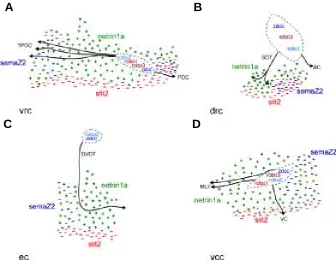
Much of what we do understand about the molecular basis of axon guidance in the rostral brain is based largely upon the expression pattern of known guidance receptors and/or their ligands (Fig. 2). These include the chemoattractive receptor zdcc (zebrafish deleted in colon cancer) (Hjorth et al., 2001) and it ligand
Fig. 2. Expression patterns of some zebrafish homologues of known axon guidance molecules in the developing brain. Rostral is to the right and dorsal is to the top in all panels. (A) Schematic representation of the first neuronal clusters (stippled lines) and the simple axon scaffold at approximately 24 hpf. (B-H) Some axon guidance molecules are expressed in a dynamic pattern at 24 hpf, which includes subpopulations of cells in the first neuronal clusters. (B) zdcc; (C) netrin1a; (D) robo1;
(E) robo2; (F) robo3; (G) slit2; and (H) semaZ2. AC, anterior commissure; drc, dorso-rostral cluster; DVDT, dorso-ventral diencephalic tract; ec, epiphyseal cluster; MLF, medial longitudinal fasciculus; nTPC, nucleus of the tract of the posterior commissure; POC, post-optic commissure; SOT, supra-optic tract; TPC, tract of the posterior commissure; TPOC, tract of the post-optic commissure; vcc, VC, ventral commissure; ventro-caudal cluster; vrc, ventro-rostral cluster.
Our analysis has also revealed that while regulatory genes form distinct domains in the rostral neural tube these domains do not simply define the phenotype of the early neuronal clusters (Hjorth and Key, 2001). These nuclei are a mosaic of neurons expressing different complements of regulatory genes. This raises the possi-bility that axons arising from these nuclei express very different guidance receptors and hence respond uniquely to the multitude of cues that they confront as they navigate to their target. The importance of early patterning of the rostral brain in axon guidance is highlighted by the widespread disruption of the forebrain axon
A
B
C
D
E
F
netrin-1a (Lauderdale at al., 1997); the chemorepulsive receptors robo1-3 (Challa et al., 2001; Fricke et al., 2001) and their ligand slit2 (Yeo et al., 2001); as well as the chemorepulsive ligand semaz2 (Halloran et al., 1999). These receptors and ligands form a com-plex overlapping array of putative signals for neurons arising from the major neuronal clusters in the rostral brain. Despite the lack of empirical data testing the role of these molecules the topology of signals provides a useful insight into putative influences on axon pathfinding (Fig. 3). An understanding of this complex spatiotem-poral pattern of expression of signals provides the first step towards deciphering the unique and redundant roles for each of these molecules in establishing the template of axon tracts in the rostral brain. For example, epiphyseal neurons appear to express the chemorepulsive robo receptors (although their expression patterns remain to be fully described) as well as the chemoattractive zdcc receptor. Although the axons of these neurons grow ventrally through a patch of neuroepithelium expressing zfEphL3 (not shown in Fig. 3) the significance of this chemorepulsive receptor remains to be determined since it appears that ligands for this receptor are not expressed by epiphyseal neurons (Brennan et al., 1997). The epiphyseal axons may be attracted ventrally towards Netrin-1a (a ligand for Dcc) as in the spinal cord (see below). While netrin-1b is expressed throughout the length of the ventral brain it does not appear to play a role in the ventral growth of axons. Ectopic expression of this chemotropic factor has no effect on the formation
Fig. 3. Schematic representation of putative guidance cues at major choice points associated with (A) the ventro-rostral cluster (vrc), (B) the dorso-rostral cluster (drc), (C) the epiphyseal cluster (ec) and (D) the ventro-caudal cluster (vcc). Each neuronal cluster is indicated by a stippled line. Known axon guidance receptors which have been reported to be expressed in these clusters are indicated inside the clusters. Guidance factors present in the surrounding environment are represented by their putative effect on axon growth as either as either repulsion (-) or attraction (+). AC, anterior commissure; DVDT, dorso-ventral diencephalic tract; MLF, medial longitudinal fasciculus; POC, post-optic commissure; SOT, supra-optic tract; TPOC, tract of the post-optic commissure; VC, ventral commissure. Rostral is to the right and dorsal is to the top in all panels.
of the axon scaffolding in the brain (Strähle et al., 1997). The ventral midline of the brain expresses slit2 (a ligand for Robo) (Yeo et al., 2001) which may prevent the growth of these axons to the midline. The epiphyseal axons instead turn at the level of the TPOC within a domain of cells expressing semaz2 (Halloran et al., 1999) and then grow rostrally. In mutants with reduced levels of this chemorepulsive ligand epiphyseal axons exhibit increased errors with inappropriate caudal turns (Halloran et al., 1999). It is interest-ing that the epiphyseal neurons express both robo and zdcc since ROBO-SLIT interactions inhibit the chemoattractive activity trans-duced by NETRIN-DCC interactions (Stein and Tessier-Lavigne, 2001). Thus, the chemoattractiveness of ventrally located Netrin-1a may be attenuated by the ventrally expressed Slit2. Similar hypothetical scenarios as outlined here for the epiphyseal axons in the DVDT can be envisaged for the other major clusters of neurons and the trajectories of their axons in the rostral brain (Fig. 3).
Axonogenesis in the Hindbrain
The first evidence of segmentation in the zebrafish hindbrain is the appearance of morphological swellings or rhombomeres (r) in the neural tube (Fig. 1B). Later, discrete sets of neuronal clusters more clearly define these segments (Fig. 1 B,C). To date, only two neuronal subclasses have been characterized in zebrafish: the reticulospinal and the branchiomotor neurons. The Mauthner neu-ron is the first neuneu-ron to develop in the hindbrain at approximately 18 hpf (Mendelson, 1985; Hanneman et al., 1988). This large reticulospinal neuron emerges in the centre of r4 and its appearance is followed shortly after by differ-entiation of MiM1, an identified reticulospinal neuron which is located on the medial surface in the middle (along the rostrocaudal axis) of the hindbrain. Subsequently, a pair of bilater-ally symmetrical reticulospinal neurons develop in each rhombomere.
The embryonic hindbrain contains two longi-tudinal axons tracts: the medial longilongi-tudinal fascicle (MLF) and the dorsal longitudinal fas-cicle (DLF). The hindbrain MLF is continuous with the MLF of the midbrain and the MLF of the spinal cord. Between 21-23 hpf the rostral portion of the hindbrain MLF is pioneered by midbrain descending axons from the MLF while the caudal portion of the MLF is pioneered by unidentified caudal hindbrain interneurons (Mendelson, 1986). The DLF is pioneered by trigeminal sensory axons and by ascending axons from the spinal cord Rohon-Beard pri-mary sensory neurons (Mendelson, 1986).
The Mauthner neuron is the first reticulospinal neuron in the hindbrain to extend an axon. Beginning at approximately 21 hpf this axon courses ventrally and crosses the midline, after which it turns caudally and grows among axons of the already established contralateral MLF (Mendelson, 1986; Metcalfe et al., 1986). The MiM1 neuron of r4 also extends its axon ven-trally but it does not cross the midline, rather, it turns caudally and grows among cells of the
A
B
ipsilateral MLF. The pattern of Mauthner and MiM1 neuron axon outgrowth is reiterated in other rhombomere compartments. That is, a lateral reticulospinal neuron projects an axon ventrally into the contralateral MLF, while a medial reticulospinal neuron projects its axon ventrally into the ipsilateral MLF (Mendelson, 1986; Metcalf et al., 1986). By 24 hpf two additional neuronal clusters are established at the borders of each rhombomere. These neurons project axons ventrally forming commissural axon bundles be-tween each rhombomere. The formation and subsequent develop-ment of these commissures appears to depend on expression of L1-related cell adhesion molecules (Weiland et al., 1997) as well as polysialic acid present on the neural cell adhesion molecule NCAM (Marx et al., 2001).
Branchiomotor nuclei of the hindbrain develop in specific rhombomeres and extend axons out of the CNS at stereotypical locations. These axons innervate muscles which differentiate in the pharyngeal arches. In zebrafish, the two most anterior arches, the mandibular and hyoid, give rise to the lower jaw and jaw associated structures. The remaining pharyngeal arches give rise to the gills (Kimmel et al., 1995). In vertebrates, the development of cranial ganglia has been well characterised and shown to be dependent on the rhombomere specific expression of hox genes. In zebrafish, cranial ganglia have been less studied although the location and development of branchiomotor neurons has been described.
Chandrasekhar et al. (1997) first described the arrangement of branchiomotor neurons in the zebrafish hindbrain using a combi-nation of anatomical landmarks, DiI labelling of axon trajectories, immunohistochemistry and in situ hybridisation. They discovered that the arrangement of branchiomotor neurons in the hindbrain was remarkably similar to their arrangement in the rodent and chick. In zebrafish, the first branchiomotor neurons develop at approximately 21 hpf and extend axons at 24 hpf. The trigeminal (V) motor neurons are located in r2 and r3 and extend axons as a tight fascicle exiting the hindbrain via r2 to innervate muscles of the mandibular pharyngeal arch. The abducens (VI) branchiomotor neurons occupy r5 and r6, but the precise location of exit of their axons from the hindbrain has not been reported. Branchiomotor neurons of the facial (VII) nerve develop in r4 and 5 but migrate to a position in r6 and r7. These neurons project axons which exit the CNS through r4 and synapse with muscles of the hyoid pharyngeal arch. The glossopharyngeal (IX) branchiomotor neurons are lo-cated exclusively in r7 and project axons which exit the hindbrain via r6 to innervate the first gill arch. The vagus (X) cranial branchiomotor neurons are located in a region caudal to r7 known as the caudal-most hindbrain. These motor neurons extend axons out of the hindbrain near the caudal border of r7 which synapse with targets in gill arches 2-5. Transgenic zebrafish expressing green fluorescence protein under the control of the isl-1 promoter have been used to examine the projection pattern of cranial motor axons (Higashijima et al., 2000). Trigeminal branchiomotor neurons in r2 extended axons to synapse with the abductor mandibulae muscle of the jaw. Trigeminal branchiomotor neurons in r3 project axons which innervated four other jaw muscles: levator arcus palatini, dilator operculi, intermondibularis anterior and intermondibularis posterior. These results demonstrated that specific branchiomotor nuclei in the hindbrain innervate precise muscle groups.
While reticulospinal and branchiomotor neurons have been characterized identification of the visceral or somatic motor neu-rons in the hindbrain has not been reported. This is probably because of the complexity of cranial nuclei as well as to the lack of
specific molecular markers for these neurons (Trevarrow et al., 1990). Nevertheless, what is apparent in the hindbrain, as in other regions of the zebrafish CNS, is that initially a simple stereotypical pattern of neurons and axons develops. While studies have pro-vided insight into development of neural circuits associated with late emerging locomotor behaviours (Metcalfe et al., 1986; Lorent et al., 2001) our understanding of generation of the early hindbrain pathways is poor. Future studies coupling the simplicity of the hindbrain and the versatility of the zebrafish as a vertebrate model, will further enhance our understanding of axon tract formation in this region of the nervous system.
Axonogenesis in the Spinal Cord
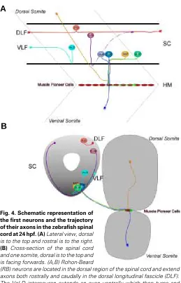
Primary neurons in the zebrafish spinal cord have been characterised and subdivided into three classes: sensory neurons, interneurons and motor neurons (Bernhardt et al., 1990; Kuwada et al., 1990; Eisen, 1991). These neurons are segmentally re-peated along the anteroposterior axis and are identified based on the location of their cell body, the projection pattern of their axon and their position relative to somite boundaries (Fig. 4). At 18-20 hpf the spinal cord is segmented and each hemisegment contains approximately 11 differentiated neurons: three primary motor neurons, three primary sensory Rohon-Beard neurons, and five interneurons (Kuwada and Bernhardt, 1990). The interneurons are individually identified as one primary commissural ascending neu-ron (CoPA), two secondary commissural ascending neuneu-rons (CoSA), one dorsal longitudinal ascending interneuron (DoLA) and one ventral longitudinal descending interneuron (VeLD). These different classes of neurons contribute axons to the four of the five principal axon pathways of the early spinal cord: the dorsal longi-tudinal fascicle (DLF); the ventral longilongi-tudinal fascicle (VLF); ventral commissure; and the ventral motor nerve. The fifth axon pathway, the MLF, is located ventral to the VLF, directly apposed to the lateral margin of the floor plate cells and contains descending axons from the midbrain and hindbrain spinoreticular neurons (Kimmel et al., 1982). The DLF is the first tract to form in the spinal cord (17 hpf), followed by the VLF (17-24 hpf) and then the MLF (24-28 hpf) (Kimmel et al., 1982). It should be noted that there is a rostrocaudal wave of development of these pathways; at any one stage the pathways are further developed in rostral segments than in more caudal regions of the spinal cord (Kimmel et al., 1982).
A
B
While it is not clear why axons from different neuronal subtypes pioneer different longitudinal pathways in the spinal cord it is possible that chemorepulsive molecules may play a role in defin-ing the pathway. For instance, semaz7 (a transmembrane class IV Semaphorin; Halloran et al., 1998) is expressed at the dorsal border of the DLF and the ventral border of the MLF and may
assist in restricting axon growth to defined tracts. An observation made over a decade ago provided an interesting insight into one of the fundamental organizing principles of the vertebrate spinal cord. Kuwada et al. (1990) revealed that pioneer axons of the longitudinal tracts in the spinal cord were spatially organized according to their sub-type. For example, the Rohon-Beard axons were located dorsally in the DLF while DoLA and ascending commissural axons were positioned progressively more ventral in this tract. Moreover, subsequent follower axons preferentially fasciculated into “like” bundles; that is, later growing Rohon-beard axons preferentially grew on Rohon-beard pioneer axons while later growing ascending commissural axons preferentially grew on earlier ascending commissural axons. Despite this remarkable specificity in sorting of axons we have little understanding about how it is achieved. The most obvious explanation is the expres-sion of different cell surface recognition molecules by the various axon sub-types, an idea consistent with selective fasciculation of axons expressing the same cell surface glycoforms of NCAM in the rostral brain of Xenopus (Anderson and Key, 1999).
Analysis of the cyc and no tail zebrafish mutants revealed that the notochord and floor plate were both important for the success-ful ventral midline crossing of commissural axons in the spinal cord (Greenspoon et al., 1995). Interestingly the lack of either the floor plate or notochord did not affect the ventral growth of commissural axons but rather increased the frequency of errors in midline crossing. When both notochord and floor plate were missing (following physical ablation in either mutant line) only 5% of commissural axons failed to grow ventrally towards the midline, indicating that signals governing the ventralward guidance of these commissural axons were not associated with these tissues. Sub-sequent analysis of the floating head zebrafish mutant (floating head is homologous to the homeobox transcription factor Xnot1; Talbot et al., 1995) has revealed that netrin-1a expression is important for the ventral growth of commissural axons (Lauderdale et al., 1997). In addition to the lack of notochord these mutants have missing patches of floor plate cells in the spinal cord. netrin-1a, which is normally expressed along the length of the ventral cord, is restricted to segregated patches or islands of ventral cells in the floating head mutant. These islands appear to act as sources of chemoattractant and CoPA axons aberrantly project to these cells. These results suggest that netrin-1a acts normally to stimulate growth of commissural neurons towards the ventral midline, which is consistent with the fact that these neurons express the netrin receptor, zdcc (Hjorth et al., 2001). In contrast, netrin-1b, which is expressed by the floor plate, can not be involved in the ventral guidance of these axons since it is absent in cyc mutants (Strähle et al., 1997).
Three primary motor neurons develop in the ventral spinal cord and have been individually identified as the caudal primary (CaP), middle primary (MiP) and rostral primary (RoP) neurons. In addition, a fourth motor neuron, the variable primary neuron (VaP), also develops adjacent to the CaP neuron, but only in some segments (Eisen et al., 1989). The VaP neuron dies later in development. Within the spinal cord these different motor neu-rons are located in stereotypical positions relative to the somite boundary. The RoP neuron is located at a level immediately anterior to the boundary, the MiP neuron is located immediately posterior to the boundary, while the Cap/VaP neurons are located centrally relative to the somite. The LIM homeobox genes are
Fig. 4. Schematic representation of the first neurons and the trajectory of their axons in the zebrafish spinal cord at 24 hpf. (A) Lateral view, dorsal is to the top and rostral is to the right.
(B) Cross-section of the spinal cord and one somite, dorsal is to the top and is facing forwards. (A,B) Rohon-Beard
thought to dictate a combinatorial code leading to primary motor neuron development (Appel et al., 1995; Tokumoto et al., 1995; Segawa et al., 2001). Initially, all primary motor neurons express isl-1, but later in development they express different combinations of LIM genes. The CaP and VaP neurons express isl-2 and lim3, while the MiP and RoP neurons express isl-1 and lim3. While this expression pattern may provide a molecular code for primary motor neuron differentiation there is no evidence that it is involved in axon guidance in zebrafish (Segawa et al., 2001).
The axon pathways of the primary motor neurons have been well characterised and shown to follow highly specific trajectories (Beattie, 2000). The pioneer motor neurons of the zebrafish spinal cord develop at 15 hpf and extend axons at approximately 17 hpf (Eisen et al., 1989). The CaP axon pioneers the common pathway of motor axons to the horizontal myoseptum separating the dorsal and ventral myotomes. Initially, the RoP and MiP axons project posteriorly to the level of the CaP axon and then they follow this axon out of the spinal cord and into the periphery. Here the axons contact specialised muscle pioneer cells, the first to express muscle specific markers (Melancon et al., 1997). The axons pause at this choice point before growing along separate path-ways to their final targets. The RoP axon does not grow much further and innervates muscle cells around the horizontal myo-septum. The MiP axon first contacts muscle pioneer cells and then sends a collateral branch into the dorsal somite. The initial muscle pioneer cell axon is subsequently retracted leaving the MiP axon innervating the dorsal myoseptum. After the CaP and VaP axons contact the muscle pioneer cells, only the CaP axon extends into the ventral somite, while the VaP neuron dies. The death of the VaP neuron appears to be dependent on interactions with the muscle pioneer cells (Eisen and Melancon, 2001). Following ablation of muscle pioneer cells the VaP survives and its axons extends into the ventral somitic tissue. The ventral extension of the CaP axons appears to be dependent on the restricted expression of the chemorepulsive ligand semaZ1b within the posterior portion of the somite since ectopic expression of this ligand throughout the somite stunts the development of the ventral motor nerve branches (Roos et al., 1999).
Zebrafish mutant lines exhibiting defects in primary motor axon guidance have provided insight into the mechanisms of axon pathway selection in this system. In the diwanka zebrafish mutant, primary motor axons navigate normally through the spinal cord but fail to project along the common path out of the spinal cord towards the somites (Zeller and Granato, 1999). After axons have entered the common peripheral pathway their subsequent growth appears to be independent of each other (Eisen et al., 1989; Eisen et al., 1990; Pike and Eisen, 1990) but dependent on contact with the horizontal myoseptum separating the dorsal and ventral myo-tomes. Ablation of muscle pioneer cells in the horizontal myosep-tum results in subtle changes to the cell specific pathway choices by primary motor neurons, although they still innervate their correct target tissue (Melancon et al., 1997). NETRIN-1a-DCC interac-tions may be mediating these recognition events since netrin-1a is expressed by cells in the vicinity of the myoseptum (Lauderdale et al., 1997) while zdcc is expressed by primary motor neurons (Hjorth et al., 2001). In the unplugged zebrafish mutant primary motor axons navigate correctly out of the spinal cord to the horizontal myoseptum. However, at this choice point CaP and RoP axons grow aberrantly (Zhang and Granato, 2000). The unplugged
gene activity is restricted to cells located dorsally, adjacent to muscle pioneer cells. In the stumpy mutant embryo CaP axons stall at the muscle pioneer cell choice point (Beattie et al., 2000). The identification of the proteins encoded by stumpy and unplugged are sure to provide further insight into axon guidance in this system.
Conclusion
Studies examining neuronal specification and axon guidance in the zebrafish CNS have provided a unique perspective into the dynamic formation of axon trajectories in vivo. The fundamental basis of these studies is the simplicity of the zebrafish nervous system and its early anatomical characterisation. Studies have begun to decipher at the single cell level the role of cell-cell interactions and molecular cues in guiding pioneer axons in the spinal cord. The accessibility of the zebrafish spinal cord to such investigations has provided an insight into axon guidance that was previously only possible in invertebrates. The challenge ahead is to now examine mechanisms of axon growth and guidance in the rostral brain at a single cell level using identified neurons as in the spinal cord. An obvious starting point is the DVDT since this tract is pioneered by a single epiphyseal neuron. Moreover, we know that the epiphyseal cluster expresses zdcc (Hjorth et al., 2001) and that the DVDT projects to a pathway rich in netrin-1a (Lauderdale et al., 1997). However, understanding the interplay of the numerous guidance cues that establish this and other brain tracts will be difficult. We are only beginning to come to terms with the extent of heterogeneity of axons types even within a single axon tract. The task is made more difficult by the fact that altering a single component within a complex network of regulatory genes may only affect the pathfinding of a minor subpopulation of axons. Nonetheless, the ease with which gene expression can be manipulated by microinjection of either syn-thetic mRNA or anti-sense oligonucleotide analogues such as morpholinos provides an opportunity to understand the formation of axon tracts in the embryonic vertebrate brain.
Acknowledgements
This work was supported by an Australian Research Grant to BK. JH was supported by an Australian Research postgraduate student award.
References
ANDERSON, R.A. and KEY, B. (1999). Guidance cues during neuronal pathfinding in the early scaffold of axon tracts in embryonic Xenopus brain. Development 126: 1859-1868.
APPEL, B., KORZH, V., GLASGOW, E., THOR, S., EDLUND, T., DAWID, I. B. and EISEN, J. S. (1995). Motoneuron fate specification revealed by patterned LIM homeobox gene expression in embryonic zebrafish. Development 121: 4117-4125.
BEATTIE, C.E. (2000). Control of motor axon guidance in the zebrafish embryo. Brain Res. Bull. 53: 489-500.
BEATTIE, C. E., MELANCON, E. and EISEN, J. S. (2000). Mutations in the stumpy gene reveal intermediate targets for zebrafish motor axons. Development 127: 2653-2662.
BERNHARDT, R.R., CHITNIS, A.B., LINDAMER, L. and KUWADA, J.Y. (1990). Identification of spinal neurons in the embryonic and larval zebrafish. J. Comp. Neurol. 302: 603-616.
BRENNAN, C., MONSCHAU, B., LINDBERG, R., GUTHRIE, B., DRESCHER, U., BONHOEFFER, F., and HOLDER, N. (1997). Two Eph receptor tyrosine kinase ligands control axon growth and may be involved in the creation of the retinotectal map in the zebrafish. Development 124: 655-664.
CHALLA, A.K., BEATTIE, C.E. and SEEGER, M.A. (2001). Identification and charac-terization of roundabout orthologs in zebrafish. Mech. Dev. 101: 249-253.
CHANDRASEKHAR, A., MOENS, C.B., WARREN, J.T. JR., KIMMEL, C.B. and KUWADA, J.Y. (1997). Development of branchiomotor neurons in zebrafish. Development 124: 2633-2644.
CHITNIS, A.B. and KUWADA, J.Y. (1990). Axonogenesis in the brain of zebrafish embryos. J. Neurosci. 10: 1892-1905.
DORNSEIFER, P., TAKKE, C. and CAMPOS-ORTEGA, J.A. (1997). Overexpression of a zebrafish homologue of the Drosophila neurogenic gene Delta perturbs differentiation of primary neurons and somite development. Mech. Dev. 63: 159-171.
EISEN, J.S. (1991). Determination of primary motoneuron identity in developing zebrafish embryos. Science 252: 569-572.
EISEN, J.S. and MELANCON, E. (2001). Interactions with identified muscle cells break motoneuron equivalence in embryonic zebrafish. Nat. Neurosci. 4: 1065-1070.
EISEN, J.S., PIKE, S.H., and DEBU, B. (1989). The growth cones of identified motoneurons in embryonic zebrafish select appropriate pathways in the absence of specific cellular interactions. Neuron 2: 1097-1104.
EISEN, J.S., PIKE, S.H., and ROMANCIER, B. (1990). An identified motoneuron with variable fates in embryonic zebrafish. J. Neurosci. 10: 34-43.
FRICKE, C., LEE, J.S., GEIGER-RUDOLPH, S., BONHOEFFER, F. and CHIEN, C.B. (2001). astray, a zebrafish roundabout homolog required for retinal axon guid-ance. Science 292: 507-510.
GREENSPOON, S., PATEL, C.K., HASHMI, S., BERNHARDT, R.R. and KUWADA, J.Y. (1995). The notochord and floor plate guide growth cones in the zebrafish spinal cord. J. Neurosci. 15: 5956-5965.
GRUNWALD, D.J., KIMMEL, C.B., WESTERFIELD, M., WALKER, C. and STREISINGER, G. (1988). A neural degeneration mutation that spares primary neurons in the zebrafish. Dev. Biol. 126: 115-128.
HALLORAN, M.C., SEVERANCE, S.M., YEE, C.S., GEMZA, D.L. and KUWADA, J.Y. (1998). Molecular cloning and expression of two novel zebrafish semaphorins. Mech. Dev. 76: 165-168.
HALLORAN, M.C., SEVERANCE, S.M., YEE, C.S., GEMZA, D.L., RAPER, J.A. and KUWADA, J.Y. (1999). Analysis of a zebrafish semaphorin reveals potential functions in vivo. Dev. Dyn. 214: 13-25.
HANNEMAN, E, TREVARROW, B., METCALFE, W.K., KIMMEL, C.B. and WESTERFIELD, M. (1988). Segmental pattern of development of the hindbrain and spinal cord of the zebrafish embryo. Development103: 49-58.
HAUPTMANN, G. and GERSTER, T. (2000). Regulatory gene expression patterns reveal transverse and longitudinal subdivisions of the embryonic zebrafish fore-brain. Mech. Dev. 91: 105-118.
HATANAKA, Y. and JONES, E.G. (1998). Early region-specific gene expression during tract formation in the embryonic rat forebrain. J. Comp. Neurol. 395: 296-309.
HIGASHIJIMA, S., HOTTA, Y. and OKAMOTO, H. (2000). Visualization of cranial motor neurons in live transgenic zebrafish expressing green fluorescent protein under the control of the islet-1 promoter/enhancer. J. Neurosci. 20: 206-218.
HJORTH, J., GAD, J., COOPER, H. and KEY, B. (2001). A zebrafish homologue of deleted in colon cancer (zdcc) is expressed in the first neuronal clusters of the developing brain. Mech. Dev. 109: 105-109.
HJORTH, J. and KEY, B. (2001). Are pioneer axons guided by regulatory gene expression domains in the zebrafish forebrain? High resolution analysis of the patterning of the zebrafish brain during axon tract formation. Dev. Biol. 229: 271-286.
KANKI, J.P. and KUWADA, J.Y. (2000). Growth cones utilize both widespread and local directional cues in the zebrafish brain. Dev. Biol. 219: 364-72.
KATZ, M.J., LASEK, R.J., and NAUTA, H.J. (1980). Ontogeny of substrate pathways and the origin of the neural circuit pattern. Neurosci. 5: 821-33.
KIMMEL, C.B. (1993). Patterning the brain of the zebrafish embryo. Annu. Rev. Neurosci. 16: 707-732.
KIMMEL, C.B., POWELL, S.L. and METCALFE, W.K. (1982). Brain neurons which project to the spinal cord in young larvae of the zebrafish. J. Comp. Neurol. 205: 112-127.
KIMMEL, C.B., BALLARD, W.W., KIMMEL, S.R., ULLMANN, B. and SCHILLING, T.F. (1995). Stages of embryonic development of the zebrafish. Dev. Dyn. 203: 253-310.
KORZH, V., EDLUND, T. and THOR, S. (1993). Zebrafish primary neurons initiate expression of the LIM homeodomain protein Isl-1 at the end of gastrulation. Development 118: 417-425.
KRAUSS, S., JOHANSEN, T., KORZTH, V. and FJOSE, A. (1991a). Expression pattern of zebrafish pax genes suggest a role in early brain regionalisation. Nature 353: 267-270.
KRAUSS, S., JOHANSEN, T., KORZH, V., MOENS, U., ERICSON, J. U. and FJOSE, A. (1991b). Zebrafish pax[zf-a]: a paired box-containing gene expressed in the neural tube. EMBO J. 10: 3609-3619.
KUWADA, J.Y. and BERNHARDT, R.R. (1990). Axonal outgrowth by identified neurons in the spinal cord of zebrafish embryos. Exp. Neurol. 109: 29-34.
KUWADA, J.Y., BERNHARDT, R.R. and NGUYEN, N. (1990). Development of spinal neurons and tracts in the zebrafish embryo. J. Comp. Neurol. 302: 617-628.
LAUDERDALE, J.D., DAVIS, N.M. and KUWADA, J.Y. (1997). Axon tracts correlate with netrin-1a expression in the zebrafish embryo. Mol. Cell. Neurosci. 9: 293-313.
LORENT, K., LIU, K.S., FETCHO, J.R., and GRANATO, M. (2001). The zebrafish space cadet gene controls axonal pathfinding of neurons that modulate fast turning movements. Development 128: 2131-2141.
MACDONALD, R., SCHOLES, J., STRÄHLE, U., BRENNAN, C., HOLDER, H., BRAND, M., and WILSON, S.W. (1997). The pax protein Noi is required for commissural pathway formation in the rostral forebrain. Development 124: 2397-2408.
MACDONALD, R., XU, Q., BARTH, K.A., MIKKOLA, I., HOLDER, N., FJOSE, A., KRAUSS, S. and WILSON, S.W. (1994). Regulatory gene expression boundaries demarcate sites of neuronal differentiation in the embryonic zebrafish forebrain. Neuron 13: 1039-1053.
MARCUS, R.C., SHIMAMURA, K., SRETAVAN, D., LAI, E., RUBENSTEIN, J.L. and MASON, C.A. (1999). Domains of regulatory gene expression and the developing optic chiasm: correspondence with retinal axon paths and candidate signaling cells. J. Comp. Neurol. 403: 346-358.
MARX, M., RUTISHAUSER, U. and BASTMEYER, M. (2001). Dual function of polysialic acid during zebrafish central nervous system development. Development 128: 4949-4958.
MASTICK, G.S., DAVIS, N.M., ANDREWS, G.L. and EASTER, S.S.JR. (1997). Pax6 functions in boundary formation and axon guidance in the embryonic mouse forebrain. Development 124: 1985-1997.
MELANCON, E., LIU, D.W., WESTERFIELD, M. and EISEN, J.S. (1997). Pathfinding by identified zebrafish motoneurons in the absence of muscle pioneers. J. Neurosci. 17: 7796-7804.
MENDELSON, B. (1985). Soma position is correlated with time of development in three types of identified reticulospinal neurons. Dev. Biol. 112: 489-493.
MENDELSON, B. (1986). Development of reticulospinal neurons of the zebrafish. II. Early axonal outgrowth and cell body position. J. Comp. Neurol. 251: 172-184.
METCALFE, W.K., MENDELSON, B. and KIMMEL, C.B. (1986). Segmental homolo-gies among reticulospinal neurons in the hindbrain of the zebrafish larva. J. Comp. Neurol. 251: 147-159.
METCALFE, W.K., MYERS, P.Z., TREVARROW, B., BASS, M.B. and KIMMEL, C.B. (1990). Primary neurons that express the L2/HNK-1 carbohydrate during early development in the zebrafish. Development 110: 491-504.
NGUYEN BA-CHARVET, K.T., VON BOXBERG, Y., GUAZZI, S., BONCINELLI, E., and GODEMENT, P. (1998). A potential role for the OTX2 homeoprotein in creating early ‘highways’ for axon extension in the rostral brain. Development 125: 4273-82
PAPAN, C. and CAMPOS-ORTEGA, J.A. (1994). On the formation of the neural keel and neural tube in the zebrafish Danio (Brachydanio) rerio. Roux’s Arch. Dev. Biol. 203: 178-186.
PATEL, C.K., RODRIGUEZ, L.C., and KUWADA, J.Y. (1994). Axonal outgrowth within the abnormal scaffold of brain tracts in a zebrafish mutant. J. Neurobiol. 25: 345-360.
PIKE, S.H. and EISEN, J.S. (1990). Identified primary motoneurons in embryonic zebrafish select appropriate pathways in the absence of other primary motoneu-rons. J. Neurosci. 10: 44-49.
ROSS, L.S., PARRETT, T. and EASTER, S.S.JR. (1992). Axonogenesis and morpho-genesis in the embryonic zebrafish brain. J. Neurosci. 12: 467-482.
SAMPATH, K., RUBINSTEIN, A.L., CHENG, A.M.S., LIANG, J.O., FEKANY, K., SOLNICA-KREZEL, L., KORZH, V., HALPERN, M. E. and WRIGHT, C.V.E. (1998). Induction of the zebrafish ventral brain and floor plate requires Cyclops/ Nodal signalling. Nature 395: 185-189.
SEGAWA, H., MIYASHITA, T., HIRATE, Y., HIGASHIJIMA, S., CHINO, N., UYEMURA, K., KIKUCHI, Y. AND OKAMOTO, H. (2001). Functional repression of Islet-2 by disruption of complex with Ldb impairs peripheral axonal outgrowth in embryonic zebrafish. Neuron 30: 423-436.
SHANMUGALINGAM, S., HOUART, C., PICKER, A., REIFERS, F., MACDONALD, R., BARTH, A., GRIFFIN, K., BRAND, M. and WILSON, S.W. (2000). Ace/Fgf8 is required for forebrain commissure formation and patterning of the telencephalon. Development 127: 2549-2561.
SINGER M., NORDLANDER, R.H. and EGAR, M. (1979). Axonal guidance during embryogenesis and regeneration in the spinal cord of the newt: the blueprint hypothesis of neuronal pathway patterning. J. Comp. Neurol. 185: 1-21.
SIMEONE, A., ACAMPORA, D., GULISANO, M., STORNAIUOLO, A. and BONCINELLI, E. (1992). Nested expression domains of four homeobox genes in developing rostral brain. Nature 358: 687-690.
STEIN, E. and TESSIER-LAVIGNE, M. (2001). Hierarchical organization of guidance receptors: silencing of netrin attraction by slit through a Robo/DCC receptor complex. Science 291: 1928-1938.
STRÄHLE, U., FISCHER, N. and BLADER, P. (1997). Expression and regulation of a netrin homologue in the zebrafish embryo. Mech. Dev. 62: 147-160.
TALBOT, W.S., TREVARROW, B., HALPERN, M.E., MELBY, A.E., FARR, G., POSTLETHWAIT, J.H., JOWETT, T., KIMMEL, C.B. and KIMELMAN, D. (1995). A homeobox gene essential for zebrafish notochord development. Nature 378: 150-157.
TOKUMOTO, M., GONG, Z., TSUBOKAWA, T., HEW, C.L., UYEMURA, K., HOTTA, Y. and OKAMOTO, H. (1995). Molecular heterogeneity among primary motoneu-rons and within myotomes revealed by the differential mRNA expression of novel islet-1 homologs in embryonic zebrafish. Dev. Biol. 171: 578-589.
TREVARROW, B., MARKS, D.L. and KIMMEL, C.B. (1990). Organization of hindbrain segments in the zebrafish embryo. Neuron 4: 669-679.
VARGA, Z.M., AMORES, A., LEWIS, K.E., YAN, Y.L., POSTLETHWAIT, J.H., EISEN, J.S., and WESTERFIELD, M. (2001) Zebrafish smoothened functions in ventral neural tube specification and axon tract formation. Development 128: 3497-3509.
WEILAND, U.M., OTT, H., BASTMEYER, M., SCHADEN, H., GIORDANO, S. and STUERMER, C.A. (1997). Expression of an L1-related cell adhesion molecule on developing CNS fiber tracts in zebrafish and its functional contribution to axon fasciculation. Mol. Cell Neurosci. 9: 77-89.
WILSON, S.W. and EASTER, S.S.JR. (1991). A pioneering growth cone in the embryonic zebrafish brain. Proc. Natl. Acad. Sci., USA. 88: 2293-2296.
WILSON, S.W., PLACZEK, M., and FURLEY, A.J. (1993). Border disputes: do boundaries play a role in growth-cone guidance? TINS 16: 316-322.
WILSON, S.W., ROSS, L.S., PARRETT, T. and EASTER, S.S.JR. (1990). The development of a simple scaffold of axon tracts in the brain of the embryonic zebrafish, Brachydanio rerio. Development 108: 121-145.
YEO, S.Y., LITTLE, M.H., YAMADA, T., MIYASHITA, T., HALLORAN, M.C., KUWADA, J.Y., HUH, T.L. and OKAMOTO, H. (2001). Overexpression of a slit homologue impairs convergent extension of the mesoderm and causes cyclopia in embryonic zebrafish. Dev. Biol. 230: 1-17.
ZELLER, J. and GRANATO, M. (1999). The zebrafish diwanka gene controls an early step of motor growth cone migration. Development 126: 3461-72