Skin field formation: morphogenetic events
DANIELLE DHOUAILLY*, ISABEL OLIVERA-MARTINEZ
α, INGRID FLINIAUX
β, SYLVAIN MISSIER,
JEAN P. VIALLET and JACQUES THELU
Equipe Biologie de la Différenciation Epithéliale, UMR CNRS 5538, Institut Albert Bonniot, Université Joseph Fourier, Grenoble, France
ABSTRACT This chapter is mostly a review of the pioneering work of the Philippe Sengel school in Grenoble carried out in the late sixties and the seventies. The questions raised concerning the morphogenesis of feather tracts were approached by means of microsurgery on chick embryos. P. Sengel and his wife M. Kieny had the feeling that proteins synthesized by the neural tube were required for the formation of feather fields. It was my pleasure to carry on the story from the beginning. Although some clarifications concerning this morphogenesis have been contributed by my group and by a few other laboratories interested in this subject, the most important contributions to recent research have been the elucidation of the nature of the required messages, which will be explored further in other papers in this Issue.
KEY WORDS:
chick, cutaneous appendage, dermis, differentiation, epidermis, feather, neural tube, pteryla
0214-6282/2004/$25.00 © UBC Press
Printed in Spain www.ijdb.ehu.es
*Address correspondence to: Dr. Danielle Dhouailly. BDE-LEDAC, UMR CNRS 5538, Institut Albert Bonniot, Domaine de la Merci, 38706, La Tronche Cedex, France. Fax: +33-4-76-54-94-25. e-mail: [email protected]
Note α α α α α Present address: Division of Cell and Developmental Biology, School of Life Sciences, University of Dundee, Wellcome Trust Biocentre, Dow St., Dundee, DD1 5EH, U.K.
Note βββββPresent address: Atelier de Transgenèse, CEA, Grenoble, DRDC/AT 17 rue des martyrs, 38054 Grenoble cedex, France.
Introduction
One of the significant steps during skin morphogenesis in birds and mammals is the establishment of the cutaneous appendage fields, these are initially homogeneous and develop into heterogeneous feather- or hair-bearing fields. In the avian embryo, the different feather tracts, or pterylae, arise sequentially following a dorso-lateral and a latero-ventral morphogenic wave (Fig. 1, and Mayerson and Fallon, 1985). The way in which the distinct pterylae are laid out is called the macropattern (Sengel, 1976). In chick, the dorsal trunk feather macropattern is composed of the spinal and scapular pterylae (Fig. 2A), while that of the ventral side is comprised of the pectoral and ventral pterylae (Fig. 2B). Each tract, in addition to its location and time of appearance, is characterized by its contour, the size and number of feathers. In each tract the design formed by the feathers is called the micropattern (Sengel, 1976). The same distinction between skin macropattern and micropattern occurs for mammals. In mouse, four main types of skin fields can be distinguished. The first hair follicles to develop are those of the tactile hairs or vibrissae. They arise in sequence along five rows on right and left pads formed by upper-lip skin. At a later stage, primary pelage hairs differentiate almost concomitantly and cover most of the body surface. The two last skin fields to differentiate are those of the tail, where pelage hairs are ranged and intermingled with “scales”, and the plantar surface, where sparse hairs are present between the foot pads which have sweat glands.
Numerous experiments have been conducted using the technique of heterotopic transplantations (Kieny and Brugal, 1977; Mauger, 1972a and b) and dermal-epidermal heterotopic and heterospecific recombinations (Dhouailly, 1973; Dhouailly, 1977; Dhouailly and Sengel, 1975; Dhouailly, unpublished data on heterospecific mouse/ chick limbs), and these showed that the information relating to the formation of chick and mouse macropattern and micropattern resides first in the mesoderm and then in the dermis.
In chick, the different pterylae are separated by semi-apteria, which are characterized by unorganized and scarce feathers. In the trunk, except for a tiny middorsal featherless region, the only true glabrous area is the midventral apterium, which forms a ribbon on each side of the midventral closure. This apterium is surrounded by the ventral pteryla and is contiguous to the amnion via the umbilical cord. The first morphological indication of the formation of a pteryla as opposed to an apterium, or semi-apterium, is the early densification of the upper part of the predermal mesenchyme to form what is called a dense superficial dermis (2.6 nuclei/1000µm3) (Sengel, 1976;
Wessels, 1965), and the subsequent differentiation of its overlying ectoderm into an epidermis (Wessells, 1965). This dermal densification occurs by day 6 in the spinal pteryla and the ventral pteryla, but only several days later in the semi-apteria. In the midventral apterium, by contrast, the dermal fibroblasts remain sparse (1.98 nuclei/1000 µm3), and extra-cellular material accumulates (Sengel et al., 1969).
A
B
A
C
B
D
these condensations being required for the development of feathers. By contrast, the loose midventral dermis is comparable to adult scar dermis, in the fact that it is unable to induce and to participate to cutaneous appendage morphogenesis (Sengel et al., 1969).
The questions which arise are: when and how are the different skin fields determined? The first questions that needed to be resolved were, what is the origin of the mesodermal component, the next what are the cellular interactions required for their determination, and finally the elucidation of the molecular nature of the swapped messages. Thirty years ago, these questions were approached by carbon markings, localized X-irradiations, heterotopic transplantations, and by chick/ quail chimeras. The results obtained
Fig. 1 (Left). The different chick skin pterylae. Modified from Mayerson and Fallon (1985). Diagram by I. Fliniaux.
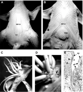
Fig. 2 (Right). Dorsal (A) and ventral (B) skin of 11-day chick embryo. Abbreviations: cp, cervical part of the spinal pteryla; dfp, dorsal part of femoral pteryla; mva, medioventral apterium; pp, pectoral pteryla; sa, semiapterium; scp, scapular pteryla; spp, spinal pteryla; u, umbilical cord; vfp, ventral part of the femoral pteryla; vp, ventral pteryla. Dissections and photographs by A. Mauger (A) and I. Fliniaux (B).
Martinez et al., 2000 and 2002). Recent results (Ben-Yair et al., 2003), obtained by measurements of cell proliferation, nuclear density, cellular rearrangement and lineage tracing experiments confirm that progenitors of the dorsal dermis reside in the three parts, medial, center and lateral, of the dermomyotome. Somites appear as epithelial structures that bud off from the presomitic mesoderm. Soon after their segmentation they become patterned along the dorsoventral and mediolateral axes. The ventral part forms a mesenchyme called the sclerotome, which is known to give rise to the vertebrae, whereas their dorsal epithelial part, called the dermomyotome, give rise to striated muscles and dermis.
The dorsal dermal cells appear progressively as part of a loose sub-ectodermal mesenchyme that forms between days 3 (E3) and 5
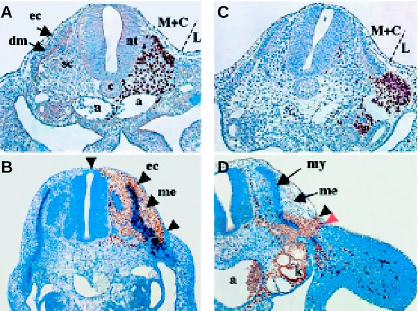
Fig. 3. Almost the entire dorsal mesenchyme derives from the medial and central somitic compartment and only a minor portion derives from the lateral somitic compartment. Grafts of medial and central presomitic mesoderm (M+C) portions (A,C) or lateral portions (B,D) from quail embryo in a chick host. Sections stained with the QCPN antibody (brown) to reveal quail nuclei. Chimeras were analyzed 15 hours (A,B) and 2 days (C,D) after grafting. Experiments by M. Coltey; Photographs by I. Olivera-Martinez.
by the P. Sengel’s school established the basis on which current research on the signals required for the determination of cutaneous fields is based.
Dermomyotomal origin of the dorsal dermis
(Olivera-A
C
B
D
E
G
F
H
A
(E5) of incubation in the chick, and days 9.5 (E9.5) and 13 (E13) of gestation in mouse. At E3 in chick (Fig. 4A), and E9.5 in mouse (Fig. 4B) in the thoracic region, the somites are differentiated into a ventral part, the sclerotome and a dorsal epithelial part, the dermomyotome, which lies directly under the ectoderm. The space between the two layers is invaded progressively by a fibrous lattice, which also penetrates between the ectoderm and the neural tube in the case of chick embryo. It has been suggested (Weiss, 1958) that this fibrous network serves the purpose of providing the incoming mesenchymal cells with a conductive substratum, through a mechanism of contact guidance.
In chick, starting by E3, mesodermal cells begin to migrate from the dermomyotome to colonize the subectodermal space over the dorsal neural tube, while in the dorsolateral region, the epithelial structure of the central dermomyotome is transformed into dermal progenitors that are directly under the ectoderm. Until E5, no distinct dermis is recognizable, with the mesodermal cells forming a loose mesenchyme of noticeable thickness between the ectoderm, the neural tube and the differentiating myotome (Fig. 4C). By the sixth day, the upper part of this mesenchymal population gives rise to the dense dermis (Fig. 4E,F), which by E7/ 7.5 in turn forms the dermal condensations that are involved in the morphogenesis of cutaneous appendages. While the dermis is differentiating, the corresponding ectoderm undergoes its transformation into an epidermis comprising of one layer of columnar cells covered with a well distinguishable periderm that forms a pavement epithelium. The dense dermis initially forms only in the dorsal area which corresponds to the future spinal pteryla. Its formation, as well that of its overlying epidermis, spreads symmetrically on both sides of the mid-dorsal line. It should be noted that the formation of epidermal placodes precedes that of the corresponding dermal condensations by the time taken to develop one row (Sengel and Rusaouïn, 1969; Dhouailly, 1984). When the first mid-dorsal row of feather primordia is completed, new lateral rows are added successively on both sides, and in order to optimally occupy the space, lead to the formation of an hexagonal micropattern.
In mouse embryo, by contrast, both the formation of the dense dermis and of the primary hair buds appear first laterally, and, only two day later, over the neural tube. This is correlated to the fact, that by E11.5, the mesenchymal cells accumulate between the mediodorsal lip of the dermomyotome and the lateral part of the neural tube (Fig. 4D). By E13, the dense dermis has formed only in the lateral part of the trunk, while no mesenchymal population is present between the dorsal neural tube and the ectoderm (Fig. 4 G,H). Mouse/chick chimeras were recently performed by orthotopically implanting mouse somites into a chick host (Houzelstein et al., 2000). The results show that the predermal mesenchyme is comprised of a first population of dermal cells which originates from the dermomyotome mediodorsal lip, and a second one that delaminates directly from the central dermomyotome.
The interaction events between the dermomyotome and
the organ axial structures in chick
In the absence of the neural tube and notochord, the medial somitic cells die, resulting in the absence of the vertebrae, dorsal muscles and ribs (Teillet and Le Douarin, 1983), and also of the dorsal feather field. Indeed, transverse glabrous bands of
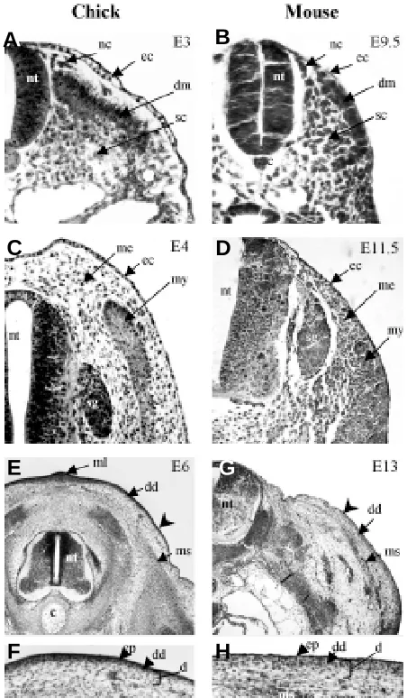
Fig. 4. Formation of dorsal chick and mouse skin. Transverse sections through the thoracic region. In (A) the 3-day chick embryo and (B) the 9.5-day mouse embryo, the dermomyotome (dm) lies directly under the ectoderm (ec). Some isolated cells are probably neural crest cells (nc) originating from the neural tube (nt). In (C) the 4-day chick and (D) the 11.5-day mouse embryos, the formation of a sub-ectodermal mesenchyme occurs. Note their different distribution: over the neural tube in chick and adjacent to it in mouse. In (E,F) the 6-day chick embryo and (G,H) the 13-day mouse embryo, the feather- and hair-forming areas become differentiated. In the chick, a dense dermis (dd) starts to form in the mediodorsal region first, and in the mouse, in contrast, this occurs in the lateral region first. The ectoderm overlying the dense dermis differentiates into an epidermis (ep). Arrow indicates the limit of the dense dermis. Abbreviations: c, cord; d, dermis; ec, ectoderm; ml, middorsal line; ms, muscles; nc, neural crest cells; nt, neural tube; sc, sclerotome. Histology by G. Chevalier, Photographs by I. Olivera-Martinez (chick embryos) and S. Missier (mouse embryos).
different effects on the somite: survival and induction of differentiation. It has been known for a long time (Strauss and Rawles, 1953) that the chick somitic mesoderm is already endowed with feather-forming capacities by E2.5, if it is grafted on the chorioallantoic membrane together with its overlying ectoderm. Heterotopic transplantations between two prospective regions of the spinal pteryla demonstrate that not only the differentiated somites but also the presomitic mesoderm are already regionally determined (Mauger, 1972b). Thus, for instance, when somitic mesoderm from the posterior cervical region was transplanted into the thoracic region, a portion of easily identifiable cervical feather tract developed into the thoracic region on the operated side (Fig. 6A). Not only is the prospective width and number of feather rows determined in the somitic cells, but also the timing of their growth.
Somatopleural origin of the chick ventral dermis
By heterospecific transplantation experiments between chick and quail embryos it has also been shown that the ventral dermis, as well as the limb dermis, originates from the somatopleural mesoderm (Mauger, 1972a). The morphogenesis of the ventral integument occurs concomitantly with the lateral expansion of the somatopleure. By E5, the somatopleural mesoderm forms a thick and loose mesenchyme into which myotomal and sclerotomal cells from the somites, which will give rise to the wall striated muscles and to the sternum respectively migrate (Fig. 7A). By E7, somatopleural mesodermal cells condense under the ectoderm to form a dense dermis which corresponds to the future pectoral pteryla (Fig. 7B). The ventral pteryla dense dermis forms one day later, at E8. By E9, the first ventral feather primordia, composed of a placode and a dermal condensation, are forming, whereas the midventral apterium
Fig. 5. Lack of feathered skin formation in the thoracic region of the spinal pteryla (spp) after the removal of the corresponding region of both neural tube and cord. (A,C) Dorsal views and (B,D) transversal thoracic sections of 11-day chick embryos. In contrast to the control embryo (A,B), the myel- and cord-ectomised embryo (C,D) shows the absence of feather buds (f) and of a dense dermis (d), as well as almost no dorsal muscles (ms) and no vertebra (v). Note that the scapula (sc) and the scapular pteryla (scp) are brought close to the midline in the absence of axial organs. nt, neural tube. Histology by G. Chevalier, experiments and photographs by I. Olivera-Martinez.
normal ventral pteryla by a semi-apterium, and the feathers were arranged either as a single more or less richly populated tuft or formed a central field surrounded by a peripheral field. Supplementary pterylae were also obtained in the amnion and even in the chorionic somatopleure with a living implant of mouse dermis (Dhouailly, 1978). In the later case, blocks of 12.5-day mouse upper-lip dermis were introduced under the ectoderm of the right extra-embryonic area of 2- to 3-day chick or duck embryos. Two kinds of ectopic cutaneous appendages were produced in the amnion or even in the chorion (Fig. 8 C-E): arrested feathers above the implanted mouse dermis and full-grown feathers made exclusively of avian epidermal and dermal cells. The micropattern at E14 corresponded to the species of the avian host: each main feather, or praepenna, was surrounded respectively by praefiloplumae and mesenchyme remains loose (Fig. 7C). This midventral subectodermal mesenchyme will progressively accumulate extra-cellular material and become unable to participate to cutaneous appendage morphogenesis (Sengel et al., 1969). When a piece of somatopleural mesoderm from the prospective region of the midventral apterium is implanted in place of thoracic presomitic mesoderm at E2, a patch of glabrous skin develops inside the territory of the thoracic spinal pteryla (Fig. 6B) (Mauger, 1972a). At that time, this result was interpreted as a predetermination of the presumptive territory of the midventral apterium.
Changing the avian midventral and even the
extra-embryonic somatopleure into a feather-bearing skin
Interestingly, experimental manipulation of the distal somatopleure of 2 day chick embryos can lead to the induction of a circular supplementary pteryla in the mid-ventral apterium (Sengel and Kieny, 1967a; Sengel and Kieny, 1967b). This has been achieved either by implanting a living piece of neural tube or an inert foreign body, such as agar or paraffin, into the presumptive right half territory of the ventral body. The highest percentage of positive results were obtained with neural tube implantation or agar implants impregnated with brain extract. Professeur Sengel and his wife, Dr. Kieny, noted that the implanted pieces caused extensive fusion between the somatopleure and the splanchopleure, and that the bigger the fused mesodermal areas, the higher the frequency of the supplementary feather tracts (from 13% to 49%). The supplementary pterylae were characterized by several typical features in their pattern (Fig. 8 A,B). They were produced by the right half of the midventral skin, as the implants were done in the right somatopleure at the limit of the embryonic and extraembryonic areas. They were separated from the
A
C
Fig. 6. Origin-specific differentiation of dorsal skin at 11 days in chick embryos, following grafting of ectopic mesoderm at 2 days of incubation. Replacement of thoracic presomitic mesoderm on the right side by (A) cervical presomitic mesoderm and by (B) distal somatopleural mesoderm leads (arrows) to the formation a cervical half tract and the formation of a patch of glabrous skin on the right side of the spinal pteryla (spp) respectively. cp, cervical region of the spinal pteryla. Experiments and photographs by A. Mauger.
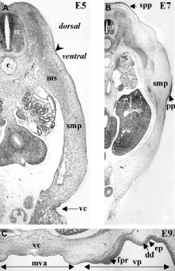
Fig. 7. Formation of chick ventral skin. At E5 (A), the somatopleural mesoderm (smp) is still homogeneous, except for the migration of muscle cells (ms) originating from the somites. In 7-day embryo (B), a dense dermis forms in both the presumptive dorsal spinal pteryla (spp) and pectoral pteryla (pp). At 9 days (C), the first feather primordia (fpr) appears in the ventral pteryla (vp). The midventral apterium (mva) extends on each side of the ventral closure (vc). Note that the mesenchyme remains loose in the mva, in contrast with the dense dermis (dd) of the forming feather tracts. Arrow indicates the limit between dorsal and ventral regions. ec, ectoderm; ep, epidermis. Histology by G. Chevalier, photographs by I. Fliniaux.
praeplumulae, in the case of a duck host, or by praefiloplumae alone, in the case of a chick host. The length of the praepennae that had developed from the extra-embryonic somatopleure was equivalent to that of the corresponding feathers of the host. By contrast, the small, arrested, feathers possessed a dermal core formed exclusively of mouse cells, similar to the arrested feathers that were obtained in chick epidermis/ mouse dermis recombinants (Dhouailly, 1973), and resulted from the messages originating from the mouse dermis. On the other hand, the long feather filaments resulted from an autonomous ability of the extra-embryonic somatopleural mesoderm and ectoderm. The mouse implant caused perturbations of the morphogenetic movements of the expanding somatopleure, as well as its fusion with the splanchnopleure. In all cases that led to the formation of a supplementary pteryla, a connection with the yolk sac was observed, which was stretched and very long in case of the chorion. In spite of this connection, these results were interpreted as implying that the mere densification of the abutting sub-ectodermal mesenchyme above a certain threshold, against the implant, entails the acquisition of feather-forming dermal tract specificity.
Comments involving some recent explanations
The formation of a dense dermis defines the future pteryla and occurs even in the case of the scaleless mutant
In amniotes, a constant feature concerning the morphogenesis of cutaneous appendage fields is the formation of a sub-ectodermal dense dermis. This dense dermis covers a loose deeper dermis that will not participate to the formation of cutaneous appendages. By contrast, the dense dermis will be redistributed into dermal condensations overlaid by epidermal placodes (Viallet et al., 1998). This step of dense dermis formation, that corresponds to the pteryla determination is also reached by the scaleless mutant (sc/ sc) chick embryos. The scaleless defect, characterized by the
absence of scales and the formation of only a few feathers, is determined by the epidermis, while the sc/sc dermis differentiates and functions normally (Goetinck and Abbott, 1963; Sengel and Abbott, 1963; Dhouailly and Sawyer, 1984; Viallet et al., 1998). In fact, transverse sections of the sc/sc trunk at E6.5/7 cannot be distinguished from those of a wild type embryo: the dense dermis corresponding to the dorsal and ventral pterylae are present, but at E7 the first placodes do not appear. Fgf-2 treatment rescues the mutant phenotype, where the epidermis is deprived of FGF4 expression, and allows the redistribution of the dense dermis in conformity with its regional origin. Scaleless skin explants, treated with an appropriate dose of FGF-2, express different wild phenotypes: feathers arranged in an hexagonal pattern, reticulate
A
B
A
C
scales, scutate scales and glabrous epidermis respectively, when they originate from dorsal, plantar, tarsometatarsal and midventral apterium skin (Dhouailly et al., 1998).
It should be noted that there is a delayed formation of a dense dermis in the semi-apteria that form between the different pterylae, while a dense dermis never forms in the true apteria, as the midventral apterium. Semi-apteria and apteria thus need absolutely to be distinguished to compare the results of different laboratories. In many recent papers, semi-apteria have been called apteria, which leads to a misinterpretation of the results. The dense dermis of a semi-apteria is able to participate in feather formation, and the formation of supernumerary feathers, added to the normal sparse feathers formed can be easily obtained.
Another point to be discussed is the formation of the semi-apterium itself between the different normal pterylae. This formation appears to result at the intersection between two adjacent waves of feather buds. This type of interaction, and the production of a semi-apterium, also occurs when an ectopic pteryla is obtained by an experimentat that initiates a new wave of feather rows. This question will thus be resolved by understanding the molecular basis of the feather waves.
Regional specification of the dermal progenitors
Numerous experiments performed in P. Sengel’s laboratory in the seventies showed that the replacement of a piece of somatic mesoderm by one from another level of the cephalo-caudal axis, led to the development of a portion of the spinal pteryla in conformity with the origin of the graft. The non-segmented somitic mesoderm is thus already regionally specified (Mauger, 1972a). Dr. Kieny and Dr. Weydert tried without success to find what were the different messenger RNAs present in the different somites. We know now that the regionalisation of the somites is based on the expression of different sets of Hox genes, and that these transcription factors are present in a tiny amount. These were discovered at the end of the eighties (among others: Duboule, 1992; Duboule and Dolle, 1989). For instance, cHoxC-8 is involved in the specification of somitic cells which give rise to the dorsal thoracic dermis by E6 (Kanzler et al., 1997).
The neural tube and the specification of the dermal progenitors
A question that remained unanswered was the specification of the dermal progenitors from the dermomyotome, and the somatopleure. An intriguing fact was the positive role of the neural tube, not only in the formation of the dorsal pteryla (Mauger, 1972b), which was easily understandable by the proximity of the two structures, but also in the initiation of supplementary pteryla from the midventral apterium (Sengel and Kieny, 1967a and b). In the last ten years, many different laboratories have identified different types of diffusible molecules synthesized by the neural tube. Thanks to this the molecular basis of the specification of the dorsal and ventral pterylae has been recently resolved in my laboratory (Olivera-Martinez et al., 2001 and 2002; Fliniaux et al., submitted). Moreover, the mechanisms of formation of a feather-forming dermis appear to be different between the dorsal and ventral sides of the chick embryo, not necessarily in the type of molecules involved, but in their role, the timing of their expression, and in the signaling cascades implicated. We can now therefore offer a better explanation than thirty years ago why a patch of glabrous skin was obtained
Fig. 8. Production of a supplementary pteryla in the midventral apterium and in the amnion of chick embryo. (A) Ventral view of an 11-day control embryo showing the midventral apterium (mva) on each side of the ventral closure (vc), contiguous with the umbilical cord (u). (B) At the same stage, an embryo implanted at 2 days with a fragment of neural tube in the right part of the distal somatopleural embryonic mesoderm shows the formation of a supplementary pteryla (sup). (C-E) Supplementary pteryla formed at 16 days by chick amniotic somatopleural ectoderm and mesoderm, next to an implanted 12.5-day mouse dermis. (C) Note the connexion with the yolk sac (ys). (D-E) The long feather filaments (f) (praepennae) of the supplementary pteryla are formed exclusively by chick (ch) cells, while arrested feathers (af) are formed by the chick ectoderm above the implanted mouse (mo) dermis. The small buds at the base of each feather filament are praefiloplumae (pl).Photographs and experiments by M. Kieny and P. Sengel (A,B) and D. Dhouailly (C-E).
A
B
E
after the graft of a piece of the presumptive territory of the midventral apterium in place of somites (Mauger 1972a). In contrast to the previous interpretation, we know at present that the somatopleure, even at its more distal part, is still malleable at E2/ E3 (Fliniaux et al., 2004). However, when this piece of somatopleure is transplanted close to the neural tube, it does not received the required signals in the correct time. Likewise, the formation of a supplementary pteryla from the somatopleure of the midventral apterium, and even from the extra-embryonic somatopleure, was recently understood. Forty years ago it was noticed the occurrence of merging between the somatopleure and the splanchnopleure in all positive cases. Recent results from my laboratory (Fliniaux et al., 2004) show that the molecular signal which arises from the splanchnopleure is not in fact irrelevant for the increase in mesodermal cell density. This fact was already noted, but previously explained only by a mechanical point of view. Moreover, the formation of feathers by the extra-embryonic somatopleure demonstrates also that feather morphogenesis is the basic program for avian ectoderm. Morphogenesis of scales requires some additional modifications mostly to prevent their growth (Kanzler et al., 1997, and see Prin et al., 2004).
Finally, most experiments were done in chick embryo. Thus defining the precise origin and the migratory behavior of the somitic and splanchnopleural derived dermis still remains an open issue in the mouse, one which cannot be easily resolved by mouse/chick chimeras.
Acknowledgments
The authors are indebted to Dr. D.J. Pearton for critical reading of the manuscript and to Mrs. B. Peyrusse for iconography.
References
BEN-YAIR, R., KAHANE, N. and KALCHEIM, C. (2003). Coherent development of dermomyotome and dermis from the entire lateral extent of the dorsal somite.
Development 130: 4325-4336.
DHOUAILLY, D. (1973). Dermo-epidermal interactions between birds and mammals: differentiation of cutaneous appendages. J. Embryol. Exp. Morphol. 30: 587-603.
DHOUAILLY, D. (1977). Dermo-epidermal interactions during morphogenesis of cutaneous appendages in amniotes. In Frontier Matrix Biology (ed. L. Robert), Creteil. 4: 86-121.
DHOUAILLY, D. (1978). Feather-forming capacities of the avian extra-embryonic somatopleure. J. Embryol. Exp. Morphol. 43: 279-87.
DHOUAILLY, D. (1984). Specification of feather and scale patterns. In Pattern formation
(Eds. G.M. Malacinski and S.V. Bryant) MacMillan publishing company, New York, 581-601.
DHOUAILLY, D. and SENGEL, P. (1975). Feather- and hair-forming properties of dermal cells of glabrous skin from bird and mammals. C. R. Acad. Sci. Hebd. Seances Acad. Sci. D. 281: 1007-10.
DHOUAILLY, D. and SAWYER, R.H. (1984). Avian scale development. XI. Initial appearance of the dermal defect in scaleless skin. Dev. Biol. 105: 343-350.
DHOUAILLY, D., PRIN, F., KANZLER, B. and VIALLET, J.P. (1998). Variation of cutaneous appendages: Regional specification and cross-species signals. In
Molecular basis of Epithelial appendage morphogenesis, Vol. 1 (Ed. C. M. Chuong), R.G. Landes Company, Georgetown, Texas, USA pp. 45-56.
DUBOULE, D. (1992). The vertebrate limb: a model system to study the Hox/HOM gene network during development and evolution. Bioassay, 14: 375-383.
DUBOULE, D. and DOLLE, P. (1989). The structural and functional organization of the murine HOX gene family resembles that of the Drosophila homeotic genes.
EMBO J. 8: 1497-1505.
FLINIAUX, I., VIALLET, S.P. and DHOUAILLY, D. (2004). Ventral vs. dorsal chick dermal progenitor specification. Int. J. Dev. Biol. 48: 103-106.
GOETINCK, P.F. and ABBOTT, U.K. (1963). Tissue interactions in the scaleless mutant and the use of scaleless as an ectodermal marker in studies of normal limb diffentiation. J. Exp. Zool. 154: 7-19.
HOUZELSTEIN, D., CHERAUD, Y., AUDA-BOUCHER, G., FONTAINE-PERUS, J. and ROBERT, B. (2000). The expression of the homeobox Msx1 reveals two populations of dermal progenitor cells originating from the somites. Development
128: 107-116.
KANZLER, B., PRIN, F., THELU, J. and DHOUAILLY, D. (1997) cHoxC-8 and cHox-D13 expression in embryonic chick skin and cutaneous appendage specification.
Dev.Dyn. 210: 274-287.
KIENY, M. and BRUGAL, M. (1977). Morphogenesis of the chick embryo limb. Competence of the embryonic and extra-embryonic ectoderm. Arch. Anat. Microsc. Morphol. Exp. 66: 235-52.
MAUGER, A. (1972a). The role of somitic mesoderm in the development of dorsal plumage in chick embryos. I. Origin, regulative capacity and determination of the plumage-forming mesoderm. J Embryol. Exp. Morphol. 28: 313-41.
MAUGER, A. (1972b). Rúle du tube neural dans le développement du plumage dorsal de l’embryon de poulet. Wilhelm Roux Archiv. 170: 244-266.
MAYERSON, P. L. and FALLON, J. F. (1985). The spatial pattern and temporal sequence in which feather germs arise in the white Leghorn chick embryo. Dev. Biol. 109: 259-67.
OLIVERA-MARTINEZ, I., COLTEY, M., DHOUAILLY, D. and POURQUIÉ, O. (2000). Mediolateral somitic origin of ribs and dermis determined by quail-chick chimeras. Development 127, 4611-4617.
OLIVERA-MARTINEZ, I., THÉLU, J., TEILLET M.A. and DHOUAILLY, D. (2001). Dorsal dermis development depends on a signal from the dorsal neural tube, which can be substituted by Wnt-1. Mech. Dev. 100: 233-244.
OLIVERA-MARTINEZ, I., MISSIER, S., FRABOULET, S., THELU, J. and DHOUAILLY, D. (2002). Differential regulation of the chick dorsal thoracic dermal progenitors from the medial dermomyotome, Development 129: 4763-4772.
PRIN, F. and DHOUAILLY, D. (2004). How and when the regional competence of chick epidermis is established: feather vs. sutate and reticulate scales, a problem en route to a solution. Int. J. Dev. Biol. 48: 137-148.
SENGEL, P. (1976). Morphogenesis of skin. In Developmental and Cell Biology series (Ed. M. Abercrombie, D. R. Newth and J. G. Torrey), pp. 1-269. Cambridge University Press.
SENGEL, P. and ABBOTT, U.K. (1963). In vitro studies with the scaleless mutant: interaction during feather and scale differentiation. J. Hered. 54: 254-262.
SENGEL, P., DHOUAILLY, D. and KIENY, M. (1969). Aptitude of the skin constituents of the mid-ventral apterium of the chicken for forming feathers. Dev. Biol. 19: 436-46.
SENGEL, P. and KIENY, M. (1967a). Production of a supplementary pteryla in the chick embryo. I. Morphologic study. Arch. Anat. Microsc. Morphol. Exp. 56: 11-29.
SENGEL, P. and KIENY, M. (1967b). Production of an additional feather tract in the chick embryo. II. Experimental analysis. Dev. Biol. 16: 532-63.
SENGEL, P. and RUSAOUEN, M. (1968). Aspects histologiques de la différenciation précoce des ébauches plumaires chez le poulet. C.R. Acad. Sci., D, 266:795-797.
STRAUSS, W.L. and RAWLES, M.E. (1953). An experimental study of the origin of the trunk musculature and ribs in the chick. Am. J. Anat. 92: 471-509.
TEILLET, M. A. and Le DOUARIN, N.M. (1983). Consequences of neural tube and notochord excision on the development of the peripheral nervous system in the chick embryo. Dev. Biol. 98: 192-211.
VIALLET, J.P., PRIN, F., OLIVERA-MARTINEZ, I., HIRSINGER, E., POURQUIÉ, O. and DHOUAILLY, D. (1998). Chick Delta-I gene expression and the formation of the feather primordia. Mech. Dev. 72: 159-168.
WEISS, P. (1958). Cell contact. Int. Rev. Cytol. 7: 391-423.