A Novel Algorithm for Enhancing an Image of Brain Tumor
S. Shri Tharani, Dr. B. Sumathi
Department of Computer Science, CMS College of Science and Commerce, Coimbatore, India
ABSTRACT
The process that deals with digital images to make some changes for user needs with some computer algorithm is called digital image processing. There are various computer algorithms available for picture/ image processing. Image Enhancement is one of the easiest technique applied on images for extracting hidden details for analysis highlight some features by changing brightness and contrast. Processing on Biomedical imaging for better diagnostic and treatment can be done using image enhancement. MRI images and CT are the commonly used biomedical images that helps in evaluating the present condition of an organ or tissue and observe a patient for diagnostic and treatment. Among various other algorithms like SMQT, Histogram Equalization, V Transform, the V transform algorithm is used to enhance a brain tumor image. The proposed system improves the accuracy, exactness, computational speed of segmentation. The diagnosing is not properly maintained only with the use of MRI images. So the computer based diagnosis are used. In the future work this method can be used for voice processing also.
Keywords: Biomedical Imaging, Brain Tumor, MRI Image, Image Enhancement, SMQT, Histogram, V transform
I.
INTRODUCTION
The digital image processing consists of processes whose inputs and outputs are images from which it extracts attributes from images which also includes object recognition. Enhancement refers to extracting concealed details for analysis and highlighting some features by changing brightness and contrast. Various image enhancement algorithms are available to process an image. In order to enhance an image, extract the highlighted features and apply the methodology to improve the quality.
II.
LITERATURE REVIEW
The author Kimmi Verma proposed that the point processing methods are most primitive, yet essential image processing operations and are used primarily for contrast enhancement. The authors Hamid Hassanpour and Najmeh Samadiani, elaborates attempt to detection & classification of tumour in
beginning stage. Snehal O.Mundhada and Prof. V. K. Shandilya, the authors state that, Image enhancement technique is to improve the image quality so that the resultant image is better than the original image. An enhancement technique such as alpha rooting operates on the transform domain. The transform domain enables operation on the frequency content of the image”. Manglesh khandelwal et.al, proposed a new algorithm using fuzzy logic by removing the Gaussian noise and compared and concluded that the new method is faster more accurate and more responsive comparing with the images having many details.
III.
METHODOLOGY
and analysis. Removing the noise and improving or refining image quality is known as Pre-processing. The pre-processing steps consists of Noise Removal, Image Sharpening. Image enhancement can be performed both in the spatial and frequency domain. Spatial domain method operates directly on pixels.
( ) ( )
Where i and j are image coordinates, in is used for input function, o is used for output function T is used to operation on output
Besides this it can be grouped into various types. Like, Point operation, Spatial operation, Transform operation, Pseudo coloring. The enhancement doesn't increase the inherent information content of the data, but it increases the dynamic range of the chosen features so that they can be detected easily.
a. SMQT
The objective of this algorithm is to have a whole dimensional range, but achieves it in very different way as Histogram Equalization technique. The difference between sensors due to loss and gain can be eliminated by SQMT. The basic unit of the SMQT is the MQU (Mean quantization unit). It consists of calculating the mean value of all the pixels in the image, and then the mean is used to quantize the value of data into 0 or 1, depending on the lower or higher mean value of the every pixel. Figure 2 shows the stepwise SMQT algorithm.
Algorithm:
Step 1: Input image: these images can be medical images, blur images etc.
Step 2: Pre-processing an image: pre-processing methods has been performed on those input images like blur image or noisy image before applying enhancement technique.
Step 3: convert the image into HSV.
Step 4: get the Brightness. Since it is a vertical vector.
Step 5: Sort the Vector and get the Size of the Interval and Assign a Start and a Stop Value.
Step 6: Apply linear transformation to each segment. Step 7: Reverse the sorting with transformed value. Step 8: Resize the vector into a matrix assigning new component V.
Step 9: Output image: the enhanced image is an output image.
Figure 2. SMQT Algorithm Steps.
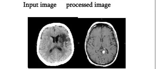
Input image processed image
Figure 3. Sample data set (SMQT).
Figure 3 is the input and the processed enhanced image using the SMQT algorithm. Here the contrast of the image is increased to differentiate between the tumor and the other part.
b. HISTOGRAM EQUALIZATION
Algorithm:
Step 1: Input image: these images can be medical images, blur images etc. the original image is considered as the input image.
Step 2: Pre-processing an image: pre-processing methods has been performed on those input images like blur image or noisy image before applying enhancement technique.
Step 3: specify the user defined function for calculating the Histogram Equalization.
Step 4: Output image: the enhanced image is an output image.
Figure 4. Histogram Equalization steps
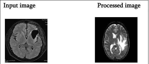
Input image Processed image
Figure 5. Sample dataset (Histogram Equalization).
Figure 5 is the input and the processed enhanced image using the Histogram Equalization. Here the contrast of the image is increased to differentiate between the tumor and the other part. And it brings some strange effects so that the image can be used for the purpose of diagnosing the tumor.
c. V TRANSFORM
V transform is advantageous over HSV model. The V transforms contains information about the brightness, so it can be modified in color images without changing the color. It is an advantage because it means that it changes color images operating only in one component, so the computational cost is lower than operating in the three channels. Apart from this, transform requires less computational power than SMQT for its simplicity. Figure 6 shows an algorithm to represent V transform.
Algorithm
Step 1: Input image: these images can be medical images, blur images etc.
Step 2: Pre-processing an image: pre-processing methods has been performed on those input images like blur image or noisy image before applying enhancement technique.
Step 3: convert the image into HSV.
Step 4: Extracting the V component and making a sorted vector with the V values.
Step 5: The sorted vector is divided in N segments of the same length. For each interval a start and a stop value is defined.
Step 6: In order to spread the value brightness from start to stop value; a linear transformation is performed on every segment.
Step 7: output image: the enhanced image is an output image
Figure 6. V Transform Algorithm Steps.
V transform algorithm can also be applied to grayscale image, if the image is considered as the V component itself. This algorithm is good for n=1 where n is the number of vector, but it doesn’t come handy for multiple images. For higher values of n, the transformed image has more contrast and gets strange effects, because the algorithm does not consider the initial composition of the picture.
Input image Processed image
between the tumor and the other part. The tumor part is in white and the rest of the parts are left in black so that the affected area is taken for the tumor diagnosis.
IV.
RESULTS AND DISCUSSIONSHistogram equalization of color image results shows strange colours. It increases the contrast and enhance gray-scale images, it is simple, requires low computational power and obtains good results. The SMQT and HSV transform (with n=1) gives better result for color images it only adjusts its contrast.
The SMQT method works on images that has good light, but the transformation of the V component in HSV algorithm gives good results in images with low light. The disadvantages are it is time consuming and needs more manual work.
The proposed technique is experimented using SMQT, Histogram Equalization and V Transform algorithms for enhancement and various image processing techniques for medical imaging. Image enhancement using V transform algorithm is applied for improving the accuracy, exactness and computational speed.
V.
CONCLUSION AND FUTURE WORKMRI image is one of the best methods in brain tumor detection, by observing only MRI images the specialists are unable to keep up with diagnosing. Hence, the computer based diagnosis is necessary for the correct brain tumor classification. For such implementation, image enhancement algorithm is used to recognize the class of the tumor as there is diagnostic error will be minimized in medical industry. This method can be applied for 3D images and other diseases for diagnosing and tracking. It can also be applied for voice processing.
VI.
REFERENCES[1]. Manglesh Khandelwal, Shweta Saxena, and Priya Bharti, "An Efficient Algorithm for Image Enhancement", Manglesh Khandelwal et al. / Indian Journal of Computer Science and Engineering (IJCSE), Vol. 2 No.1, pp. 118- 123. [2]. Lucy Balian Rorke, Md, Floyd H. Gilles, Md , T Richard L. Davis, Md, And Laurence E. Becker, Md, "Revision of the World Health Organization Classification of Brain Tumors for Childhood Brain Tumors", CANCER October1 Supplement 1985 Vol. 56.
[3]. Ed-Edily Mohd. Azhari1 Muhd. Mudzakkir Mohd. Hatta, Zaw Zaw Htike and Shoon Lei Win, "Brain Tumor Detection and Localization in Magnetic Resonance Imaging" (IJITCS) Vol.4, No.1, February 2014, pp.1-11.
[4]. Sudipta Roy, Sanjay Nag, Indra Kanta Maitra, Prof. Samir Kumar Bandyopadhyay, "A Review on Automated Brain Tumor Detection and Segmentation from MRI of Brain".
[5]. Raman Maini and Himanshu Aggarwal, "A Comprehensive Review of Image Enhancement Techniques", JOURNAL OF COMPUTING, VOLUME 2, ISSUE 3, MARCH 2010, pp.8-13. [6]. Kimmi Verma, Aru Mehrotra, Vijayeta Pandey,
and Shardendu Singh, "Image Processing Techniques for The Enhancement of Brain Tumor Patterns", International Journal of Advanced Research in Electrical, Electronics and Instrumentation Engineering Vol. 2, Issue 4, April 2013, pp. 1611- 1615.
[7]. Pratibha Sharma, Manoj Diwakar and Sangam Choudhary, "Application of Edge Detection for Brain Tumor Detection", International Journal of Computer Applications (0975 – 8887) Volume 58– No.16, November 2012, pp.21-25. [8]. Snehal O.Mundhada and Prof. V. K. Shandilya
Software Engineering, Volume 2, Issue 4, April 2012, pp. 370-372.
[9]. Aziz Makandar and Bhagirathi Halalli, "Image Enhancement Techniques using Highpass and Lowpass Filters" International Journal of Computer Applications (0975 – 8887) Volume 109 – No. 14, January 2015, pp. 12-15.
[10]. Roopali D Pai, Prof. Srinivas Halvi, Prof. Basavaraj Hiremath, "Medical Color Image Enhancement using Wavelet Transform and Contrast Stretching Technique", International Journal of Scientific and Research Publications, Volume 5, Issue 7, July 2015,pp.1-7.
[11]. Pragati G. Patil, Dr. K. J. Karande," Brain tumor detection techniques: A Survey", International Research Journal of Engineering and Technology (IRJET), Volume: 03 Issue: 10- Oct -2016, pp. 396-399.
[12]. Nikita V. Chavan, B.D. Jadhav, and P.M. Patil," Detection and Classification of Brain Tumors", International Journal of Computer Applications (0975 – 8887) Volume 112 – No. 8, February 2015, pp48-53.
[13]. Hamid Hassanpour, Najmeh Samadiani, "Using morphological transforms to enhance the contrast of medical images", the Egyptian journal of radiology and nuclear medicine, 2015, 46, 481-489.