A Novel Image Segmentation Method for Early Detection and
Analysis of Melanoma
L.Anjali Devi
1& Dr.M.Roja Rani
21.M.Tech student,Amara Institute of Engineering&Technology,JNTUK,NRT,AP. 2.Professor, Amara Institute of Engineering&Technology,JNTUK,NRT,AP.
Abstract:
Skin cancer through all types of cancers, is the least frequent and most common form of
human cancers. Skin cancer may be
Melanoma, Basal and Squamous cell among which Melanoma is the most precarious. It is compulsory to recognize it in its early stage for its proper cure. In this paper, image segmentation method is implemented in MATLAB followed by STOLZ and TDS algorithm.
An image segmentation method is used to classifying the current state of melanoma lesions. It further includes various steps for analysis; pre-processing which is used for image enhancement, removal of noise, hair.
Then the boundary of the skin lesion is detected with the help of initial segmentation. The lesion is investigated to find the parameters using feature extraction property. The fetched parameter values are therefore used to calculate STOLZ algorithm and by feeding the output of the STOLZ algorithm in TDS,
Detection of the current stage of melanoma can be identifying.
Keywords: Image segmentation; Feature
extraction; STOLZ algorithm; TDS.
I. INTRODUCTION
The Skin biology is a vast field. Body’s largest organ is Skin. It saves us from various infections, sunlight and from many other
problems and helps us to maintain body temperature [1]. As, Skin itself is a very complicated structure consists of different types of tissues.
These tissues can go through cancerous conversion during their development process resulting in cancer with different biological attitude. Cancer is defined as disease which is caused due to abnormal growth of cells and covers others body parts with the help of blood and lymph system. The expansion of the cancer tissues is very much different from normal cell expansion. The important factor is rather than vanishing the cancer cells it increase continuously and produce abnormal cells. The DNA cause cell to become cancer cells [3].Figure 1.1 differentiates cancer cell growth from normal growth of cell .Above 100 types of cancer are possible on the skin which are clinically proven and are having genetic components with many other properties. Melanoma is a type of Skin Cancer and it originates in Melanocytes [1]. It can be in any part of the body and can hit anyone. Every year, approximately 55,000 peoples found suffering from this kind of cancer. As compared to all other types of cancer,
Figure 1.1: Normal and Cancer cell growth
II. Cancer Directory
Epidermis: - Epidermis is top most
layer of the skin. It is made of dead cells and contains Kerotinocytes and Melocytes
[1]. Dermis: - It is the lower layer of the skin
contains fats. The main function of this layer is to feed, excrete and exchange of heat. In this layer, Fat cells act as the most important cells [1].
Incidence: - Incidence rate of cancer is the
division of number of new cases to total number of persons multiplied by 100,000 .Numerical representation is (same type of cancer is considered at one time) [4].
IR=(New cases/Total persons) X 100,000.
Mortality: - Mortality rate of cancer division
of no. of deaths cases to the total no. of persons multiplied by 100,000 .Numerical representation is (same type of cancer is considered at one time) [4].
MR=(Cancer Deaths/Total persons) X
100,000.
Prevalence: - Number of live person on
specific date who is already being diagnosed of the disease. It contains the new and already existing cases. This factor is very useful for the planning of health and resource [4].
Survival: - Survival rate is basically expressed
as the ratio of a live patient to the analysis of tumor. Relative survival is a percentage estimate of the patients who are expected to survive Observed survival is the actual percentage of patient’s still alive after examination [4].
Lifetime Risk: - It is defined as the maximum
chances of growing of tumor. It also helps in the calculation of the certain age wise development of tumor [4].
III. Design Flow
By considering figure 1.2 the flow chart is mainly shown in two main blocks i.e. Image
analysis block which contain image
segmentation procedure and second is
melanoma detection block which consist of STOLZ algorithm and TDS parameter value that gives detection result. For early detection of melanoma, the very first and basic requirement of the diagnosis is the acquisition of digital images of melanoma.
Figure 1.2: Design flow of proposed method . The constructive view of proposed method is shown in figure 1.3
Figure 1.3: Constructive view of proposed method
IV. Results
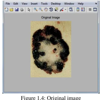
The software tool used is MATLAB R2013a (8.1.0.604) version. The original image is in jpeg format & having RGB color model which is then converted to grayscale image.
Figure 1.4: Original image
In order to fetch the features of the image, regionprops command is used. The need of area, perimeter and Minor axis length is to calculate the asymmetry, border irregularity and diameter of the mole.
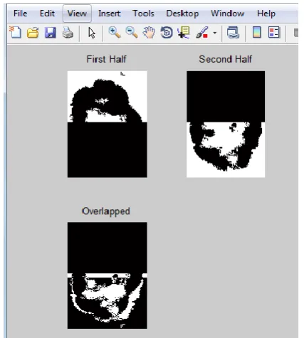
Figure 1.5: Bisection of the image in upper and lower part with an overlap image to get asymmetry.
The mathematical expression used to calculate asymmetry is:-
Asymmetry = (ΔT/ T) * 10 Where, ΔT = Pixel difference
T =Total Pixel count of lesion
A = 0.73233 (approx) pixel or 0.194mm Border : - The border is irregular in case of Melanoma. So, the edge or the border are first recognized and then fetched from the image excluding the inner, outer parts of the mole
Figure 1.6: Edge and border detection of an
The border irregularity is calculated using B = [(perimeter) 2 / 4πA] Where,
A= Area of the lesion
π = 22.7 After calculation the border irregularity value is
B = 0.47774pixel or B= 0.13 mm
Colour: - The third dimension of the skin mole is colour. If the mole is having more than one colour
Figure 1.7: Colors of Skin mole
Dermoscopic structure it contains one or more than one structure inside itself. Dermoscopic structure = 2 structures.
B= 0.13 * 0.1 C = 3 *0.5 D= 2 * 0.5
TDS = [(A* 1.3) + (B* 0.1) +(C* 0.5) + (D* 0.5)] --- [Equation 1]
TDS = 3.44 (using Equation 1)
For final diagnosis result, classification is done using TDS table (refer table 1) By getting the TDS value the result comes out is benign skin lesion. It is an early stage of Melanoma
TDS is < 4.75 i.e. 3.44 therefore it is Benign Skin Lesion
V. Conclusion
In this paper, Melanoma image is processed using image segmentation method in MATLAB and then the features of the same is implemented on STOLZ Algorithm and verified according to TDS parameter in order to analysis the stage of melanoma, here the Melanoma is in Benign stage so that it can be cure in early stage.
References
[1] D J Gawkrodger,Dermatology an
illustrated colour text. Elsevier health science, 2003.
[2] Chudnovsky , Yakov,Paul A.Khavari, and Amy E. Adams.“Melanoma genetics and the development of rational therapeutics.” Journal of clinical Investigation 115.4 (2005): 813-824
[3] National Cancer Institute, “Surveillance, Epidemiology, and End Result program,” http://seer.cancer.gov/statfacts/html/melan.htm l, April 15, 2014.
[4] “American Cancer Society,”
http://www.cancer.org/cancer/skincancer/inde x, Oct 10, 2013.
[5] “Cancer council
Victoria,”http://www.cacervic.org.au/about-cancer/cancer_types/melanoma, Oct 31, 2012.
[6] Ignazio Stanganelli, MD Skin Cancer Unit, IRST, Meldola, and Ospedale Niguarda, Milan, Italy,“ Melanoma Molecular Map
Project ,”
http://www.mmmp.org/MMMP/import.mmmp ?page=dermoscopy.mmmp , Oct , 2008
[7] Madhankumar, K., and P. Kumar, “Characterization of skin lesions”, Pattern
Recognition, Informatics and Medical
Engineering (PRIME), 2012 International Conference on. IEEE, 2012.
[8] Saini, Harpreet Kaur, and Onkar Ch. "Skin Segmentation Using RGB Color Model and Implementation of Switching Conditions." Skin 3.1 (2013): 1781-1787.
[9] Silveira, Margarida, et al. “Comparison of segmentation methods for melanoma diagnosis in dermoscopy images.” Selected Topics in Signal Processing, IEEE Journal of 3.1, pp. 35-45, 2000.
[10] Pham, Dzung L., Chenyang Xu, and Jerry L. Prince. “Current methods in medical image segmentation 1.” Annual review of biomedical engineering 2.1, pp. 315-337, 2000.
[11] Abercrombie, M., and E. J. Ambrose. “The surface properties of cancer cells: a review.” Cancer research 22.5 Part 1, pp. 525-548, 2003.
Biomedicine, IEEE Transactions on 16.6, pp. 1239-1252, 2012.
[13] Kosovsky, Marguerite. “Cancer stem cell research.”,pp. 1-8, 2012.
[14] Narayanan, Deevya L., Rao N. Saladi, and Joshua L. Fox. “Review: Ultraviolet radiation and skin cancer.” International journal of dermatology 49.9 (2010): 978-986.
[15] Blum, A., et al. “Digital image analysis
for diagnosis of cutaneous melanoma.
Development of a highly effective computer
algorithm based on analysis of 837
melanocytic lesions.” British Journal of Dermatology 151.5 (2004): 1029-1038.