Article
1
Comparison of Quantitative PCR (qPCR)
2
Paenibacillus Larvae
Targeted Assays and Definition
3
of Optimal Conditions for Its
4
Detection/Quantification in Honey and Hive Debris
5
Franca Rossi 1,*, Carmela Amadoro 2, Addolorato Ruberto 1 and Luciano Ricchiuti 1
6
1 Istituto Zooprofilattico Sperimentale dell’Abruzzo e del Molise “G. Caporale”, Via Campo Boario 1, 64100
7
Teramo; [email protected]
8
2 Medicine and Health Science Department “V. Tiberio”, University of Molise, Via de Santis, Campobasso,
9
Italy; [email protected]
10
* Correspondence: Istituto Zooprofilattico Sperimentale dell’Abruzzo e del Molise “G. Caporale”, Via
11
Campo Boario 1, 64100 Teramo, Diagnostic Laboratory, Isernia, Italy; [email protected]; Tel.: +39-0861-332664
12
Abstract: The application of quantitative PCR (qPCR) as a routine method to detect and
13
enumerate Paenibacillus larvae in honey and hive debris could greatly speed up the estimation of
14
prevalence and outbreak risk of the American foulbrood (AFB) disease of Apis mellifera. However,
15
none of the qPCR tests described so far has been officially proposed as a standard procedure for P.
16
larvae detection and enumeration for surveillance purposes. Therefore, in this study inclusivity,
17
exclusivity and sensitivity in detection of P. larvae spores directly in samples of honey and hive
18
debris were re-evaluated for the previously published qPCR methods. To this aim recently
19
acquired P. larvae sequence data were considered to assess inclusivity in silico and more
20
appropriate non-target species were used to verify exclusivity experimentally. This led to the
21
modification of one of the previously described methods resulting in a new test capable to allow
22
the detection of P. larvae spores in honey and hive debris down to 1 CFU/g. The application of the
23
qPCR test optimized in this study can allow to reliably detect and quantify P. larvae in honey and
24
hive debris, thus circumventing the disadvantages of late AFB diagnosis based on clinical
25
symptoms and possible underestimation of spore numbers that is the main drawback of
culture-26
dependent procedures.
27
Keywords: Paenibacillus larvae; optimized qPCR; quantification; honey; hive debris
28
29
1. Introduction
30
Paenibacillus larvae is the causative agent of American foulbrood (AFB), the most destructive
31
and highly contagious disease of the honey bee (Apis mellifera) that infects larvae during the first 48
32
h after egg etching [1]. Notification of AFB to the veterinary authority is mandatory in many
33
countries and its diagnosis and official outbreak registration is based on the observation of clinical
34
symptoms [2].
35
P. larvae endospores are the infective form of the bacterium that resist to high temperatures
36
and antimicrobial agents and can persist in hives for decades [3]. Their spread occurs via bee
37
products, e.g. honey, equipment from infected hives and the robbing behavior of bees [4,5].
38
Diagnosis based on clinical symptoms does not efficiently prevent AFB spread since the
39
bacterium might have already been transmitted through the above mentioned routes.
40
Therefore, the application of diagnostic procedures allowing to early detect and quantify the
41
bacterium in substrates like honey and hive debris could help to identify apiaries with a high risk of
42
infection, thus allowing the prevention of clinical manifestation of the disease and further spread of
43
P. larvae spores.
44
The usefulness of detecting and enumerating P. larvae in honey is justified by the existence of a
45
positive correlation between the presence and number of spores in honey and the prevalence of
46
AFB outbreaks in apiaries. Pernal and Melathopoulos [6] associated a prevalence of 1-5% in apiaries
47
to beekeepers whose honey samples contained approximately 1000 CFU/g of spores, while 500
48
CFU/g spores or lower were not always associated to AFB outbreaks.
49
One study regarding the correlation between the number of P. larvae spores in hive debris and
50
AFB clinical manifestations was carried out by Carpana [7], who found that the number of P. larvae
51
spores in hive debris and the percentage of AFB cases were strongly correlated and clinical
52
symptoms ranged between 8% of hives for apiaries with less than 1,000 CFU/g spores and 78% of
53
hives for apiaries with 100,000 CFU/g of spores in hive debris. Forsgren and Laugen [8] observed
54
that samples of debris can reveal the AFB infection in course in the bee colony. Moreover, in the
55
debris P. larvae spores accumulated during time, thus allowing the a posteriori diagnosis of acute
56
infection episodes and the identification of hives more at risk of spreading the infection.
57
Therefore, not just presence but the number of P. larvae spores in honey and hive debris is an
58
indicator of AFB prevalence and outbreak risk. Consequently, its determination by rapid methods
59
would be of great support in AFB containment.
60
Cultural methods used to enumerate P. larvae spores are time consuming, not completely
61
selective and need confirmation by isolate identification. Moreover, differences among biotypes in
62
resistance to the heat treatments used to kill vegetative cells prior to enumeration and in the
63
germination rate determines underestimation of spore numbers [9]. Therefore, qPCR can be the
64
only reliable method to quantify P. larvae in hive associated samples.
65
However, despite different qPCR methods were developed to this purpose, none of them has
66
been still recommended for the direct detection and enumeration of P. larvae in hive associated
67
materials [10,11]. Four qPCR tests targeted on the P. larvae 16S rRNA gene were described for rapid
68
identification and early detection of this bacterium. Han et al. [12] developed an ultra-rapid
69
amplification method and applied it to enumerate P. larvae vegetative cells in AFB infected larvae
70
for early diagnosis. Chagas et al. [13] proposed a method for the unequivocal identification of
71
presumptive P. larvae isolates. The qPCR test designed by Martínez et al. [14] allowed to detect as
72
little as 2 P. larvae spores/g in honey and 103 CFU/g in hive debris [8]. Quintana et al. [15] designed a
73
qPCR test able to detect as little as 28 P. larvae spores in larval scales. In addition, a P. larvae-specific
74
Real Time PCR assay was included in a triplex test aimed at the qualitative detection of the
75
microorganism in brood samples [16]. Quantification of P. larvae by qPCR was not applied to honey
76
and hive debris so far.
77
The aim of this study was to select the most suitable P. larvae-specific qPCR method among
78
those described, considering that, since only a few gene sequences were available for P. larvae and
79
strictly related microorganisms when most of those primers were designed, their inclusivity and
80
exclusivity needed to be re-assessed. These aspects were evaluated in silico and experimentally in
81
this study. Based on the results obtained, it was deemed opportune to modify or design new
82
primers and optimize amplification conditions to make qPCR detection/quantification of P. larvae in
83
honey and hive debris more sensitive and accurate.
84
2. Materials and Methods
85
2.1.Bacterial strains and culture conditions
86
Reference bacterial strains used in this study were P. larvae ATCC 9545, P. naphthalenovorans
87
DSM 14203, P. glucanolyticus DSM 5162 and P. chitinolyticus DSM 11030. In addition 48 P. larvae
88
isolates previously identified with the end point PCR test with primers AFB-F/AFB-R (unpubl. data)
89
recommended by OIE [10], were used to experimentally confirm the inclusivity of the new test. All
90
the strains were grown on Paenibacillus larvae agar (PLA), in which all the Paenibacillus species tested
91
grew well, prepared as described by Schuch et al. [17] with components from Sigma Aldrich (Milan,
92
Italy), or on Sheep Blood Agar (Biolife Italiana, Milan, Italy) incubated at 37°C for 2-5 days in the
93
presence of 9% CO2. All the reference strains were checked for purity by streaking on Sheep Blood
94
Agar plates before extracting DNA.
3
of11
To prepare qPCR standards from known numbers of P. larvae spores, colonies were harvested
96
by adding 2 mL of phosphate buffered saline (PBS, 8.0 g/L NaCl, 0.2 g/L KH2PO4, 2.9 g/L Na2HPO4,
97
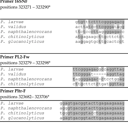
0.2 g/L KCl, pH 7.4) and scraping with an “L” shaped sterile plate spreader from Sheep Blood Agar
98
plates kept at room temperature for 30 days after growth. At this time no vegetative cells were
99
visible in the suspensions by microscope observation of slides stained with a 3 g/L crystal violet
100
(BioMerieux Italia, Bagno a Ripoli, FI, Italy) water solution. The spore suspensions were
heat-101
shocked at 80°C for 10 min to kill the remaining vegetative cells and soon cooled down by
102
incubation at -20°C for 5 min. The heat treated spore suspensions were centrifuged at 10,000 rpm
103
for 5 min, washed twice with 2 mL of sterile PBS and serially diluted to determine their number on
104
PLA medium and artificially inoculate honey and hive debris. Spore suspension dilutions were
105
stored at -20°C for six months prior to sample inoculation if not used immediately.
106
The types of inoculated samples were 0.5 g/mL honey suspensions and 100 µg/mL hive debris
107
suspensions in deionized water sterilized by autoclaving at 121°C for 15 min.
108
109
2.2. DNA extraction
110
Crude DNA extracts were prepared from Paenibacillus spp. bacteria by re-suspending a single
111
colony picked with a sterile loop in 100 µl of sterile 10 mM Tris/HCl buffer, pH 8.0 and heating at
112
100°C for 5 min. The suspension was centrifuged for at 8,000 rpm for 5 min and the clear
113
supernatant was used in qPCR reactions.
114
DNA extraction from honey and hive debris samples, artificially inoculated with decimal
115
dilutions of spore suspension to obtain final spore numbers in the range 0.1 – 106 CFU/g for honey
116
and in the range 1 – 107 CFU/g for hive debris, was carried out from 2 mL of sterilized honey
117
suspension or 1 mL of hive debris suspension. The DNA extraction was carried out with the
118
NucleoSpin Tissue kit (Macherey-Nagel GmbH & Co. KG, Düren, Germany) as follows: the
119
inoculated honey and hive debris suspensions were centrifuged at 14,000 rpm for 2 min and the
120
pellets were resuspended in 90 µL of T1 buffer µL added with 10 of µL proteinase K. The samples
121
were incubated for 1 h at 56°C. To the suspensions T1 buffer was added to reach the volume of 205
122
µL and these were centrifuged at 12,000 rpm for 10 min. The supernatant was transferred in a new
123
sterile tube and the extraction was prosecuted according to the NucleoSpin Tissue kit instructions
124
that follow proteinase K treatment. DNA was finally re-suspended in 20 µL of elution buffer.
125
126
2.3. In silico analysis of primer specificity and inclusivity and primer design
127
The exclusity of the oligonucleotides previously proposed for P. larvae detection by qPCR
[12-128
15] was verified as follows: (i) the bacterial species with highest identity of the 16S rRNA gene
129
sequence with P. larvae were identified by BLAST analysis (https://blast.ncbi.nlm.nih.gov) run in
130
“megablast” mode and excluding the “Paenibacillus larvae” taxon, (ii) the 16S rRNA genes of the
131
identified species and of Paenibacillus species known to be associated to hive matrices were aligned
132
by Clustal Omega (http://www.ebi.ac.uk/Tools/msa/clustalo/), (iii) the positions of the previously
133
designed primers were determined in the aligned sequences to analyze matching with the
134
corresponding region in P. larvae.
135
To determine primer inclusivity, the 16S rRNA gene sequences of 90 P. larvae isolates available
136
in the nucleotide database (https://www.ncbi.nlm.nih.gov/nucleotide) and in the Ribosomal
137
Database Project (RDP; https://rdp.cme.msu.edu/), plus all the 16S rRNA genes found (eight in each)
138
in the eight P. larvae completely assembled genomes and six 16S rRNA genes of a not completely
139
assembled genome of strain P. larvae DSM 25719 (Acc. N. NZ_ADFW00000000), were aligned by
140
Clustal Omega (https://www.ebi.ac.uk/Tools/msa/clustalo/). The target gene region of primers PL-F
141
and PL-R designed by Dainat et al. [16] was defined by BLAST analysis.
142
143
2.4. PCR amplification
144
PCR was carried out in 20 µL reactions with the KapaSybr Fast qPCR Master Mix
145
(KapaBiosystems, Sigma-Aldrich, Milan, Italy). Two µL of DNA and of each primer were added to
146
the reaction and nuclease-free water was added to reach the reaction volume. The qPCR programs
were run in a QuantStudio 5 thermal cycler (Applied Biosystems, Thermofisher Scientific, Rodano,
148
MI, Italy).
149
PCR with primer pair Pltr-F/Pltr-R was carried out as previously described [13]. Moreover, the
150
method was modified to be more specific by using primers in 0.25 µM concentration, decreasing the
151
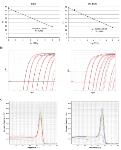
number of cycles from 40 to 36 and increasing the annealing temperature from 60°C to 64°C, while
152
the annealing time was decreased from 1 min to 13 s.
153
Primers PL2-Fw/PL2-Rev, were used in the conditions described by Martínez et al. [14].
154
Forward primers PLAup and PLAup2, and reverse primer PLAdw were designed in this study
155
and are reported in Table 1.
156
Table 1. Oligonucleotides designed in this study and respective positions in the 16S rRNA gene of
157
the P. larvae type strain ATCC 9545, GenBank acc. CP019687, locus tag BXP28_01730.
158
Label Sequence 5’→3’ Nucleotide positions
PLAup TTCGGGAGACGCCAGGTTA 323279-323297
PLAup2 KKTYYYTTCGGGAGACGCCA*
323273-323292
PLAdw CTTTCATGACTTCTTCATGCGAAG 323387-323410
*according to the IUPAC code, the ambiguous primer positions have the following meaning: Y (C,
159
T), K (T, G)
160
161
In the optimized PCR test primers PLAup and PLAup2 were used in 0.25 µM concentration,
162
while PLAdw was used in 0.15 µM concentration to avoid primer-dimer formation. The PCR
163
program comprised initial denaturation at 94°C for 4 min, 40 cycles of denaturation at 95°C for 15 s
164
and annealing at 56°C for 10 s followed by melting curve analysis.
165
166
2.5. 16S rRNA Sequencing
167
All the DNA extracts from single colonies of the reference strains were submitted to species
168
confirmation by sequencing of the 16S rRNA gene.
169
The 16S rRNA gene amplification was carried out as described by Weisburg et al. [18] with
170
primers fD2/rD1 re-designed without 5’ linker sequence.
171
Amplification products were purified by the Wizard SV Gel and PCR Clean-Up System
172
(Promega, Madison, USA) and sequenced on both directions with the same primers by GATC
173
Biotech (Constance, Germany).
174
3. Results
175
3.3. In silico analysis of primer exclusivity
176
The oligonucleotide pairs previously proposed for the detection of P. larvae by qPCR were
re-177
assessed in silico for exclusivity. The primer pair designed by Dainat et al. [16] was not included in
178
the analysis since BLAST alignment showed that it is targeted on phage DNA present in all P. larvae
179
genomes in a very variable and high copy number and is therefore unsuitable for quantification.
180
The first step was identifying the bacterial species most closely related to P. larvae at the 16S
181
rRNA gene sequence level. These were identified by BLAST analysis using as query the 16S rRNA
182
gene locus BXP28_01730 of P. larvae ATCC 9545, GenBank Acc. N. CP019687. The species most
183
closely related to P. larvae resulted to be P. naphthalenovorans and P. chitinolyticus with 95% identity
184
of the 16S rRNA sequence with P. larvae. These species and other sharing 94% identity of the 16S
185
rRNA sequence with P. larvae, as well as Paenibacillus spp. ubiquitous or found to occur in hive
186
matrices, namely P. glucanolyticus, P. alvei and P. apiarius [19], were aligned by Clustal Omega to
187
analyze the sequence identities at the annealing sites of the qPCR P. larvae targeted primers
188
previously described.
189
It appeared that, with no exception, the previously reported PCR tests used reverse primers
190
annealing at sites either identical or differing at most for two nucleotides in internal sites between P.
5
of11
larvae and the other species considered, while the forward primers were specific for P. larvae.
192
Moreover, among the reverse primers, 16SNR [12], was found to lack a “C” nucleotide
193
corresponding to position 323502 of the P. larvae ATCC 9545 genome GenBank acc. CP019687, 16S
194
rRNA locus tag BXP28_01730 and present in all the P. larvae 16S rRNA gene sequences analyzed.
195
The forward primers showed different degrees of identity with the corresponding regions in
196
other species. Figure 1 shows all the different types of sequence matching of the forward primers
197
observed with representative non target species.
198
The forward primer PL 167 fw [15] was not reported in Figure 1 since it is identical to primer
199
PL2-fw but with three more nucleotides at the 5’ terminus, and one nucleotide less at the 3’
200
terminus. The three first nucleotides at the 5’ terminus are identical in all the species compared
201
except for some P. larvae strains in which the first nucleotide is “T”.
202
203
Primer 16SNF
positions 323271 – 323290*
P. larvae gtgtttctttcgggagacg
P. validus acttatccttcgggatagg
P. naphthalenovorans ttctcccttaggggagacc
P. chitinolyticus atgagaagcttgcttctct
P. glucanolyticus aaggagtgcttgcactcct
Primer PL2-Fw
positions 323279 – 323298*
P. larvae ttcgggagacgccaggttag
P. validus ttcgggat---aggttag
P. naphthalenovorans taggggagac-ctcctggag
P. chitinolyticus cttgcttctctgatggttag
Primer Pltr-F
positions 323682– 323706*
P. larvae ggagtgacggtacttgagaagaaag
P. naphthalenovorans agggtgacggtacttgagaagaaag
P. chitinolyticus tgggtgacggtacctgagaagaaag
P. glucanolyticus agagtgacggtacttgagaagaaag
204
Figure 1. Sequence alignments of the annealing sites of forward primers from P. larvae targeted
205
identification and detection qPCR assays in P. larvae and closely related or hive associated
206
Paenibacillus species. All the types of matching observed are shown and positions matching between
207
P. larvae and at least one of the other species are shadowed. The aligned sequences have accession
208
numbers NR_112053, AB073189, NR_028817 and AB073203 for P. chitinolyticus, P. glucanolyticus, P.
209
naphthalenovorans and P. validus, respectively.
210
*positions in the P. larvae ATCC 9545 genome GenBank Acc. n. CP019687, locus tag BXP28_01730.
211
3.2. In silico analysis of primer inclusivity
212
A BLAST alignment of all the 16S rRNA gene sequences available for P. larvae was carried out
213
to analyze the intra-species variability at the annealing sites of the primers considered, in order to
214
define their inclusivity for all P. larvae strains.
215
To this aim, all the eight 16S rRNA genes found in each P. larvae genome and other 90 P. larvae
216
16S rRNA gene sequences available in the public domain database were aligned by Clustal Omega.
217
For one of those primers, i.e. 16SNF [12], an intra-genome and intra-species 16S rRNA gene
218
sequence variability was observed. One mismatch at position 8 of the primer, consisting in a “C” to
219
“T” transition was observed in most cases. Moreover the insertion of a “T” nucleotide was observed
220
at the same position for two strains. Strain P. larvae Ymb1 (Acc. N. EF187246) has two mismatches
221
with the primer 16SNF, while P. larvae PL75 (Acc. n. KU682820) has a deletion corresponding to
222
position 6 of the primer. Concerning intra-genome variability, for P. larvae Eric_I (Acc. n. CP019651)
223
three 16S rRNA genes vary in one position and one in two positions of the 16SNF primer annealing
site, for P. larvae ATCC 9545, ATCC 13537 (Acc. n. CP019794), CCM 38 (Acc. n. CP020327),
225
Eric_III(Acc. n. CP019655) and Eric_IV(Acc. n. CP019659) five 16S rRNA genes vary in one position,
226
for P. larvae SAG 10367 (Acc. n. CP020557) all 16S rRNA genes vary in one position, while strain
227
DSM 25430 (Acc. n. NC_023134) has one mismatch with the primer in only one 16S rRNA gene.
228
The above described mismatches appeared to be frequent in P. larvae strains since they were
229
found in about 35% of the 16S rRNA genes analyzed. Moreover intra-genome variability in this
230
region was also high. Notably, the annealing site of primer 16SNF is contained in or overlapping to
231
the annealing sites of forward primers used in conventional PCR test designed by Govan et al. [20]
232
and Dobbelaere et al. [21] that are currently considered the gold standard for P. larvae detection and
233
identification [10] and in the conventional PCR test designed by Piccini et al. [22]. The presence of
234
mismatches in the annealing sites of these primers could reduce the PCR efficiency, an effect that
235
increases with the number of mismatches [23].
236
The other forward primers analyzed (Figure 1) did not present mismatches with any P. larvae
237
16S rRNA gene and therefore were experimentally evaluated for specificity against Paenibacillus
238
species not previously tested and closely related to P. larvae, namely P. naphthalenovorans and P.
239
chitinolyticus, and against P. glucanolyticus as a representative of the ubiquitous Paenibacillus species
240
with best matches of the primer annealing sites with P. larvae.
241
242
3.3. Experimental evaluation of exclusivity and sensitivity the qPCR tests
243
Exclusivity was re-evaluated by using crude DNA extracts obtained from single colonies of all
244
the bacterial strains used in this study.
245
The qPCR tests proposed by Chagas et al. [13] gave amplification products at low Ct values,
246
e.g. 18-22, from the non-target species even when PCR conditions were made as stringent as
247
possible by using primers at 0.25 µmol/L concentration, much lower than indicated by the authors,
248
and by increasing the annealing temperature from 60°C to 64°C. Moreover, all the non-target
249
species presented a melting peak at the same temperature of that given by P. larvae ATCC 9545, and
250
therefore could generate false positives in isolate identification and in the direct detection of P.
251
larvae from hive associated matrices.
252
Primers PL2-Fw/PL2-Rev, when used in the conditions described by Martínez et al. [14], gave
253
primer dimers in the no template control and in reactions with non-target species, according to
254
what reported also by the authors. Moreover amplification with Ct 38 and a melting peak that
255
could be confused with the amplification product from P. larvae, appeared for P. naphthalenovorans
256
and P. chitinolyticus. This could generate uncertain results when colonies of bacterial isolates are
257
analyzed for identification.
258
Specificity was improved by using PL2-Fw in pair with a new reverse primer, PLAdw (Table
259
1), designed in this study to be specific for P. larvae in order to improve exclusivity, and increasing
260
the annealing temperature to 60°C. To avoid primer-dimer formation, the primer PLAdw was used
261
at 0.25 µM concentration. In these conditions 102 and 10 CFU/g of P. larvae spores could be detected
262
in artificially inoculated hive debris and honey, respectively.
263
The reverse primer PLAdw, designed in this study to be specific for P. larvae, can present
264
mismatches consisting in a “G” to “A” transition in two positions that correspond to nucleotides
265
323391 and 323407 in the genome of P. larvae ATCC 9545 and located at 6 and 21 nucleotides from
266
its 3’ terminus, respectively. These transitions never occur together in the same gene. Only one
267
strain was found to have the mutation at the position corresponding to nucleotide 6 of primer
268
PLAdw in one of the eight 16S rRNA gene copies. The mutation at the position corresponding to
269
nucleotide 21 of primer PLAdw was found for four strains in two 16S rRNA gene copies and for
270
one strain in three 16S rRNA gene copies. These mutations were not observed in all other available
271
P. larvae 16S rRNA sequences. Therefore the primer PLAdw was designed without degenerated
272
positions, considering that the above described mutations are not frequent, being observed
273
respectively in 0.01 and 0.15% of the 16S rRNA gene sequences in P. larvae genomes.
3.4. Design of modified P. larvae specific forward primers
276
To ensure exclusivity, primer PL2-Fw was shortened of one nucleotide at the 3’ terminus and
277
the resulting primer was labeled as PLAup (Table 1).
278
Moreover, considering the good specificity for P. larvae of the 16SNF primer annealing site
279
(Figure 1), a second forward primer, PLAup2, with annealing site overlapping to that of 16SNF, but
280
with degenerated positions corresponding to the variable nucleotides, was designed in this study
281
(Table 1).
282
283
3.5. Optimization of new qPCR tests for P. larvae
284
PCR cycle and primer concentration were optimized for the two primer pairs PLAup/PLAdw
285
and PLAup2/PLAdw. Maximum sensitivity was reached for both primer pairs when an annealing
286
temperature of 56°C and a concentration of 0.25 µM of the forward primer and 0.15 µM of reverse
287
primer were used. The Ct values obtained for the same samples inoculated with known P. larvae
288
ATCC 9545 spore numbers was found to be comparable for the two primer pairs and the lowest
289
number of P. larvae spores detected was 1 CFU/g in honey and hive debris for both. However, the
290
latter primer pair gave amplification at Ct 37 from the non-target species P. naphthalenovorans and P.
291
glucanolyticus of PCR products with melting peaks that could be confused with the P. larvae specific
292
peak. For this reason the primer pair PLAup/PLAdw was selected for the detection of P. larvae
293
directly in samples and for the construction of calibration curves for its quantification in honey and
294
hive debris.
295
296
3.6. Quantification of P. larvae in honey and hive debris
297
Calibration curves were constructed by plotting Ct values against CFU/g in samples of honey
298
and hive debris artificially inoculated with spores of P. larvae ATCC 9545 in known numbers.
299
Examples of those curves constructed by using three replicates of DNA extracts for each point are
300
given in Figure 2, with the corresponding amplification and melting curves. The linearity range
301
encompassed the whole set of spore numbers tested and the “R” coefficient was high for both
302
honey and hive debris.
303
Therefore demonstration was achieved that the PCR test with PLAup/PLAdw optimized in
304
this study would allow a rapid, quantitative screening of apiaries in AFB monitoring plans.
Figure 2. a) Calibration curves used for the quantification of P. larvae spores in honey and hive
307
debris by the qPCR test with primers PLAup/PLAdw: Ct values, defined on the automatic threshold,
308
are the average of those from three replicate reactions; b) corresponding amplification curves; c)
309
melting curves of the amplification products obtained from one series of standards for each sample
310
type.
311
4. Discussion
312
The choice of qPCR tests that are fully inclusive for the target species and exclusive for closely
313
related microorganisms is crucial for obtaining reliable results from analytical procedures applied
314
in pathogen detection and quantification directly from samples.
3
of11
Based on the results of this study, the verification of previously proposed methods by a
316
preliminary analysis of primer specificity using BLAST is necessary and can allow to select the best
317
performing tests among those available that can be further optimized. In particular, it was put in
318
evidence that some of the primers used in the available P. larvae targeted tests did not sufficiently
319
discriminate other Paenibacillus species or had mismatches with the respective annealing site in all
320
or in some P. larvae strains, thus making some of the available protocols unsuitable for adoption as
321
a qPCR test for P. larvae detection/quantification.
322
It is also opportune to verify if the non-target organisms used to assess the specificity of PCR
323
methods were chosen according to correct criteria that are taxonomical relatedness, degree of
324
sequence matching at the primer annealing site and occurrence in the same ecological niche.
325
A BLAST analysis of the 16S rRNA gene of P. larvae put in evidence that strictly related
326
microorganisms possibly present in hive associated matrices and that could generate false positives
327
in direct analysis of samples, were not tested as non-target species when the qPCR methods were
328
designed. Indeed, for most of the P. larvae-specific qPCR tests previously designed P. alvei was the
329
microorganism most closely related used to assess exclusivity [12-14]. However, P.
330
naphthalenovorans and P. chitinolyticus have a better matching with the P. larvae targeted primers
331
compared to P. alvei. These species can be both present in honey and pollen, as stated in the
332
description of the isolation sources for sequences with accession numbers KJ638115 and MG650019,
333
so that it was deemed more correct to use them to assess the specificity of P. larvae targeted assays.
334
Choosing the right non target species permitted the experimental verification and optimization
335
of amplification conditions suitable to guarantee reliable results in the analysis of hive associated
336
matrices. The proven exclusivity of the qPCR test optimized in this study toward these species and
337
the sensitivity reached showed the suitability of the method for direct analysis of honey and hive
338
debris for surveillance and risk assessment purposes.
339
Inclusivity had to be re-assessed since most of the qPCR methods previously proposed for P.
340
larvae, all targeted on the 16S rRNA gene, were developed before the acquisition of genome
341
sequences and numerous 16S rRNA gene sequences from many P. larvae strains isolated all over the
342
world. The alignment of all the P. larvae 16S rRNA gene sequences from the public domain database
343
allowed to identify the primers with a perfect annealing with all strains and with potential to allow
344
the detection of all field strains.
345
5. Conclusions
346
This study presents an evaluation of inclusivity and exclusivity of qPCR protocols previously
347
proposed for the identification of P. larvae and the definition of a more reliable test for
348
quantification of P. larvae spores in honey and hive debris for AFB surveillance. The in silico and
349
experimental evaluation resulted in the improvement of specificity for one of the existing qPCR
350
tests and in the design of a more sensitive method derived from the latter. The qPCR protocol
351
assessed can be adopted in standard procedures to reliably quantify P. larvae spores, thus
352
estimating AFB prevalence and outbreak risk before the manifestation of clinical signs and allowing
353
to prevent the spread of the etiological agent to other hives or apiaries from heavily infected ones.
354
Moreover, the qPCR protocol can be used in alternative to the time consuming cultural methods
355
that usually give an underestimation of P. larvae spore load.
356
Conclusions of general interest that can be drawn from this investigation are that an
357
appropriate choice of non target species is necessary to ensure the specificity of a qPCR test and that
358
inclusivity of the already described primer pairs should be re-assessed on the basis of newly
359
acquired sequence data if only few sequences from organisms belonging to the target species were
360
available when those primers were designed.
361
Author Contributions: FR planned the study, performed experiments and wrote the article; CA and AR
362
performed experiments; LR promoted and supervised the study. All authors read and approved the final
363
Funding: This research was funded by the Italian National Health Fund 2017 from the Italian Ministry of
365
Health.
366
Conflicts of Interest: The authors declare no conflict of interest.
367
References
368
1. Brødsgaard, C.J.; Ritter, W., Hansen, H. Response of in vitro reared honey bee larvae to various doses of
369
Paenibacillus larvae larvae spores. Apidologie199829, 569-578. DOI: 10.1051/apido:19980609
370
2. Italian Ministry of Health (2012). Regolamento di polizia veterinaria-Art 155 misure di controllo della
371
peste americana. Nota DGSAF 0007575-P-18/04/2012.
372
3. Heyndrickx, M., Vandemeulebroecke, K., Hoste, B., Janseen, P., Kersters, K., De Vois, P., Logan, N., Ali,
373
N., Berkeley, R.C.W. Reclassification of Paenibacillus (formely Bacillus) pulvifaciens (Namakura, 1984) Ash
374
et al., 1994, a later subjective synonym of Paenibacillus (formely Bacillus) larvae (White, 1906) Ash et al.,
375
1994, as subspecies of P. larvae, with amended description of P. larvae as P. larvae subsp. larvae and P.
376
larvae subsp. pulvifaciens. Int. J. Syst. Bacteriol. 199646, 270-279. DOI: 10.1099/00207713-46-1-270
377
4. Alippi, A.M., Reynaldi, F.J., López, A.C., De Giusti, M.R., Aguilar, O.M. Molecular epidemiology of
378
Paenibacillus larvae larvae and incidence of American Foulbrood in Argentinean honeys from Buenos Aires
379
province. J. Apic. Res. 200443, 135-143. DOI: 10.1080/00218839.2004.11101124
380
5. Lindström, A., Korpela, S., Fries, I. Horizontal transmission of Paenibacillus larvae spores between honey
381
bee (Apis mellifera) colonies through robbing. Apidologie 200839, 515-522. DOI: 10.1051/apido:2008032
382
6. Pernal, S.F., Melathopoulos, A.P. Monitoring for American Foulbrood spores from honey and bee
383
samples in Canada. Apiacta200641, 99-109.
384
7. Carpana, E. Profilassi della peste americana: dal monitoraggio preventivo al risanamento. Progetto di
385
ricerca e sperimentazione nel contesto del Piano integrato igienico sanitario dell’Emilia Romagna.
386
http://api.entecra.it/immagini/2012_peste_americana_ER.pdf
387
8. Forsgren, E., Laugen, A.T. Prognostic value of using bee and hive debris samples for the detection of
388
American foulbrood disease in honey bee colonies. Apidologie201445, 10-20. DOI:
10.1007/s13592-013-389
0225-6
390
9. Forsgren, E., Stevanovic, J., Fries, I. Variability in germination and in temperature and storage resistance
391
among Paenibacillus larvae genotypes. Vet. Microbiol.2008129, 342-349. DOI: 10.1016/j.vetmic.2007.12.001
392
10. World Assembly of Delegates of the OIE. American foulbrood of honey bees. Chapter 2.2.2. OIE
393
Terrestrial Manual 2016 pp. 1-17.
394
http://www.oie.int/fileadmin/Home/eng/Health_standards/tahm/2.02.02_AMERICAN_FOULBROO
395
D.pdf
396
11. De Graaf, D.C., Alippi, A.M., Antúnez, K., Aronstein, K.A., Budge, G., De Koker, D., De Smet, L.,
397
Dingman, D.W., Evans, J.D., Foster, L.J., Fünfhaus, A., Garcia-Gonzalez, E., Gregore, A., Human, H.,
398
Murray, K.D., Nguyen, B.K., Poppinga, L., Spivak, M., van Engelsdorp, D., Wilkins, S., Genersch, E.
399
Standard methods for American foulbrood research. J. Apicult. Res. 2013 52, 1-28. DOI:
400
10.3896/IBRA.1.52.1.11
401
12. Han, S.H., Lee, D.B., Lee, D.W., Kim, E.H., Yoon, B.S. Ultra-rapid real-time PCR for the detection of
402
Paenibacillus larvae, the causative agent of American Foulbrood (AFB). J. Invertebr. Pathol.2008 99, 8-13.
403
DOI: 10.1016/j.jip.2008.04.010
404
13. Chagas, S.S., Vaucher, R.A., Brandelli, A. Detection of Paenibacillus larvae by Real-Time PCR. Acta Sci. Vet.
405
201038, 251-256.
406
14. Martínez, J., Simon, V., Gonzalez, B., Conget, P. A real-time PCR-based strategy for the detection of
407
Paenibacillus larvae vegetative cells and spores to improve the diagnosis and the screening of American
408
foulbrood. Lett. Appl. Microbiol.201050, 603-610. DOI: 10.1111/j.1472-765X.2010.02840.x
409
15. Quintana, S., Fernández, N.J., Pagnuco, I., Medici, S., Eguaras, M.J., Gende, L.B. Report of a real-time PCR
410
assay for Paenibacillus larvae DNA detection from spores of scale samples. Rev. Arg. Prod. Anim.201737,
411
83-88.
412
16. Dainat, B., Grossar, D., Ecoffey, B., Haldemann, C. Triplex real-time PCR method for the qualitative
413
detection of European and American foulbrood in honeybee. J. Microbiol. Methods2018146, 61-63. DOI:
414
5
of11
17. Schuch, D.M.T., Madden, R.H., Sattler, A. An improved method for the detection and presumptive
416
identification of Paenibacillus larvae subsp. larvae spores in honey. J. Apic. Res. 2001 40, 59-64. DOI:
417
10.1080/00218839.2001.11101052
418
18. Weisburg, W.G., Barns, S.M., Pelletier, D.A., Lane, D.J. 16S ribosomal DNA amplification for phylogenetic
419
study. J. Bacteriol.1991173, 697-703.
420
19. Nakamura, L.K. Paenibacillus apiarius sp. nov. Int. J. Syst. Bacteriol. 1996 46, 688-693. DOI:
421
10.1099/00207713-46-3-688
422
20. Govan, V.A., Allsopp, M.H., Davidson, S. A PCR detection method for rapid identification of Paenibacillus
423
larvae. Appl. Environm. Microbiol.199965, 2243-2245.
424
21. Dobbelaere, W., De Graaf, D.C., Peeters, J.E., Jacobs, F.J. Development of a fast and reliable diagnostic
425
method for American foulbrood disease (Paenibacillus larvae subsp. larvae) using a 16S rRNA gene based
426
PCR. Apidologie200132, 363-370. DOI: 10.1051/apido:2001136
427
22. Piccini, C., D’Alessandro, B., Antúnez, K., Zunino, P. Detection of Paenibacillus larvae subspecies larvae
428
spores in naturally infected bee larvae and artificially contaminated honey by PCR. World J. Microbiol.
429
Biotechnol.200218, 761-765.
430
23. Lefever, S., Pattyn, F., Hellemans, J., Vandesompele, J. Single-nucleotide polymorphisms and other
431
mismatches reduce performance of quantitative PCR assays. Clin. Chem. 2013 59, 1470-1480. DOI: