1 Article
The Apoptosis Induction of Oleandrin on
Osteosarcoma Cells through Regulating
Mitochondrial- and Death Receptor-Dependent
Apoptotic Pathways
in Vitro
Yunlong Ma 1,†, Bin Zhu 2,†, Lei Yong 1,†, Chunyu Song 3, Xiao Liu 1, Huilei Yu 1, Peng Wang 1 , Zhongjun Liu 1, Xiaoguang Liu 1,*
1 Department of Orthopedics, Peking University Third Hospital, No. 49, North Garden Street,
Haidian District, Beijing 100191, China. [email protected] (Yunlong Ma);
[email protected] (Lei Yong); [email protected] (Xiao Liu); [email protected] (Huilei Yu);
[email protected] (Peng Wang.); [email protected] (Zhongjun Liu)
2 The Center for Pain medicine, Peking University Third Hospital, No. 49, North Garden Street,
Haidian District, Beijing 100191, China. [email protected] (Bin Zhu)
3 Department of Anesthesiology, Peking University Third Hospital, No. 49, North Garden Street,
Haidian District, Beijing 100191, China. [email protected] (ChunYu Song)
† These authors contributed equally to this work.
* Correspondence: [email protected]; Tel: +86-010-82267368
Abstract: Our previous study has found the anti-tumor activity of oleandrin in osteosarcoma cells in vitro, but the signal transduction process of cell apoptosis induced by oleandrin is uncertain, which is explored in this study. Fluorescence staining and flow cytometry (FCM) was performed to detect the cell apoptosis, intracellular reactive oxygen species (ROS) and mitochondrial membrane potential (MMP). Caspase-3 activity was detected using a commercial kit. The protein expression of cytoplasmic cytochrome c, mitochondrial cytochrome c, bcl-2, bax, caspase-9, Fas, FasL, caspase-8 and caspase-3 was detected using western blot. A pan-caspase inhibitor, z-VAD-fmk, was applied to block the apoptotic pathway and the apoptosis status were re-tested.We found that oleandrin significantly induced the increased apoptosis of U2OS cells. Meanwhile, the intracellular ROS was elevated, but the MMP decreased. The cytochrome c in mitochondria was notably decreased but increased in cytoplasm. The caspase-3 activity was also enhanced with the increase of drug concentration and treatment time. Oleandrin also down-regulated the level of bcl-2, but remarkably up-regulated the expression of bax, cleaved caspase-9, Fas, FasL, cleaved caspase-8 and cleaved caspase-3. Furthermore, the pre-treatment with z-VAD-fmk almost completely reverted the oleandrin-induced apoptosis. The results suggested that oleandrin induces the apoptosis of osteosarcoma cells via mitochondrial- and death receptor-dependent pathways.
2 1. Introduction
Oleandrin, a polyphenolic component of cardiac glycosides, has been reported recently to play an anti-tumor effect on many kinds of tumor cells. Considerable evidences have suggested that oleandrin is a perfect agent for tumor chemotherapy due to its selective cytotoxicity and chemoradiation-sensitization [1-4]. Recently, the result of a clinical Phase I trial suggested that it may become a new antitumor drug for the treatment of patients with refractory solid tumors [5]. The anti-tumor effect of oleandrin involves in the regulation of diverse molecular biology processes, including inhibition of nuclear factor kappa B (NF-κB) / c-Jun NH2-terminal kinase (JNK) signaling pathway [6], activation of death receptor / Apo2L / TRAIL apoptosis signaling pathway [7], suppression of interaction between fibroblast growth factor-2 (FGF-2) and Na+,
K+-ATPase pump [8], induction of intracellular reactive oxygen species production [9], and
stimulation of intracellular Ca2+ increases and activation of caspase cascade apoptotic signals
[10].
Osteosarcoma is still an intractable bone malignance with high mortality due to the frequent recurrence, early metastasis and chemotherapeutic toxicity, even though the improvement of modified surgical techniques and novel chemotherapies has reduced patients’ mortality to a certain extent [11, 12]. The heterogeneity and the genomic complexity also challenge its chemotherapy and molecular targeted therapy [11]. Thus, it is of importance to identify a novel agent with selective cytotoxicity to osteosarcoma cells.
Our previous study has identified the anti-proliferation and anti-invasion effect of oleandrin in osteosarcoma cells, which reveals that its potential value in the osteosarcoma treatment [13]. Meanwhile, we have also observed that oleandrin can modulate the expression of certain apoptosis-related genes including c-myc, cyclin D1 and survivin, which ultimately promotes cells apoptosis through enhancing signal transduction pathway of cell apoptosis [13-15]. However, the detailed signal transduction mechanism of cell apoptosis induced by oleandrin in osteosarcoma is still uncertain. So, we focus on exploring these issues in current study.
2. Results
2.1. Oleandrin induces the apoptosis of U2OS cells
Both DAPI and Hoechst 33342 staining indicated that cell nuclei presented an uneven morphology and appeared obvious pyknosis or even karyolysis in response to oleandrin with the increase of treatment time, which revealed a typical characteristics of cell apoptosis (Figure 1a). In addition, the flow cytometry (FCM) analysis also showed that different concentration of oleandrin induced cell apoptosis (Figure 1b), and the total apoptosis rate significantly increased from 7.13 ± 1.78 % (control) to 15.47 ± 2.04 % (25nM, P < 0.01), and to 21.97 ± 2.10 % (50 nM, P < 0.001) (Figure 1c).
2.1. Oleandrin causes the generation of intracellular reactive oxygen species (ROS)
3
result of FCM also verified that oleandrin increased ROS level to nearly 1.5-fold at 25 nM (P < 0.05) and 2.0-fold at 50 nM (P < 0.01) when compared with the control group (Figure 1e).
Figure 1. (a) DAPI and Hoechst staining of U2OS cells with oleandrin treatment; (b) Cells apoptosis detected by FCM; (c) Statistical analysis of cell apoptosis detected by FCM; (d) Intracellular ROS level detected by fluorescence staining; (e) Semi-quantitative analysis of intracellular ROS level detected by FCM; Bar scale: 30 μm; n = 3, Mean ± SD; *P < 0.05, **P < 0.01, ***P < 0.001, compared to the control group.
2.3. Oleandrin decreases intracellular mitochondrial membrane potential (MMP) level
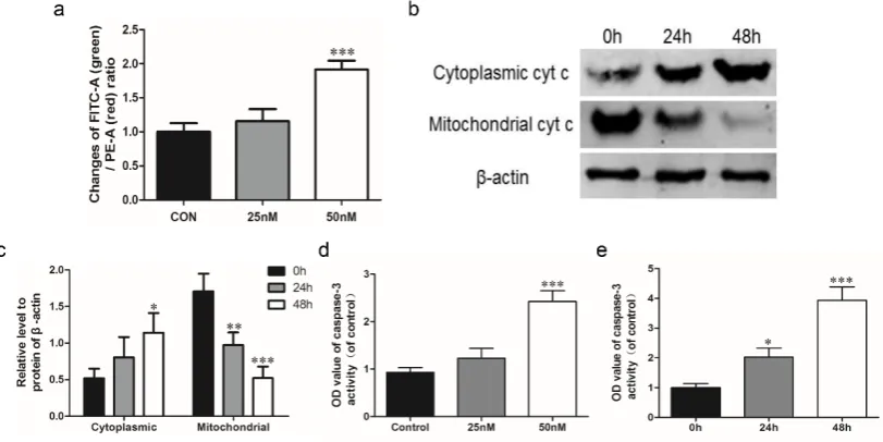
The intracellular MMP level is determined using FCM and the result was represented by the FITC-A (green) / PE-A (red) ratio. Actually, the intracellular MMP level is negatively correlated with FITC-A / PE-A ratio, so the increase of this ratio represented a descending MMP level and the occurrence of apoptosis. Our result suggested that the FITC-A / PE-A ratio was gradually increased with oleandrin treatment in a concentration-dependent manner, and the significance was observed in 50 nM oleandrin-treated group (P < 0.001) (Figure 2a).
2.4. Oleandrin induces cytochrome c released from mitochondria to cytoplasm
After treating with oleandrin for different time, the cytochrome c was down-regulated in mitochondria, but up-regulated in cytoplasm (Figure 2b). Compared with the 0 h group, the cytoplasmic cytochrome c in U2OS cells gradually increased and the significance was occurred at 48 h (P < 0.05). On the contrary, the mitochondrial cytochrome c remarkably reduced after treating for 24 h (P < 0.01) and 48 h (P < 0.001) (Figure 2c).
2.5. Oleandrin enhances caspase-3 activity
4
Figure 2. (a) Statistical analysis of intracellular MMP level detected by FCM; (b) Protein expression of cytoplasmic and mitochondrial cytochrome c detected by western blot; (c) Semi-quantitative analysis of western blot; (d) Caspase-3 activity with the extension of drug concentration; (e) Caspase-3 activity with the extension of treatment time; n = 3, Mean ± SD; *P < 0.05, **P < 0.01, ***P < 0.001, compared to the control group or the 0 h group.
2.6. Oleandrin regulates apoptosis-related proteins in mitochondrial- and death receptor-dependent apoptotic pathways
5
Figure 3. (a) Proteins expression of bcl-2, bax, caspase-9, Fas, FasL, caspase-8 and caspase-3 detected by western blot; (b) quantitative analysis of the bax / bcl-2 ratio; (c) Semi-quantitative analysis of the proteins including Fas, FasL, caspase-9, caspase-8 and caspase-3; n = 3, Mean ± SD; *P < 0.05, **P < 0.01, ***P < 0.001, compared to the 0 h group.
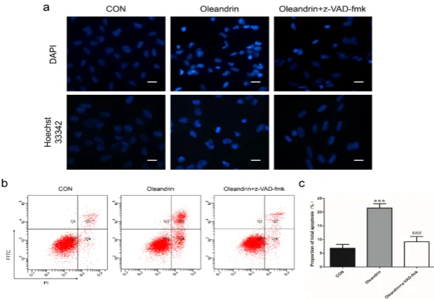
2.7. The pro-apoptosis effect of oleandrin is mitochondrial- and death receptor-dependence
In order to confirm the pro-apoptotic effect of oleandrin was mitochondrial- and death receptor- dependence, furthermore, we pretreated cells with a pan-caspase inhibitor named z-VAD-fmk to suppress caspases cascade activation in both pathways and re-tested the pro-apoptotic effect of oleandrin. The results showed that pro-pro-apoptotic effect of oleandrin was significantly restrained when both pathways were suppressed by z-VAD-fmk. The number of pyknotic or karyolysic nuclei in apoptosis cells descended both in the DAPI and Hoechst 33342 staining with the addition of z-VAD-fmk (Figure 4a). Likewise, the number of apoptotic cells (Figure 4b) and total apoptosis rates (Figure 4c) were also nearly reduced to normal level in this group (P < 0.001).
Figure 4. (a) DAPI and Hoechst staining of U2OS cells re-tested after pretreating with z-VAD-fmk; (b) Changes of cells apoptosis after pretreating with z-VAD-fmk detected by FCM; (c) Statistical analysis of FCM; Bar scale: 30 μm; n = 3, Mean ± SD; ***P < 0.001, compared to the control group; ###P < 0.001; compared to the oleandrin group.
3. Discussion
Our previous study has identified that oleandrin has antitumor effect on osteosarcoma cells and regulates the expression of certain apoptosis-related genes [13]. However, the following signal transduction process of apoptosis induced by oleandrin in osteosarcoma is still unclear. In this study, we re-confirmed that oleandrin-treated cells presented a typical apoptotic morphology with obvious nuclei pyknosis or karyolysis and the cell apoptosis rate was remarkably increased. Besides, we also explored the detailed apoptosis regulation of oleandrin in osteosarcoma cells.
6
(intrinsic) and death receptor (extrinsic) pathways [17].
In the mitochondria-associated apoptosis pathway, B-cell lymphoma 2 (bcl-2) family proteins including the anti-apoptotic proteins (bcl-2 and bcl-xl) and the pro-apoptotic proteins (bax, bak, bad and bim), are primary molecules of the up-stream that respond to apoptosis signals. A decrease of the bax / bcl-2 ratio is commonly observed in various tumors, and closely correlated to the inhibition of cell apoptosis, insusceptibility to undergo apoptosis, tumor recurrence and patients’ poor prognosis [18, 19]. These proteins are mostly localized to the mitochondrial membrane and involve in the mediation of mitochondrial membrane function. Mitochondria are the crucial production sites of ROS, which regulates the intracellular redox status. Physiologically, low concentration of ROS can be used as the second messenger to participate in multiple signaling pathways to maintain homeostasis. When cells are stimulated by apoptosis signals, the aforementioned apoptosis-associated proteins rapidly react to the stress and induce ROS generation dramatically [20]. Meanwhile, high concentration of ROS in mitochondria may reduce the MMP that enhances the mitochondrial permeability, and induces certain mitochondrial factors (such as cytochrome c) released from mitochondria to cytoplasm [20, 21]. In cytoplasm, cytochrome c involves in the formation of cytochrome c / Apaf-1 / caspase-9-containing apoptosome, which subsequently result in the activation of caspase-9 and caspases-3 [21]. Caspases, the cytoplasmic aspartate-specific cysteinic proteases, play a central role in cell apoptosis to transfer apoptosis signals and their cascade activation is the hallmarker of apoptosis [22]. Caspase-3, serves as the final performer, leads to the degradation of the cellular specific substrates and induces cells apoptosis.
Our results showed that oleandrin significantly up-regulated the bax expression but down-regulated the bcl-2 level with the extension of treatment, which caused a significant increase of bax / bcl-2 ratio. In addition, the intracellular ROS level of oleandrin-treated U2OS cells was also substantially elevated in a concentration-dependent manner. However, the intracellular MMP level presented a decreasing trend according to the result of an increase of the FITC-A / PE-A ratio with drug concentration. Furthermore, we determined the levels of cytoplasmic and mitochondrial cytochrome c by separating the cytoplasmic and mitochondrial proteins. The results demonstrated that oleandrin treatment resulted in an intensive down-regulation of the mitochondrial cytochrome c, but a strong up-down-regulation of the cytoplasmic cytochrome c, which suggested that oleandrin could induce the cytochrome c released from mitochondria to cytoplasm. Besides, oleandrin treatment also caused the increased expression of cleaved caspase-9 and cleaved caspase-3, and simultaneously enhanced the caspase-3 activity in a time- and concentration-dependence. These evidences revealed that oleandrin initiated the activation of mitochondria-dependent apoptosis pathway in U2OS cells through the up-regulation of pro-apoptosis protein bax and the down-regulation of anti-apoptosis protein bcl-2. And then, the extensive generation of intracellular ROS and the reduction of MMP level were also induced by oleandrin and lead to the release of cytochrome c from mitochondria to cytoplasm. Thus, caspase cascade reaction including the activation of caspase-9 and caspase-3 was initiated, which ultimately led to the occurrence of cells apoptosis.
7
downstream through directly cleaving caspase-3 or indirectly cleaving Bid to tBid, which further translocate to the mitochondrial membranes and induce the membrane permeabilization [23, 24]. As well, these events eventually cause the occurrence of apoptosis. Likewise, we also determined the expression of Fas, FasL and cleaved caspase-8 were all significantly increased after incubating with oleandrin for different time. These results demonstrated that oleandrin also prompted the activation of death receptor apoptosis pathway through up-regulating the expression of death receptor Fas and its ligand FasL, and inducing caspase cascade reaction with the activation of caspase-8.
Furthermore, z-VAD-fmk, a pan-caspase inhibitor, was also applied to test whether the oleadrin-induced apoptosis was exactly a mitochondrial- and death receptor-dependence. z-VAD-fmk induces caspase-3 alkylation and blocks its activity, which results in the inactivation of caspase cascade reaction both in mitochondria- and death receptor-dependent apoptosis pathways [25]. The result of fluorescence stain indicated that a decreasing cell number with nuclei pyknosis and karyolysis was found in the oleandrin combined with z-VAD-fmk group. What’s more, the total apoptosis rate of U2OS cells with the z-VAD-fmk pre-treatment was significantly lower than oleandrin group and also nearly reverted to the normal level.These findings illustrated that z-VAD-fmk could weaken the apoptosis induced by oleandrin and the oleandrin-induced apoptosis is associated with the caspases cascade activation in both mitochondrial and death receptor apoptosis pathways.
4. Materials and Methods 4.1. Drug preparation
Oleandrin was obtained from Sigma-Aldrich Chemical Co. (St. Louis, MO, USA). The molecular structure of oleandrin was showed in Figure S1. The purity was approximately 99 %, as analyzed by HPLC. A 1 mmol / L stock solution (Mr = 576.73) was prepared by dissolving oleandrin in 100 % DMSO (Sigma-Aldrich, USA) and was stored at - 80 °C. All subsequent dilutions were made in the medium. The final concentration of DMSO was controled less than 0.1 %.
4.2. Cell line and cell culture
U2OS human osteosarcoma cell line was kindly donated by Medical Research Center of Peking University Third Hospital and derived from the China Infrastructure of Cell Line Resources. Cells were routinely cultured in McCoy’s 5A medium (HyClone, UT, USA) contained with 1 % penicillin-streptomycin (10,000 U/mL) (Gibco, Grand Island, NY) and 10 % fetal bovine serum (FBS) (Gibco, Grand Island, NY), and maintained at 37 °C in a humidified 5 % CO2 incubator.
4.3. DAPI and Hoechst 33342 staining
8
using a fluorescence microscope (Leica DM3000, Frankfurt, Germany).
4.4. Annexin V-FITC/PI apoptosis assay
Cell apoptosis was detected using an Annexin V-FITC/PI apoptosis detection kit (BioVision, California, USA) according to the manufacturer’s protocol. Briefly, cells were seeded into 6-well plates and incubated with different concentration of oleandrin (0, 25 and 50 nM) for 24 h. After that, cells were collected and stained with Annexin V-FITC and PI. Then, they were immediately uploaded on a BD Accuri ® C6 flow cytometer (BD Biosciences, New Jersey, USA) to detect the fluorescence intensity of cells. The experiments were repeated three times and the total apoptosis rate was analyzed.
4.5. Intracellular ROS assay
After the treatment with various concentration of oleandrin, cells were washed with PBS and incubated with 100 μl PBS containing 10 μm DCFH-DA in the incubator for 30 min according to the protocol. The intracellular ROS level was detected using Reactive Oxygen Species Assay Kit (Beyotime Biotechnology, Beijing, China). The result was observed under fluorescence microscope and also analyzed with FCM. Before using fluorescence microscope to observe cells, cell nuclei were also stained with Hoechst 33342. The experiment was performed in triplicate for each group.
4.6. Mitochondrial membrane potential (MMP) assay
The changes of intracellular MMP after the oleandrin treatment were determined with a commercial Mitochondrial Membrane Potential Assay Kit with JC-1 (Beyotime Biotechnology, Beijing, China) according to the protocol. Briefly, cells were treated with diverse concentration of oleandrin (0, 25 and 50 nM) for 24 h and the treated cells were incubated with 1 ml JC-1 dye for 20 min at 37 °C in the dark, and then they were washed twice with 1 × JC-1 staining buffer. In healthy cells, JC-1 was selectively accumulated in the intact mitochondria to form multimer J-aggregates that emitting at 590 nm (PE-A, red fluorescence), but in apoptotic cells JC-1 enters the cytoplasm as a monomer, emitting at 527 nm (FITC-A, green fluorescence). The result was detected and analyzed using FCM. The increase of FITC-A (green) / PE-A (red) ratio was represented the occurrence of cell apoptosis. The experiment was repeated three times.
4.7. Caspase-3 activity assay
The oleandrin-treated cells were digested by trypsin and thoroughly lysed on ice for 30 min accompanied by vortexing every 10 min. The concentration of protein was detected using a BCA Protein Assay Kit according to the protocol (Applygen Technologies Inc., Beijing, China). A total 150 μg protein was detected using a Caspase-3 Colorimetric Assay kit (Beyotime, Biotechnology, Beijing, China) and the absorption at 405 nm was determined using an Thermo ScientificTM automatic ELISA microplate reader (Thermo Fisher Scientific Inc., Waltham, MA,
USA). The caspase-3 activity was represented by the ratio of OD405 (experiment group) / OD405
(control group). The assay was performed three times.
4.8. Western blot assay
9
time point, total protein of all groups was isolated using Total Protein Extraction Kit (Applygen Technologies Inc., Beijing, China), and the cytoplasmic and mitochondrial proteins were separately extracted using a Cytoplasmic and Mitochondrial Protein Extraction Kit (Beyotime Biotechnology, Beijing, China). Protein concentration was determined as described above. The same amount proteins were resolved on SDS-PAGE and transferred to a nitrocellulose membrane. The blots were blocked with 5 % BSA at room temperature for 1 h and were incubated with the following primary antibodies that purchased from Cell Signaling Technology (CST, Massachusetts, USA): bcl-2 rabbit monoclonal antibody (1 : 1000), bax rabbit monoclonal antibody (1 : 1000), caspase-3 rabbit monoclonal antibody (1 : 1000), caspase-9 rabbit polyclonal antibody (1 : 1000), Fas rabbit monoclonal antibody (1 : 1000), FasL rabbit polyclonal antibody (1 : 1000) and caspase-8 mouse monoclonal antibody (1 : 1000) as well as
β-actin mouse monoclonal antibody (1 : 3000, CWBIO, Beijing, China). Cytochrome c in cytoplasm and mitochondria were determined with cytochrome c rabbit polyclonal antibody (1 : 500, Santa Cruz Biotechnology, CA, USA). IRDye 800CW conjugated goat (polyclonal) anti-rabbit and anti-mouse IgG secondary antibody (1 : 10000 dilution) (LI-COR® Biosciences,
Nebraska, USA) were used as secondary antibodies. The fluorescent blots were detected with an OdysseyCLx infrared imaging system (LI-COR® Biosciences, Nebraska, USA), and the gray
values were analyzed using Odyssey V3.0 software. The expression of all proteins was quantified with respect to the expression of β-actin.
4.9. Statistical analysis
All data were analyzed with IBM SPSS statistics 20.0 software and represented as the mean ± standard deviation (SD). For the comparisons with the control group, statistical analyses were performed using one-way analysis of variance (ANOVA) with post hoc Dunnet analysis. Pairwise comparison was applied using ANOVA with post hoc Tukey’s test. A value of P < 0.05 was considered statistically significant.
5. Conclusions
Based on the results above, we can conclude that oleandrin induces the apoptosis of osteosarcoma cells via the activation of mitochondrial apoptosis pathway and death receptor pathway, and also induces the caspases cascade in both apoptosis pathways.
Acknowledgements: This work was supported by the National Natural Science Foundation of China (81472041). Thanks to Medical Research Center of Peking University Third Hospital for the technical guidance.
Author Contributions: Yunlong Ma, Bin Zhu, Lei Yong and Xiaoguang Liu conceived and designed the experiments; Yunlong Ma, Bin Zhu, Lei Yong and Chunyu Song performed the experiments; Xiao Liu, Huilei Yu and Peng Wang analyzed the data; Yunlong Ma wrote the paper; Xiaoguang Liu and Zhongjun Liu revised the manuscript.
Conflicts of interest: The authors declare no conflict of interest
Abbreviations
MMP Mitochondrial membrane potential ROS Reactive oxygen species
10 References
1. Nasu, S.; Milas, L.; Kawabe, S.; Raju, U.; Newman, R., Enhancement of radiotherapy by oleandrin is a caspase-3 dependent process. Cancer letters 2002, 185, (2), 145-51.
2. Pathak, S.; Multani, A. S.; Narayan, S.; Kumar, V.; Newman, R. A., Anvirzel, an extract of Nerium oleander, induces cell death in human but not murine cancer cells. Anti-cancer drugs 2000, 11, (6), 455-63.
3. Raghavendra, P. B.; Sreenivasan, Y.; Manna, S. K., Oleandrin induces apoptosis in human, but not in murine cells: dephosphorylation of Akt, expression of FasL, and alteration of membrane fluidity. Molecular immunology 2007, 44, (9), 2292-302.
4. Sreenivasan, Y.; Sarkar, A.; Manna, S. K., Oleandrin suppresses activation of nuclear transcription factor-kappa B and activator protein-1 and potentiates apoptosis induced by ceramide. Biochem Pharmacol 2003, 66, (11), 2223-39.
5. Mekhail, T.; Kaur, H.; Ganapathi, R.; Budd, G. T.; Elson, P.; Bukowski, R. M., Phase 1 trial of Anvirzel in patients with refractory solid tumors. Invest New Drugs 2006, 24, (5), 423-7.
6. Manna, S. K.; Sah, N. K.; Newman, R. A.; Cisneros, A.; Aggarwal, B. B., Oleandrin suppresses activation of nuclear transcription factor-kappaB, activator protein-1, and c-Jun NH2-terminal kinase. Cancer research 2000, 60, (14), 3838-47.
7. Frese, S.; Frese-Schaper, M.; Andres, A. C.; Miescher, D.; Zumkehr, B.; Schmid, R. A., Cardiac glycosides initiate Apo2L/TRAIL-induced apoptosis in non-small cell lung cancer cells by up-regulation of death receptors 4 and 5. Cancer research 2006, 66, (11), 5867-74.
8. Smith, J. A.; Madden, T.; Vijjeswarapu, M.; Newman, R. A., Inhibition of export of fibroblast growth factor-2 (FGF-2) from the prostate cancer cell lines PC3 and DU145 by Anvirzel and its cardiac glycoside component, oleandrin. Biochem Pharmacol 2001, 62, (4), 469-72.
9. Newman, R. A.; Yang, P.; Hittelman, W. N.; Lu, T.; Ho, D. H.; Ni, D.; Chan, D.; Vijjeswarapu, M.; Cartwright, C.; Dixon, S.; Felix, E.; Addington, C., Oleandrin-mediated oxidative stress in human melanoma cells. J Exp Ther Oncol 2006, 5, (3), 167-81.
10. McConkey, D. J.; Lin, Y.; Nutt, L. K.; Ozel, H. Z.; Newman, R. A., Cardiac glycosides stimulate Ca2+ increases and apoptosis in androgen-independent, metastatic human prostate adenocarcinoma cells. Cancer research 2000, 60, (14), 3807-12.
11. Anderson, M. E., Update on Survival in Osteosarcoma. The Orthopedic clinics of North America 2016, 47, (1), 283-92.
12. Luetke, A.; Meyers, P. A.; Lewis, I.; Juergens, H., Osteosarcoma treatment - where do we stand? A state of the art review. Cancer treatment reviews 2014, 40, (4), 523-32.
13. Ma, Y.; Zhu, B.; Liu, X.; Yu, H.; Yong, L.; Liu, X.; Shao, J.; Liu, Z., Inhibition of oleandrin on the proliferation show and invasion of osteosarcoma cells in vitro by suppressing Wnt/beta-catenin signaling pathway. Journal of experimental & clinical cancer research : CR 2015, 34, (1), 115. 14. Suzuki, A.; Ito, T.; Kawano, H.; Hayashida, M.; Hayasaki, Y.; Tsutomi, Y.; Akahane, K.; Nakano, T.;
Miura, M.; Shiraki, K., Survivin initiates procaspase 3/p21 complex formation as a result of interaction with Cdk4 to resist Fas-mediated cell death. Oncogene 2000, 19, (10), 1346-53.
15. Cao, X.; Bennett, R. L.; May, W. S., c-Myc and caspase-2 are involved in activating Bax during cytotoxic drug-induced apoptosis. The Journal of biological chemistry 2008, 283, (21), 14490-6. 16. Sreenivasan, Y.; Raghavendra, P. B.; Manna, S. K., Oleandrin-mediated expression of Fas potentiates
apoptosis in tumor cells. J Clin Immunol 2006, 26, (4), 308-22.
17. Verbrugge, I.; Johnstone, R. W.; Smyth, M. J., SnapShot: Extrinsic apoptosis pathways. Cell 2010, 143, (7), 1192, 1192 e1-2.
18. Prokop, A.; Wieder, T.; Sturm, I.; Essmann, F.; Seeger, K.; Wuchter, C.; Ludwig, W. D.; Henze, G.; Dorken, B.; Daniel, P. T., Relapse in childhood acute lymphoblastic leukemia is associated with a decrease of the Bax/Bcl-2 ratio and loss of spontaneous caspase-3 processing in vivo. Leukemia 2000, 14, (9), 1606-13.
11
20. Fulda, S.; Galluzzi, L.; Kroemer, G., Targeting mitochondria for cancer therapy. Nat Rev Drug Discov 2010, 9, (6), 447-64.
21. Kroemer, G.; Galluzzi, L.; Brenner, C., Mitochondrial membrane permeabilization in cell death. Physiol Rev 2007, 87, (1), 99-163.
22. Galluzzi, L.; Lopez-Soto, A.; Kumar, S.; Kroemer, G., Caspases Connect Cell-Death Signaling to Organismal Homeostasis. Immunity 2016, 44, (2), 221-31.
23. Ashkenazi, A., Targeting the extrinsic apoptosis pathway in cancer. Cytokine & growth factor reviews 2008, 19, (3-4), 325-31.
24. Adams, J. M.; Cory, S., The Bcl-2 apoptotic switch in cancer development and therapy. Oncogene 2007, 26, (9), 1324-37.
25. Schulz, J. B.; Weller, M.; Moskowitz, M. A., Caspases as treatment targets in stroke and neurodegenerative diseases. Annals of neurology 1999, 45, (4), 421-9.