1
Virion structure and mechanism of propagation of coronaviruses including SARS-CoV 2 (COVID -19 ) and some meaningful points for drug or vaccine development
Swapan Kumar Ghosh
Molecular Mycopathology Lab, Cancer Research Unit, Ramakrishna Mission Vivekananda Centenary College (Autonomous), Rahara, Kolkata- 700118, WB, India.
Corresponding address:[email protected]
Abstract
SARS-CoV-2 or COVID-19, a new seventh human corona virus, has out-broken in Wuhan, China since 31st December 2019, and quickly escalated to take the form of pandemic which killed many human beings throughout almost all countries across continents. The rapidity of its transmission from human to human is far greater than all previous human corona viruses which came into existence like SARS-CoV, MERS-CoV, etc. The nucleotide sequence of SARS-CoV-2 (isolates Wuhan-Hu-1) is 29,875 bp in ss-RNA. Symptoms of SARS-CoV-2 infected pneumonia include from asymptomatic to high fever and/or respiratory illnesses. Coronavirus virion (spherical/round /elliptical in shape) consists of three parts- outer membrane or envelope, nucleocapsid and genome (RNA). SARS-CoV-2 was shown to use receptor, angiotensin converting enzyme 2 (ACE2) for attachment to the cells through its surface spike (S) protein (S1), and the virion enters into the host cell through two routes- direct membrane fusion and endocytotic pathway. The RNA of SARS-CoV acts directly as mRNA and here minus(-) 1 programmed ribosomal frameshift (-1PRF) is being operated by slippery sequence and pseudoknot, so it translates 16 nonstructural proteins including RNA dependent RNA replicase. Then genomic RNA replicated continuously on – strand RNA template and subgenomic RNA transcribed discontinuously on –RNA template to sgmRNA. Subgenomic RNAs/sgmRNAs synthesize all structural proteins. This article takes into consideration the details of established theories of viral structure, viral attachment, mode of entry into human cells, different models of replication and transcription of virus genome proposed by eminent scientists over the years, and makes an in depth examination highlighting meaningful points or important target cites of viral propagation or synthesis, which are conserved, for prompt development of potent drugs or vaccine to counter COVID-19 for which human race is anxiously and eagerly waiting.
KEYWORDS: SARS-CoV-2(COVID-19), + ssRNA, ribosomal – framshift, Pseudoknot, replication, model, drug, vaccine
2 INTRODUCTION
3
4
will further help in the gradual development and final discovery of appropriate drugs and vaccines as soon as possible.
VIRION STRUCTURE
5
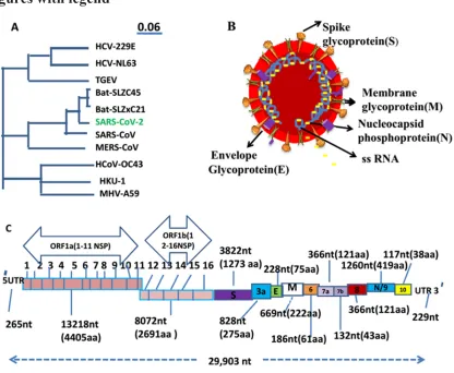
The whole genomic analysis of SARS-COV2 (isolate Wuhan-Hu-1) shows that it includes 29903 nucleotide(nt) bp (accession No NC_045512.2 ) in non-segmented ss-RNA(14), while other few isolates of this virus have been also submitted and published in genbank and they are 29881 bp (isolate nCoV WHU01, GenBank: the accession no MN988668), 29838 bp (isolate 2019-nCoV_HKU-SZ-002a_2020. the accession no MN938384), etc. The isolate Wuhan-Hu-1 of SARS-CoV2 exhibits that it contains many genes and they are ORF1ab (21,290nt), ORF1a (13,218nt), S gene (3822nt), ORF3a (828nt), E gene (228nt), M gene (669nt), ORF6 (186nt), ORF7a (366nt), ORF7b (132nt), ORF8 (366nt), N gene/ORF9 (1260nt) and ORF10 gene (117nt). At 5′ end UTR (untranscribed region) contains 265nt while at 3′ end UTR has 229nt (GenBank: accession No NC_045512.2). This polycistronic genome of SARs-CoV2 can be categorized into two parts: the first 2/3rd of the genome (ORF1ab & 1a genes) can translate to 16 non-structural proteins and while the remaining part of the genome(3-terminal) encodes the structural proteins (S, M, E and N) and some accessory proteins that are translated from a nested set of subgenomic mRNAs (sgmRNAs)47(GenBank: accession No NC_045512.2)(Fig 1C). Previously, same thing is also recorded in other corona viruses(8,48). Notable thing is that SARS-CoV-2 bears 79% identity with SARS-CoV and 50% identity with MERS-CoV while 88% identity to two bat-derived SARS-like bat coronaviruses [CoVZC (45) and bat-SL-CoVZXC (21)] collected and identified in 2018 in Zhoushan, eastern China. SARS-CoV-2 is much divergent from SARS-CoV and it has been recognized as a new human-infecting betacoronavirus (15), so it is known as SAR-CoV-2. In general, a coronavirus genome (CoVs) contains a non-segmented, +ssRNA genome of 30 kb having a 5′ 7mG( methylated guanine) cap structure along with a 3′ polyadenylated (A) tail, permiting its function as an mRNA for synthesis of the replicase polyproteins polyprotein 1a/1ab (pp1a/pp1ab) in the infected cells. The genome encodes five open reading frameS(ORFs) that are replicated to genomic RNA and transcribed to six + subgenomic mRNA (+sgmRNA). The 5' end of the genomic RNA has the untranslated leader (UTL) sequence with the TRS- L( Transcription Regulation Sequence) in the downstream part. The TRS-L is very similar to sequences (TRS-B) that can be found in front of each open reading frame. It is well known that almost all nsps (nsp1 ‐16) have their specific function in the replication of Coronairuses but the role of few nsps are unclear.
VIRAL PROPAGATION IN HOST CELL
Viral binding to host cell
6
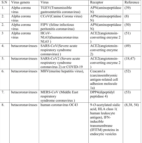
Table 1: A list of receptors used by different genera of coronavirus
S.N Virus genera Virus Receptor Reference
1. Alpha corona virus TGEV(Transmissible gastroenteritis coronavirus) APN(aminopeptidase N) (39)
2. Alpha corona virus
CCoV(Canine Corona virus) APN(aminopeptidase N)
(8)
2. Alpha corona virus
FIPV (feline infectious peritonitis coronavirus)
APN(aminopeptidase N)
(50)
3 Alpha corona virus
HCoV-NL63)(humancoronavirus NL63 )
ACE2(angiotensin-converting enzyme 2 )
(51)
4. betacoronaviruses SARS-CoV(Severe acute respiratory syndrome coronavirus) ) ACE2(angiotensin-converting enzyme 2) (49)
5. betacoronaviruses SARS-CoV2 (Severe acute respiratory syndrome
coronavirus 2) or COVID-19
ACE2(angiotensin-converting enzyme 2 )
(18,47)
6. betacoronaviruses MHV(murine hepatitis virus), Ceacam1a
(carcinoembryonic antigen-related cell adhesion molecule 1a)
(52)
7. betacoronaviruses MERS-CoV (Middle East respiratory
syndrome coronavirus )
DPP4(dipeptidyl peptidase 4)
(53)
8. betacoronaviruses human coronavirus OC43 9-O-acetylated sialic acid, HLA class 1( human leukocyte antigen), IFN-inducible transmembrane (IFITM) proteins in endocytic vesicles
(8,38, 54)
7
8
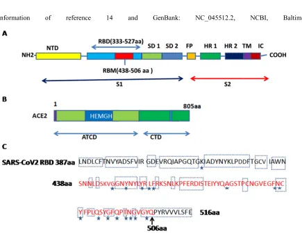
done an atomic comparison of the two viral ligands (SARS-CoV-2-CTD and SARS-RBD) binding the receptor ACE2. It revealed more interactions in SARS-CoV-2-CTD/ACE2 than in SARS-RBD/ACE2. It was also validated by other workers (57). Walls et al.(58) also reported that the SARS CoV 2-S1B receptor binding domain (residues 338-506) has a core domain and a subdomain (residues 438-498) for receptor binding and looping out from the antiparallel beta sheet core domain structure that directly engages the receptor. An irreversible conformational change of spike proteins is noted to be triggered due to receptor interaction which results membrane fusion.
The ACE2, was recorded as ACE homologue. The protein encoded by the gene is grouped to the angiotensin-converting enzyme family of dipeptidylcarboxydipeptidases. Genomic structure comparison suggests that ACE2 and ACE genes have been created by duplication of a common ancestor(67). ACE2 has 805 amino acids and is a type I transmembrane glycoprotein (metalloproteinase). The ACE2 gene is located on the X chromosome. ACE2 consists of two domains: an amino-terminal catalytic domain and a carboxy-terminal domain. The catalytic domain has an functional zone known as the zinc metallopeptidase domain (HEXH motif) and exhibits 41.8% sequence similarity with the amino domain of ACE(49) (Fig. 2B). The affinity of binding of ACE2 with the RBD (receptor binding domain) of SARS-CoV-2 is many times higher than its affinity with the RBD of the SARS-CoV(49). In addition, some workers (68) reported that the M protein also play a vital role during early stages of HCoV-NL63 infection, and that the concerted action of the two proteins (M and S) is a prerequisite for effective infection. The variation of binding capacity at different temperatures demonstrated that at both 4 and 370C, virions can bind to the surface of host cells as noted for the LPV(B-lymphotropic papovavirus)(69) while in case of HIV-170, the affinity of virion-binding at 370C is higher than that at 40C but internalization only occurs at 370C. Kuba et al.(71) using a flow cytometry assay exhibited that SARS-CoV S protein is internalized by VeroE6 cells together with ACE2 at 370C.
The correlation between receptor expression in human and infectivity of coronavirus was searched by some scientists. ACE2 expression in human tissues has a positive correlation with possibility of SARS-CoV infection, including lung and intestine (72,73). SARS-CoV has much tendency to infect ciliated epithelial cells expressing ACE2(74). Wang and Cheng (75) noted that SARS-CoV-2 upregulated the expression of ACE2 in lung tissue, a s a results viral replication and transmission becomes increase and but a negative correlation between ACE2 expression and SARS-CoV-2 (COVID-19) severity and fatality at a population level was reported by Chen et al.(47). More research is required to determine the actual receptor–ligand interaction. More than one receptor may play here. The proper understanding of this process can quicken the development of effective vaccines and antiviral drug development.
Entry into host cell
9
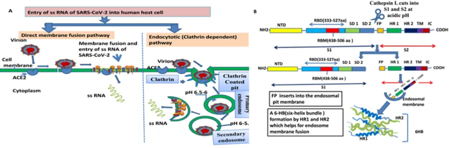
into the cytoplasm of host cell after their envelopes join or fuse with the plasma membrane at the cell surface and it is pH independent (ii) endocytotic pathway(76). It occurs through endocytic machinery. It is influenced by the acidic endosomal pH, which triggers the fusion of viral and endosomal membranes and ultimately of the viral gRNA enters into the cytoplasm.. SARS-CoV has adapted to enter into cells by direct fusion at the plasma membrane(77)(Fig 3A). In this case, virus fusogenic mechanism was described by Liu et al.(78). According to them, as soon as the S1 subunit of SARS-CoVS protein attaches to ACE2, the S2 subunit rearranges conformation by invading the fusion peptide(FP) into the plasma membrane. The HR2 domain reacts with the HR1 trimer to make 6-HB core, directing the fusion between the cell membrane and envelope of CoV(78). Further work has confirmed that entry of SARS-CoV is pH-dependent(79), and that the endosomal protease cathepsin L(80) is engaged here, advocating that this virus follows endocytosis.So, this pathway has been vigorously worked out.
Some scientists worked out by flow cytometry internalization assay to acertain that the RBD spike protein binding directs the endocytosis of SARSCoV by inected cells. So they confired that cellular endocytosis is a mechanism for SARS-CoV entry (81,82). Wang et al. (82) propose that the RBD S- protein induces the ACE2 directed cellular endocytosis signal pathway, as a result, gRNA of SARS-CoV invades the host cells. As the binding of SARS-CoV RBD spike protein triggered ACE2 internalization, now question may arise whether N-linked glycosylation on the RBD could affect ACE2 internalization or not. It was noticed that deletion of N-glycans from RBD-Fc can still induce ACE2 internalization(83). It is very interesting to note that after internalization of spike S or virus, down regulation of receptor occurred. For instance, the down-regulation of the receptor ( CD46) occurred due to attachment of measles hemagglutinin, to imbalance the complement pathways and immune systems(84). So, after utilizing the receptors for cell entry, some viruses influence down-regulation of the receptor to disturb its normal function, causing severe disease.
10
entry into VeroE6 cells(82). According to Burkard et al,(87) coronaviruses take enty into host cell through Endo/Lysosomal pathway in a proteolytic processing of fusion proteins by lysosomal protease, They proposed two models like early (e.g. HCoV NL 63 ) and late coronavirus fusion(e.g. HMV). At present, questions may arise whether CoV induces autophagy, and whether the autophagy machinery or ATG proteins are engaged for the infection and replication of CoVs. Viral attack was not arrested by the knockdown of ATG5 gene (88), so it confirmed us that the autophagy was not involved in the viral infection or replication.
Replication of genome, subgenomic RNA or m RNA formation,nsps and structural protein and RTC formation
The whole genome sequences, replication, transcription and translation to proteins or enzymes of SAR –CoV2 have been recorded since December 2019 (accession No NC_045512.2) (22, 24, 65, 56, 47) but actual mechanism of these processesof this virus have not been reported. As it belongs to same group of SARS-CoV, we assume that this virus follows same mechanism of replication, transcription and translation of SARS-CoV or other corona viruses, so, mechanism of synthesis of coronaviruses including SARs-CoV has been reviewed here. The replication or synthesis entirely happens in the cytosol of host cell. In the first step of viral synthesis, translation of pp1a and pp1ab for production 16 non-structural proteins or enzymes that are neccessary for viral further biosynthesis. As corona viruses including SARS-CoV 2 belongs to + SS RNA, their genome can be directly utilized as mRNA and they use RNA directed RNA transcription in their replication.
Translation of pp1a and pp1ab and nsps formation
After entry and uncoating, RNA genome attaches with host ribosome and exploits host RNA, amino acid pool and other factors, and initiates translation of the corona virus genome at the replicase-(RdRp) ORF1a start codon and translation of ORF1a gives polyprotein 1a (pp1a), and – 1RFS(1 ribosomal frameshifting) directs the translation of ORF1b to produce pp1ab,
ultimately through co- and posttranslational processing or cleaving by virus-encoded
proteinases like papain-like (PLpro) that reside in nsp3 and chymotrypsin-like main proteinase (Mpro) in nsp5 of pp1a, into 16 nonstructural proteins(nsps); which are utilized later for
replication of genomic RNA and translation of six structural proteins. ORF1a and 1b are linked by a -1ribosomal frameshift site (89). During synthesis, nsp1 to nsp11 are encoded in ORF1a, and nsp12 to nsp16 are encoded in ORF1b(90)( Fig 4A). Nsps 4–16 are conserved in CoVs (91). The functions of 16 nsps are listed in table 2.
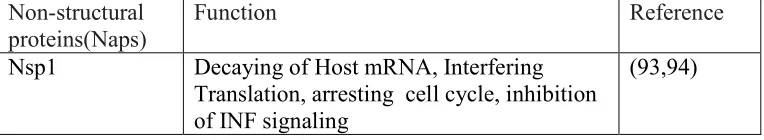
Table 2 List of non-structural proteins (Naps) and their functions with references
Non-structural proteins(Naps)
Function Reference
Nsp1 Decaying of Host mRNA, Interfering
Translation, arresting cell cycle, inhibition of INF signaling
11
Nsp2 , Nsp11 Yet to known -
Nsp3 PL1 pro and PL2pro
(papain-like proteases), in the assembly of virus and has poly (ADP-ribose) binding abilities, and deubiquitylating activity in its protease domain
(92,95)
Nsp4 and Nsp6 DMV formation, Potential transmembrane scaffold protein
(96, 97)
Nsp5 3CLpro( chymotrypsin-like Protease) or
Mpro (Main protease )
(92)
Nsp7 ssRNA binding (98,99)
Nsp 8 Primate (99)
Nsp9 Partial function of replicase enzyme (100)
Nsp10 Partial function of of replicase enzyme (100)
Nsp12 RdRpolymerase (101)
Nsp13 Nucleoside triphosphatase, RNA 5
triphosphatase, helicase activity
(102,103)
Nsp14 3-5 exoribonuclease, RNA cap formation (
guanine-N7 )-, methyltransferase , role in RNA proof reading
(104,105,91)
Nsp15 Viral endoribonuclease, NendoU (106,103)
Nsp16 RNA cap formation (2 O-transferase) (107)
It has been recently reported that, a spike mutation of CoV, which probably happened in late November 2019, directed jumping to humans. In particular, Angeletti et al (108) compared the SAR-CoV-2 gene sequence with that of SARS-CoV. An genetic analysis of SAR-CoV-2 and SAR-CoV, revealed that the transmembrane helical segments in the ORF1ab encoded 2 (nsp2) and nsp3 and exibited that at the position 723 a serine replaced a glycine residue, while in the position 1010 proline replaced isoleucine(108). The viral mutations is responsible for potential the relapse of this viral disease.
Programmed ribosomal framshifting
12
analysis by mutation. A stem - loop SL1 with a proximal ds-segment (double - stranded ) segment which exists in SARS-CoV, is absent in other characterized coronavirus 1a/1b pseudoknots(11,113). The -/+ ribosomal frameshifting is generally occurred due to presence of slippery sequence and pseudoknot but, there are alternative ways for formation of “ out-of-frame proteins”. Alternative splicing can cause out-of-frameshifting (113).
However, in programmed -1 ribosomal frame shifting, the ribosome is bound to move one nucleotide backwards into an overlapping reading frame and to translate an completely new amino acids sequence. It is very common SARS-CoVs(114). In SARS-CoVs, the stimulatory structure is to be an mRNA pseudoknot as described in the infectious bronchitis virus(IBV) where a ‘slippery’ sequence of the type UUUAAAC and a H-type pseudoknot are able to induce translation of the zone 1b of the polyprotein 1a/1b(115). The SARS-CoV genome has been found to be responsible for encoding an mRNA segment that directs −1 ribosomal frameshifting (36). Smith et al.(116) and Tu et al.(117) recorded that ribosomal movement was stalled at a pseudoknot neccessary for frameshifting (Fig 4B,C). Many models are present to analyze −1 ribosomal frameshifting operation by pseudoknots. It is found that RNA pseudoknot structures force to stall ribosomes over a slippery sequence, at which the ribosome-bound tRNAs realign in the - 1 frame, but that pausing alone is not sufficient to operates frameshifting (111,118). To discuss the in- depth mechanism of it at the gene level, Namy et al (89) introduced a method of purifying “rabbit reticulocyte lysate (RRL) ribosomes”. Cryo-EM (Cryo-electron microscopy) study of purified mammalian 80S ribosomes from rabbit reticulocytes halted at a coronavirus pseudoknot revealed an “intermediate of the frameshifting process”. The translating 80S ribosome, halted at the IBV pseudoknot (Fig 4C), contains a P-site tRNA and elongation factor 2 (eEF2). Frank, and Agrawal (119) noted that the 70S complex exhibited “a ratchet-like rearrangement of the ribosome” which is connected to the trapping of eEF2 in E. coli, and in 80 S complex, “a ratchet like rearrangement of the ribosome” connected to the trapped eEF2 in yeast by sordain antibiotic treatment (120). So, they (119,120) proposed a model where “the ratchet-like rearrangement of the ribosome” is a part of a mechanism for moving the tRNAs during the translocation. Similarly, experimental results observed by Namy et al, (89) where no antibiotic was applied, also showed a ratchet like rearrangement of the ribosome in eukaryote. Plant et al.(121) have proposed a “golden mean” model in which viruses utilize both “programmed ribosomal frameshifting” and “translational attenuation” to maintain the relative ratios of their encoded proteins.
RTC formation
13
the work of Bechill et al,(124) it was found that uPR(unfolded protein response)) may mediate DMV formation as it is promoted during coronavirus infections.Snijder et al. (122) and Snijder et al.(125) recorded that the DMVs of SARS-CoV are most likely derived from the endoplasmic reticulum (ER). The results of Knoops et al. (126) showed that DMVs are likely to originate from the part of a reticulovesicular network of modified ER membranes. Later on, these networks become a large single-membrane vesicles. gRNA ( genomic RNA) which are synthesisezed in infected cell, are packed into virions on membranes which are situated between the endoplasmic reticulum (ER) and the Golgi apparatus ( “ER–Golgi intermediate compartment (ERGIC)” (127). Proteins ( nsp3, nsp5 and nsp8) engaged in the replication of virus are situated in adjacent reticular structures but not inside of DMVs. RNA, which is either “replicative intermediates” or “dead end” double-stranded RNA, was found in DMVs. The non-ionic detergents disrupt all RTC and make SARS-CoV RNA to susceptible for breaking by nuclease, and this reminds us the intact membrane structure of RTC for RNA synthesis (128). It is found that pool of replicated genomic RNAs of SARS-CoV are connected with the RTC, while sg RNAs after synthesis come out from the RTC structures. Moreover, it has been recorded that RTC activity relies on a host factor which is present in cytosol, that indicates that crosstalk between RTC and cytosol, takes place via channels that help transport through membranes (128). The single DMV can move freely but when many are “captured” by the DMV/CM(convoluted membrane) assemblies, they are unable to move. It was also reported that disruption of microtubule-dependent transport of DMVs, did not interfered on RNA replication (129). In coronaviruses the RTC formation might be one of strategies for involving of cellular and viral proteins or enzymes for successful replication of g RNA and other parts of virus and also for creating a safe place from the attrack of host defense (129).
RNA replication and transcription of the sub-genomic RNAs (sgRNAs) sequences.
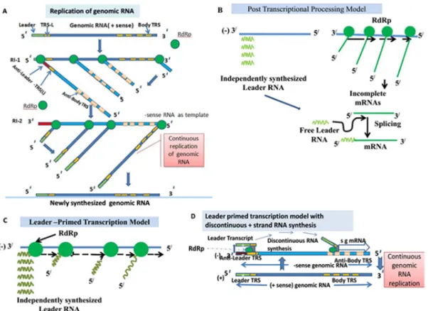
During genome replication and sgRNA synthesis, two replicative intermediate (RIs) such as RI -1 and RI- 2 are operated. From RI-1, – sense full genome strand was formed on the template on + sense ss RNA by viral encoded the RNA dependent RNA polymerase (RdRp). In RI-2, - sense RNA genome strand is used as template to generate + sense genome of virus by viral encoded RdRp ( Fig 5A) . The TIs (Transcription intermediates) and TFs(transcription forms) are main transcription structures that are responsible for synthesis of subgenomic mRNA (130). Here the exiting recent model of coronavirus RNA synthesis reveals that minus strand RNAs come from copying the + RNA continuously to form genomic templates and discontinuously to create subgenomic templates (131).
Formation of subgenomic RNA template(-) and subgenomic +stand mRNA
14
RNA template. As soon as replication of leader RNA completed, the polymerase enzyme "jumps" over to the different initiation sites for several mRNA species, so, it is probable to create "loop out" of the RNA template. This model is not supported by maximum workers, 2. Post-transcriptional processing model (Fig 5B). This model indictes that synthesis of the leader RNA and mRNAs are independant; after the finishing of synthesis, the leader RNA is then bound to the body sequences of the mRNAs by a mechanismwhich is yet to know. UV transcription inactivation studies were performed and the results indicated that the formation of mass of subgenomic RNAs were not possible post-transcriptionally by cis-splicing of a genome-length precursor molecule (132) and 3.“leader primed transcription model”(Fig 5C): The this model exhibits that the leader RNA is replicated and "falls off" from the template. Then a viral RNA dependent RNA polymerase (RdRp) binds with this free leader RNA and initiates mRNA synthesis at several initiation points. This model indicates that the leader RNA serves as a primer for RNA synthesis. Lai et al.(133) gave this model and it is known as “leader primed transcription model”(134). Out of these three models, this one was accepted by many authors by few modification.
15
but almost co-transcriptional fusion of leader and body is being reported in all models (135,141,142). The RNA-dependent RNA-polymerase has been seemed to halt after a body TRS(TRS-B) of a particular gene is replicated during (-) strand synthesis, therefore, switching to the TRS-L occurres and thus a common L sequence to each sg mRNA is added.
The gRNA and 6 sgmRNAs are formed during replication of MHV-A59. It was observed that 1stsgmRNA- 6th sgmRNA are 9.6, 7.4, 3.4, 3.0, 2.4 and 1.7 kb respectively. All together form a 3′ co-terminal nested set. The sgmRNAs, in size, ranges from 1/3rd (1stsgmRNA) to about 1/20th (6th sgmRNA) of the genomic RNA. It was also recorded that + strands (genomes and subgenomic mRNA) are formed in huge amounts but out of them about 1% minus strands of both genome- and subgenomic length act as the templates for genome and subgenomic mRNA synthesis(143.144). It was also reported that the newly synthesized viral genomic RNAs may act as template for the formation of sg-length minus strands. Experimental studies conducted by Stanley and Dorothea Sawicki and coworkers (130), where they used MHV as a model, revealed that both –strand gRNA and sg-length minus strands are formed during very early stage of infection. Each sg mRNA is formed from a corresponding transcription intermediate (TI) which bears the sg-length minus-strand template. Many sg mRNAs are synthesized from these complexes in constant amounts but in non-equimolar. It appears that the ratio of the synthesis of genomic RNA to sg mRNAs is constant in the whole replication stage (130,131).
minus-16
strand sg RNA synthesis was operated discontinuously, with attenuation of nascent strand RNA synthesis occurring in the different body TRS regions of the genomic template, and while plus strands were synthesized continuously. A small portion of the polymerase complex detaches from the +strand template when it comes to TRS to develop an -sg RNA. So, the synthesis of a longer -sg RNA, i.e., the product of the upstream TRS, is attenuated by the presence of a downstream TRS because only a little percentage of the polymerase complexes reach the upstream TRS. The nascent sg-length minus strand, bearing an anti-body TRS at its 3′ end, become moved to the leader TRS(TRS-L) in the genomic template andthe CS-L could base-pairing process procceds between CS-L and CS-B (cCS-B) as the CS is similar to the genome leader (CS-L) and all mRNA coding sequences (CS-B), and it allows for leader-body joining (Fig 6 ) and the polymerase(RdRp) would continue and dulicate the leader sequence to put an antileader (complementary to the 5′ plus-strand leader sequence) on the 3′ end of the nascent, subgenomic minus strand(Fig 6). Here the anti-TRS plays as a primer to finish transcription and duplicates the leader RNA(141). Ultimately the subgenomic minus strands are produced and they perform as templates for the transcription of subgenomic mRNAs (Fig 6). The genomic and subgenomic RNA synthesis take place in TRC/DVC. Later the RTCs employing in plus-strand synthesis became aged and released their minus-strand templates, which are then decayed, and fresh RTCs are produced in infection (131). Both genetical and biochemical researchs validated this model (130,131,148).
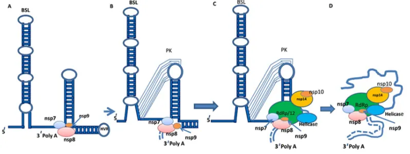
Accoding to some workers, the RNA synthesis of cononaviruses must have to involve by cis-acting RNA elements, that are situated in structured 5′ and 3′ untranslated Regions (UTRs), and into the associated coding sequences (149-151). These RNA elements have stem-loops (SL1, SL2, SL3, SL4 and SL5) structures which are conserved but degree of conservation among different coronaviruses are variable, and all have some role in replication of RNA, discontinuous synthesis of sgRNA and sgmRNA (149,151,152). Very primary research work using defective interfering RNAs from alpha-, beta-, and gamma-coronaviruses limited the 3′ cis-acting RNA elements needed for synthesis of RNA of CoVs coronavirus to the 3′ UTR plus the poly(A) tail (150) but later it was found in other gene like at down stream N gene stop codon where BSL((Bulged Stem Loop) and H-type PK(pseudoknot) are involved. So, a model for the initiation of replication of negative-strand RNA of CoVs was postulated (140,153) (Fig 7).
17
virus transcription as shown by some workers (154-156). The steps are: (i) Complex formation (ii) Base pairing scanning and (iii) Template switch (for details ref 140).
The regulation of transcription process have suggested that it is regulated by many factors through monitoring the template switch frequency during discontinuous transcription (131,149). These factors are like complementarily between the leader TRS and the body TRS-B(154), TRS secondary structure to the 3′ end, RNA-RNA or protein-RNA interactions eg N-protien RNA chaperone (149,155), cell factor RNA helicase DDX1.(158), the two polymerases nsp8 and nsp12(159), etc. It is very interesting to note that Nucleocapsid phosphorylation and RNA helicase DDX1 recruitment enable coronavirus transition from discontinuous to continuous transcription (158).
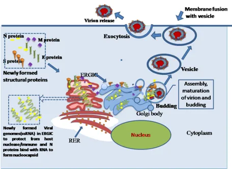
After synthesis of nested sgmRNAs (10 sgmRNSs in SARS-CoV-2), each of them undergoes translation by host ribosome, amino acids and other factors and express its gene as protein. As a results, four known structural proteins(sps) like S, E, M and N and other proteins in coronaviruses but in case of SARS-CoV2, in addition to sps, others are ORFs proteins (3a, 6, 7a,7b, 8 and 10) in SARS-CoV2 (NC_045512.2, NCBI, Baltimore) (Fig 6). Througout the discussion, we found that all RNAs and protein synthesis take place in solely cytoplasm, there is no involment of host nucleus. But researches of some workers reported that some viral proteins (e.g N, nsp 3, 6, 9b etc) have been located in nucleus. N protein enters into nucleus through nuclear pore complex and it later induces the cell cycle blockage and impediment of cytokinesis. (160,161). Protein 6 inhibits nuclear import of factors like STAT1(162) and nullifies IFN signaling pathways(163). Although viral proteins are observed in nucleus of deseased cells, its actual mechanism of entry into nucleus is unknown.
Assembly and release of virion
18
cells. E-proteins of SARS-CoV also serves a vital function on assembly and release of the virus and pathogenesis (42). Finally after assembly of viral parts and maturation of virion in ERGIC(Endoplasmic reticulum Galgi body intermediate compartment or Golgibody, buds with virions come out from Golgi body and form vesicles. These vesicles with virions approach to plasma membrane and by exocytosis virions are released from infected host cell(Fig 8).
Meaningful points to note for development of antiviral drugs and vaccines against SARS-CoV-2
Till now, no approved effective antiviral drug or vaccine has been developed against SARs-CoV2 (COVID-19). Scientists of many private companies and Government institutional laboratories of some countries putting their hard labour around the clock to develop effective remedies. After thorough search and rigorous reading of numerous research papers related to genomic structures, ligand receptor interaction, mode of entry in human cell, replication and transcription, RTC formation, assembly of viral parts, maturation and release of coronona virus, particularly human coronona viruses, such as SARS –CoV, MERS –CoV, SARs –CoV-2, we have attempted to highlight some meaningful points that may be utilized for the production of drugs or vaccines and developed drugs must target the conserved zone of virus–
i) A lot of compounds like polypeptides, peptides, antibiotics etc are able to interact with receptor ACE2 and inactivate the latter (168,169). We may use some of them against SARS-CoV2 because they block the S-protein-binding site, or change the conformation of ACE2 which is not suitable for binding or fusion. So, now we can remember the words of Li et al. (49)- “ if SARS returns as a threat to human health, these studies may contribute to its control”(49). The chloroquine or hydroxychloroquine have been considered by in vitro trial in Vero E6 cell line as effective drug against Covid-19 as it disturbs the glycosylation of a virus cell surface receptor, ACE2 on Vero E6 cell line(170). So, hydroxy chloroquine sulfate (HCQS) has been approved as one of the important drugs for the treatment of severe SARSCoV-2 infections (171).
ii)The virus utilizes TMPRSS2, host serine protease to prime S protein as a result it facilitates the fusion of viral and cellular membranes and leads to the entry of virus into the cell. Here, scientists may develop or search already existing any serine protease inhibitor for inhibiting the viral entry (172,173). We may mention here serine protease inhibitors like “camostat mesylate” and K11777 , that inhibit TMPRSS2 and partially arrest SARS-CoV infection of lung epithelial cells.(174,175). Hoffmann et al.(62) recently reported that TMPRSS2 is hindered by a protease inhibitor which has been clinically tested. Some peptide inhibitors have been formulated which are active to destabilize HR regions of S2 and they have exhibited their effectiveness in both in vitro and in vivo trial(176,177).
19
advocated that these drugs might be applied in human patients aganst this virus(171). Adedeji et al.(179) screened some of 14,000 compounds of the Maybridge Hit Finder small-molecule library. These compounds are at per with Lipinski’s rule of five(180). Out of these compounds, they found three compounds-SSAA09E2, SSAA09E1 and SSAA09E30 arrest SARS-CoV entry by the following principles: (i) checking of early SARS-S–ACE2 interactions (ii) inactivation of cathepsin-L, and (iii) hindering fusion respectively.
iv) Studies have shown that Mpro of different coronaviruses are highly conserved in terms of both sequences and 3D structures. The main protease of SARS-CoV-2 and SARS-CoV are almost similar ( 96.1%) (59). These features, together with its functional importance, have considered Mpro an important object for the design of anti-coronaviral drugs(172). Very recently, Li et al.(59) selected the structure of SARS-CoV-2 main protease as a homologous target for drug molecule screening on basis of bioinformatics analysis, and suggested that out of 8,000, four drugs such as Prulifloxacin (fluoroquinolone antibiotic), Tegobuvir, Bictegravir and Nelfinavir (anti-HIV drugs) exhibited maximum binding conformations with the main protease of virus(181). Similarly, Li et al.(182) shorted out available drugs which may be potential inactivator for SARS-CoV-2 M protease.and these drugs have higher mutation tolerance than widely used drugs lopinavir. In this respect, we may mention the review work of Ghosh et al (183) which highlighted the repurposing of drugs against SARS-CoV2,, SARS-CoV and MERS-CoV.
v) The functional domains exist in the replicase polyproteins are conserved in all CoVs, so, they must be good targets for anti viral drugs or vaccine(90). According to Amici et al.(184). Indothethacin showed antiviral activity against SAR- CoV blocking viral synthesis in early stage in Vero 6E cell line.
vi) Coronavirus RNA is synthesized in a RTC or DVC. Drugs may be developed for changing its micro-environment or degradation of this DMV(185) .
vii) The −1 ribosomal frameshifting is very essential for SARS-CoV to synthesize the replication–transcription complex and the 1a/1b ribosomal frameshift signal is conserved (111). So it can be attractive approach that this mRNA structure is an important target for drug development. ANXA2 has been suggested as an antiviral regulator which specifically binds to the frameshift signal as frameshift signal binding compounds186 and other compounds could be applied targeting this conserved framshift signal.
viii) During RNA replication of SARS-CoV, nucleic acid unwinding by the viral helicase is a critical point , so this point may be targeted for inhibition of RNA replication. Adedeji et al.(187) applied some inhibitors to this point and become successful to block RNA replication .
20
x) Chloroquine drug has a power for inhibiting proteolytic processing of the M protein and affects construction of virus and budding. Besides, this drug changing pH of cell can damage the viral protein (188) and interferes the recognition of viral antigen by dendritic cells, which operates by a Toll-like receptor-dependent pathway that needs changes of pH of endosomes to low acidic(189 ) Chloquine may be good drug against COVID-19 (171) .
xi) Viral proiens such as nsp12(RdRp), N protein, nsp14 etc have conserved zones, as these three proteins have vital role in RNA synthesis, drug designer may target their conserved sequences. As for example nsp14 of corona viruses is highly succeptible to ribavirin and 5-fluorouracil agents (116).
xii) Bloking of entry of viral proteins (N, nsp3, nsp 6, etc ) into nucleus may be a good strategy to arrest.the viral synthesis and to restore our immune system which became antagonized by viral proteins. Ivermectin, an FDA-approved anti-parasitic and broad spectrum anti-viral drug , has been found as an inhibitor of SARS-CoV-2 isolate Australia/VIC01/2020) in cell line( Vero/hSLAM). It reduced nearly 5000-fold viral RNA synthesis at 48 h. It binds with the carrier( IMPα/β1) of viral protein which carries the protein inside the nucleus. So, binding to the viral protein becomes inhibited and preventing it from entering the nucleus. As a result our antiviral responses becomes normal or more efficient antiviral response. If patients are treated by this drug early in infection, it reduces the viral load, checks severity of COVID-19 and arrests “person-person transmission” (190).
xiii) The newly replicated genomic RNA and structural proteins of SARS-CoV are assembled into virions in ERGICor endoplasmic reticular or Golgicomplex membrane.Virions are shred off from infected cells by exocytosis, so these two works ( virion assembly and shredding ) may be arrested by any inhibitor.
21
antibodies can clear virus or protect an uninfected host that is exposed to the virus. We may recall here the words of Li et al (49) “ if SARS returns as a threat to human health, these studies may contribute to its control”. Hence, this antibody application may prevent and/or treat COVID-19 (191). A recombinant SARS-CoV-2 spike protein vaccine combined with other top epitopes could be a meaningful step for development of vaccine against SARS-CoV-2. The ray of hope is coming from some laboratories for drug and vaccine production, examples may be cited here as the mRNA base vaccine (mRNA -1273) under a phase 1 clinical trial against SARS-Co-2 since 25th February, 2020 with 1st dose already applied on human on 16th March at Kaiser Permanente Washington Health Research Institute (KPWHRI) in Seattle under guidance of NIH(national Institution for Health, USA(194). Other numerous vaccines developed are being undertrial in different institutions in different countries like Ad5-nCoV in China, ChAdOx1 nCoV-19 in Oxford University, INO-4800 in Philadelphia and Kansas City, BNT162 a1, b1, b2, and c2 in Germany, bacTRL-Spike in Canada, Covid-19 S-Trimer & a nanoparticle vaccine SARS-CoV-2 rS in Australia, and BCG trial in Brasil, Germany, France, Denmark, Hungary and South Africa (195), etc. Previous antiviral vaccine like live attenuated vaccines was applied to arrest dangerous diseases caused by avian and porcine CoVs. By gaining experiences of previous vaccines, a live attenuated vaccine could be invented against COVID-19 or SARS-CoV-2, because we can multiply this virus in high titers in Vero 6E or other cell lines.
CONCLUSION
In conclusion, now the SARS-CoV-2 or COVID-19 pandemic appears to be out of control in some countries. Although the development of drugs and vaccine are very urgent, we have to remember it needs time and patience to discover the appropriate drug. In such a moment new quick tests to identify SARS-CoV- 2 patients at the earliest stages of disease are also necessary as these tests will lead quarantine and isolation procedures to arrest the transmission of this disese. We are hopeful that the elaborate discussion of virion structure , molecular mechanism of propagation and clues for drug or vaccine development embedded in this article will help us for quick proper and effective drug or vaccine discovery. The whole world is anxiously waiting to defeat the COVID-19 pandemic and win the game of survival by discovering drugs and vaccines against SARS-CoV-2.
Contribution of author
As SKG, is sole author, all works like data/literature collection, writing Ms, fig drawn, editing etc are done by myself.
ACKNOWLEDGEMENT Author is grateful to Principal, RKMVC College, Rahara. Author declares no competing financial and nonfinancial interest.
22
REFERENCES
1 Ge, X.Y. et al. Isolation and characterization of a bat SARS ‐like coronavirus that uses the ACE2 receptor. Nature503(7477):535 ‐538(2013).
2 Chen, Y, Guo, D. Molecular mechanisms of coronavirus RNA capping and methylation. Virol. Sin.31(1): 3 ‐11(2016).
3 Butler, D. SARS veterans tackle coronavirus. Nature490:20(2012).
4 Peiris, J.S. et al. Coronavirus as a possible cause of severe acute respiratory syndrome. Lancet
361:1319-1325(2003).
5 Zaki, A.M. et al. Isolation of a novel coronavirus from a man with pneumonia in Saudi Arabia. N Engl. J. Med.367:1814-1820(2012).
6 WHO. Novel coronavirus infection - update (Middle East respiratory syndrome- coronavirus). Available online: http://www.who.int/csr/don/2013_05_29_ncov/en/index.html. (accessed May 30, 2013).
7 Rahman, A, Sarkar, A. Risk factors for fatal middle east respiratory syndrome coronavirus infections in Saudi Arabia: analysis of the WHO Line List, 2013– 2018. Am. J. Public Health
109(9):1288–93(2019)
8 Fehr, A.R., Perlman S. Coronaviruses: an overview of their replication and pathogenesis. Methods Mol. Biol.1282: 1 ‐23(2015).
9 Su, S. et al. Epidemiology, genetic recombination, and pathogenesis of coronaviruses. Trends Microbiol.24(6):490 ‐502(2016).
10 Jiang, S., Lu L., Du, L. Development of SARS vaccines and therapeutics is still needed (editorial). Future Virol.8: 1-2(2013).
11 WHO Announces COVID-19 Outbreak a pandemic. http://www.euro.who. int/en /health topics/health-emergencies/coronavirus-covid-19/news/news/2020/3/who-announces-covid-19-outbreak-apandemic (accessed on 21 March 2020).
12 http://virological.org/ 13 https://www. gisaid.org/
14 Wu, F, S. Zhao, B. Yu, et al., A new coronavirus associated with human respiratory disease in China. Nature 2020, https://doi.org/10.1038/s41586-020-2008-3.
15 Lu, R, et al. Genomic characterisation and epidemiology of 2019 novel coronavirus: implications for virus origins and receptor binding Lancet 2020; 395(10224): 565-574.
16 ChenY, Liu Q, Guo D . Emerging coronaviruses: Genome structure, replication, and pathogenesis. J Med Virol. 2020; 92:418–423.
17 Zhou, P, Yang X-L, Wang, X-G, et al. A pneumonia outbreak associated with a new coronavirus of probable bat origin. Nature 2020 https://doi.org/10.1038/s41586-020-2012-7, February . 18 Yuan, M, Yin W, Tao Z, Tan W, Hu Y (2020) Association of radiologic findings with
mortality of patients infected with 2019 novel coronavirus in Wuhan, China. PLoS One 2020;
15(3): e0230548. https://doi.org/10.1371/journal.pone.0230548.
19 Wölfel, R. et al. SARS-CoV-2 Replication at Different Body Sites. Nature 2020; 581: 465–469. 20 WHO
http://www.euro.who.int/en/healthtopics/health-emergencies/coronavirus-covid-19/news/news/2020, 4th April ,2020
23
22 Chu H., et al, Comparative replication and immune activation profiles of SARS-CoV-2 and SARS-CoV in human lungs: an ex vivo study with implications for the pathogenesis of COVID-19. Clin Infect D 2020 DOI: 10.1093/cid/ciaa410, April.
23 Chan JF, et al. A familial cluster of pneumonia associated with the 2019 novel coronavirus indicating person-to-person transmission: a study of a family cluster. Lancet 2020; 395: 514–23. 24 To KK, et al. Temporal profiles of viral load in posterior oropharyngeal saliva samples and
serum antibody responses during infection by SARS-CoV-2: an observational cohort study. Lancet Infect Dis 2020; 20: 565–74.
25 Chan JF, et al. Simulation of the clinical and pathologic manifestations of Coronavirus Disease 2019 (COVID-19) in golden Syrian hamst model: implications for disease pathogenesis and transmissibility. Clin Infect D 2020. doi: 10.1093/cid/ciaa325
26 Huang C, et al. Clinical features of patients infected with 2019 novel coronavirus in Wuhan, China. Lancet 2020; 395:497–506.
27 CDCP Centers for disease control and prevention https://www.cdc.gov/coronavirus/2019-ncov/ January 28, 2020.
28 Phan LT, et al. Importation and human-to-human transmission of a novel coronavirus in Vietnam. N Engl J Med. DOI: 10.1056/NEJMc2001272
29 WHO, https://www.who.int/docs/default-source/coronaviruse/situation-reports/20200610-covid-19-sitrep-142 , 10 June,2020
30 Leung C 2020 Estimating the distribution of the incubation period of 2019 novel coronavirus (COVID-19)infection between travelers to Hubei, China and non-travelers med Rxiv preprint doi: https://doi.org/10.1101/2020.02.13.20022822
31 Li Q, et al (2020) Early Transmission Dynamics in Wuhan, China, of Novel Coronavirus– Infected Pneumonia. N Engl J Med. doi:10.1056/NEJMoa2001316
32 Backer JA, Klinkenberg D, Wallinga J. Incubation period of 2019 novel coronavirus (COVID-19) infections among travellers from Wuhan, China. Eurosurveillance. 6 Feb 2020.
doi:10.2807/1560-7917.ES.2020.25.5.2000062.
33 CDC-Centers for disease control and prevention 4/4/2020 https://www.cdc.gov/coronavirus
/2019-ncov/prevent-getting-sick/social-distancing.html), 34 Barcena M, et al. Cryo-electron tomography of mouse
hepatitis virus: insights into the structure of the coronavirion. Proc Natl Acad Sci ,USA, 2009
106:582–587
35 Neuman BW, et al . A structural analysis of M protein in coronavirus assembly and morphology. J Struct Biol 2011; 174:11–22.
36 Rota PA, et al. Characterization of a novel coronavirus associated with severe acute respiratory syndrome. Science 2003; 300:1394-1399.
37 Bosch BJ, et al. The coronavirus spike protein is a class I virus fusion protein: structural and functional characterization of the fusion core complex. J Virol 2003; 77:8801–8811
38 Collins AR, et al (1982) Monoclonal antibodies to murine hepatitis virus-4 (strain JHM) define the viral glycoprotein responsible for attachment and cell–cell fusion. Virology 1982; 119: 358– 371.
39 Delmas B, Laude H . Assembly of coronavirus spike protein into trimers and its role in epitope expression. J Virol 1990 ; 64:5367–5375
40 Armstrong J, Niemann H, Smeekens S et al . Sequence and topology of a model
intracellular membrane protein, E1 glycoprotein, from a coronavirus. Nature 1984; 308:751–752
41 Nal B, et al. Differential maturation and subcellular localization of
24
42 Nieto-Torres JL, et al. Severe acute respiratory syndrome coronavirus envelope protein ion channel activity promotes virus fi tness and
pathogenesis. PLoS Pathog 2014; 10:e1004077. doi:10.1371/journal.ppat.1004077
43 Chang C, et al. Modular organization of SARS coronavirus nucleocapsid protein. J Biomed Sci. 2006; 13(1): 59 ‐72.
44 Sturman LS, Holmes KV, Behnke J. Isolation of coronavirus envelope glycoproteins and interaction with the viral nucleocapsid. J Virol 1980; 33:449–462.
45 Cui L, et al. The nucleocapsid protein of coronavirusesacts as a viral suppressor of RNA silencing in mammalian cells. J Virol.2015; 89(17):9029 ‐9043.
46 Cui J, Li F, Shi ZL. Origin and evolution of pathogenic Coronaviruses. Nat Rev Microbiol. 2019;
17(3):181–92.
47 .Chen J, Jiang Q, Xia X, Liu K, Yu Z, Wanyu Tao , Gong W ,. Han J-D J. Individual variation of the SARS-CoV2 receptor ACE2 gene expression and regulation. 2020(www.preprints.org) 12 March .Preprint.
48 Hussain S, et al. Identification of novel subgenomic RNAs and noncanonical transcription initiation signals of severe acute respiratory syndrome coronavirus. J Virol 2005;79(9):5288 ‐5295.
49 Li W, et al., Angiotensin-converting enzyme 2 is a functional receptor for the SARS coronavirus, Nature 2003; 426: 450e454,https://doi.org/10.1038/nature02145.
50 Tresnan DB, Levis R, Holmes KV. 1996. Feline aminopeptidase N serves as a receptor for feline, canine, porcine, and human coronaviruses in sero group I. J Virol 1996;70: 8669–8674. 51 Hofmann H, Pyrc K, van der Hoek L, Geier M, Berkhout B, Pöhlmann S.Human coronavirus
NL63 employs the severe acute respiratory syndrome coronavirus receptor for cellular entry. Proc Natl Acad Sci 2005; 102:7988–7993.
52 Saeki K, Ohtsuka N, Taguchi F. Identification of spike protein residues of murine coronavirus responsible for receptor-binding activity by use of soluble receptor-resistant mutants. J Virol 1997; 71: 9024 –9031.
53 Mou H, Raj VS, van Kuppeveld FJ, Rottier PJ, Haagmans BL, Bosch BJ.. The receptor binding domain of the new Middle East respiratory syndrome coronavirus maps to a 231-residue region in the spike protein that efficiently elicits neutralizing antibodies. J Virol 2013; 87: 9379 –9383. 54 Zhao Z, et al. Coronavirus replication does not require the autophagy gene ATG5. Autophagy 3:
581-585(2007).
55 Lu, G. et al. Molecular basis of binding between novel human coronavirus MERS-CoV and its receptor CD26. Nature 500: 227–231(2013).
56 Wang, Q, et al. Structural and Functional Basis of SARS-CoV-2 Entry by Using Human ACE2, Cell 2020,https://doi.org /10.1016/j.cell.2020.03.045
57 Wrapp, D. et al. Cryo-EM structure of the 2019-nCoV spike in the prefusion conformation. Science; 367: 1260–1263(2020).
58 Walls, A. C. et al. Structure, Function, and Antigenicity of the SARS-CoV-2 Spike Glycoprotein. Cell 2020; S0092-8674(20)30262-2. https://doi.org/10.1016/j.cell. 2020.02.058
59 Li, X.. Genga M, Penga Y, Menga L , Lua, S. Molecular immune pathogenesis and diagnosis of COVID-19. J Pharmaceutical Analysis10: 102-108(2020).
60 Wan, Y., Shang, J., Graham, R., Baric, R.S., Li, F. Receptor recognition by the novel coronavirus from Wuhan: an analysis based on decade-long structural studies of SARScoronavirus. J Virol 2020; 94:e00127-20. https://doi.org/10.1128/JVI.00127-20
61 Letko M, Marzi A, Munster V. Functional assessment of cell entry and receptor usage for SARS-CoV-2 and other lineage B betacoronaviruses. Nat. Microbiol 2020:https://doi.org
/10.1038/s41564-020-0688-y
62 Hoffmann M et al. ACE2 is the SARS-CoV-2 Receptor Required for Cell Entry Cell 2020 Mar5. 63 Lu G, Wang Q, Gao GF Bat-to-human: spike features determining ‘host jump’ of coronaviruses
25
64 Li F. Structural analysis of major species barriers between humans and palm civets for severe acute respiratory syndrome coronavirus infections. J Virol 2008; 82:6984 – 6991.
65 Wu KL, Peng GQ, Wilken M, Geraghty RJ, Li F. Mechanisms of host receptor adaptation by severe acute respiratory syndrome coronavirus. J Biol Chem 2012; 287:8904 – 8911.
66 Lan J , Ge J, Yu J, et al.. Structure of the SARS-CoV-2 spike receptor binding domain bound to the ACE2 receptor Nature 2020;https://doi.org/10.1038 /s41586-020-2180-5 .
67 Donoghue M, et al. A Novel Angiotensin-Converting Enzyme-Related Carboxypeptidase (ACE2) Converts Angiotensin I to Angiotensin 1-9 Circ Res 2000; 87(5), E1–9
68 Naskalska A, Dabrowska A, Szczepanski A, Milewska A, Jasik KP, Pyrc K. Membrane protein of human coronavirus NL63 is responsible for interaction with the adhesion receptor. J Virol 2019;93:e00355-19. https://doi.org/10.1128/JVI.00355-19.
69 Haun G , Keppler OT, Bock CT, Herrmann M, Zentgraf H, And Pawlita M The Cell Surface Receptor Is a Major Determinant Restricting the Host Range of the B-Lymphotropic Papovavirus J Virol 1993; 67 (12):.7482-7492.
70 Moore JP, McKeating JA, Norton WA, Sattentau QJ. Direct measurement of soluble CD4 binding to human immunodeficiency virus type 1 virions: gp120 dissociation and its implications for virus-cell binding and fusion reactions and their neutralization by soluble CD4. J Virol 1991;
65: 1133–1140.
71 Kuba K, et al A crucial role of angiotensin converting enzyme 2 (ACE2) in SARS coronavirus– induced lung injury Nature Medicine 2005; 11(8)::875- 879.
72 Hamming I., Timens W, Bulthuis ML, Lely AT, Navis G J, van Goor H. . Tissue distribution of ACE2 protein, the functional receptor for SARS coronavirus. A first step in understanding SARS pathogenesis. J Pathol 2004; 203:631–637.
73 Harmer D, Gilbert M, Borman R, Clark KL. Quantitative mRNA expression profiling of ACE 2, a novel homologue of angiotensin converting enzyme. FEBS Lett 2002; 532:107–110. 74 Jia HP, Look DC, Shi L, et al. ACE2 Receptor Expression and Severe Acute Respiratory
SyndromeCoronavirus Infection Depend on Differentiation of Human Airway Epithelia. J Virol 2005; 79: 14614–14621.
75 Wang PH, Cheng Y. Increasing host cellular receptor—angiotensin-converting enzyme 2 (ACE2) expression by coronavirus may facilitate 2019-nCoV infection. BioRxiv 2020 Feb 27.
doi:10.1101/2020.02.24.963348. Preprint.
76 Zumla A, Chan JF, Azhar EI, Hui DS, Yuen KY. Coronaviruses – drug discovery and therapeutic options. Nat Rev Drug Discov 2016; 15: 327-47.
77 Simmons G, Reeves JD, Rennekamp AJ, Amberg SM, Piefer AJ, Bates P. Characterization of severe acute respiratory syndrome associated coronavirus (SARS-CoV) spike glycoprotein-mediated viral entry. Proc Natl Acad Sci USA; 2004; 101:4240-4245.
78 Liu S, et al. Interaction between heptad repeat 1 and 2 regions in spike protein of SARS-associated coronavirus: implications for virus fusogenic mechanism and identifiation of fusion inhibitors. Lancet 2004; 363: 938-47.
79 Yang ZY, et al. pH-dependent entry of severe acute respiratory syndrome coronavirus is mediated by the spike glycoprotein and enhanced by dendritic cell transfer through DC-SIGN. J Virol 2004; 78:5642-5650.
80 Huang I C,. et al. SARS coronavirus, but not human coronavirus NL63, utilizes cathepsin L to infect ACE2-expressing cells. J Biol Chem 2006; 281:3198–3203.
81 Inoue Y, et al. Clathrin dependent entry of severe acute respiratory syndrome coronavirus into target cells expressing ACE2 with the cytoplasmic tail deleted. J Virol 2007; 81:8722–8729. 82 Wang H, et al. SARS coronavirus entry into host cells through a novel clathrin- and
caveolae-independent endocytic pathway Cell Research 2008; 18:290-301.
26
84 Oldstone MB, et al.. Measles virus infection in a transgenic model: virus-induced immunosuppression and central nervous system disease. Cell. 1999;98:629–640. 85 Marsh M, Helenius A.Virus entry: open sesame. Cell 2006;124:729–740 , 29.
86 Pelkmans L, Helenius A. Insider information: what viruses tell us about endocytosis. Curr Opin Cell Biol 2003; 15:414-422.
87 Burkard C, et al. (2014) Coronavirus Cell Entry Occurs through the Endo-/Lysosomal Pathway in a Proteolysis-Dependent Manner. PLoS Pathog 2014;10(11): e1004502.
doi:10.1371/journal.ppat.1004502.
88 Cottam EM, et al. Coronavirus nsp6 proteins generate autophagosomes from the endoplasmic reticulum via an omegasome intermediate. Autophagy. 2011;7: 1335-47.
89 Namy O, Moran SJ,. Stuart DL, Gilbert RJ, Brierley L.. A mechanical explanation of RNA pseudoknot function in programmed ribosomal frameshifting. Nature 2006; 441:244–247. 90 Ziebuhr J, Snijder EJ, Gorbalenya AE. Virus-encoded proteinases and proteolytic processing in
the Nidovirales. J Gen Virol 2000; 81:853–879.
91 Denison MR, Graham RL, Donaldson EF, Eckerle LD, Baric RS. Coronaviruses . An RNA proofreading machine regulates replication fidelity and diversity. RNA Biol 2011; 8:270–79. 92 Snijder EJ, et al. Unique and conserved features of genome and proteome of SARS-coronavirus,
an early split-off from the coronavirus group 2 lineage. J Mol Biol 2003, 331:991-1004. 93 Huang C et al. Alphacoronavirus transmissible gastroenteritis virus nsp1 protein suppresses
protein translation in mammalian cells and in cell-free HeLa cell extracts but not in rabbit reticulocyte lysate. J Virol 2011; 85:638–643.
94 Kamitani W, et al (2009) A two-pronged strategy to suppresshost protein synthesis by SARS coronavirus Nsp1 protein. Nat Struct Mol Biol 2009;16:1134–1140.
95 Ratia K, et al. Severe acute respiratory syndrome coronavirus papain-like protease: structure of a viral deubiquitinating enzyme. Proc. Natl Acad. Sci. USA 2006;103, 5717–5722.
96 Clementz M A, Kanjanahaluethai A, O’Brien T E, Baker S C. Mutation in murine coronavirus replication protein nsp4 alters assembly of double membrane vesicles. Virology 2006; 375, 118– 129.
97 Gadlage MJ, et al Murine hepatitis virus nonstructuralprotein 4 regulates virus-induced membrane modifications and replication complex function. J Virol 2010; 284: 280–290. 98 Egloff M P, et al. The severe acute respiratory syndrome-coronavirus replicative protein nsp9 is
a single-stranded RNA-binding subunit unique in the RNA virus world. Proc. Natl Acad. Sci.
USA 2004;101 ,3792–3796.
99Zhai, Y. et al. Insights into SARS-CoV transcription andreplication from the structure of the nsp7-nsp8 hexadecamer. Nature Struct. Mol. Biol. 12, 980–986(2005).
100 Deming, D. J., Graham, R. L., Denison, M. R. & Baric, R. S. Processing of open reading frame 1a replicase proteins nsp7 to nsp10 in murine hepatitis virus strain A59 replication. J. Viro 2007;
81: 10280–10291.
101 Xu X, Liu Y, Weiss S, Arnold E, Sarafianos SG, Ding J. Molecular model of SARS
coronaviruspolymerase: implications for biochemical functions and drug design. Nucleic Acids Res 2003;31:7117–30.
102 Ivanov KA, et al. Major genetic marker of nidoviruses
encodes a replicative endoribonuclease. Proc Natl Acad Sci, U S A 2004; 101:12694–12699 103 Ivanov KA, et al. Multiple enzymatic activities associated with
severe acute respiratory syndrome coronavirus helicase. J Virol 2004; 78:5619–5632., 104 Eckerle L D, Lu X, Sperry S M, Choi L, Denison M R. High fidelity of murine hepatitis virus
replication is decreased in nsp14 exoribonuclease mutants. J. Virol.2007; 81 , 12135–12144. 105 Chen Y, et al. Functional screen reveals SARScoronavirus nonstructural protein nsp14 as a novel
cap N7 methyltransferase. Proc. Natl Acad. Sci. USA 2009; 106(9):3484-9348 106 Bhardwaj K, et al (2006) RNA recognition and cleavage by the SARS coronavirus
27
107 Decroly E, et al. Coronavirus nonstructural protein 16 is a cap-0 binding enzyme possessing (nucleoside-2′O)-methyltransferase activity. J. Virol.2008; 82: 8071–8084.
108Angeletti S, Benvenuto D, Bianchi M, Giovanetti M, Pascarella S, Ciccozzi M. COVID-2019: The role of the nsp2 and nsp3 in its pathogenesis. J Med Virol 2020; 10.1002/jmv.25719. doi: 10.1002/jmv.25719.
109 Reil H, Kollmus H, Weidle Uh, Hauser Ha. Heptanucleotide Sequence Mediates Ribosomal Frameshifting in Mammalian Cells. J Virol 1993; 67( 9): 5579-5584.
110 Dinman J, Icho DT, Wickner RB.1991. A 1 ribosomal frameshift in a double-stranded RNA virus of yeast forms a gag-pol fusion protein. Proc. Natl. Acad. Sci. USA 1991; 88:174–178. 111 Ramos FD, Carrasco M, Doyle T , Brierley I. Programmed−1 ribosomal frameshiftingin the
SARS coronavirus. Biochem Soc Transact ;2004; 32, part 6.
112 Plant EP, Sims AC, Baric RS, Dinman JD, Taylor DR. Altering SARS coronavirus frameshift efficiency affects genomic and subgenomic RNA production. Viruses. 2013; 5:279–94.
113 Ketteler R. On programmed ribosomal frameshifting: the alternative proteomes Front Gen 2012;
3(1): :Article 242 https://doi.org/10.3389/fgene.2012.00242.
114 Brierley I and Ramos FJD Programmed ribosomal frameshifting in HIV-1 and the SARS–CoV. Virus Res. 2006; 119(1): 29–42.
115 Brierley I., Digard P, Inglis SC. Cell (Cambridge, Mass.) 1889; 57, 537–5478. 116 Smith EC, Blanc H, Vignuzzi M, Denison MR. Coronaviruses lacking exoribonuclease
activityare susceptible to lethal mutagenesis: evidence for proofreading and potential therapeutics. PLOS Pathog 2013; 9: e1003565.
117 Tu, C., Tzeng, T.-H. & Bruenn, J. A. Ribosomal movement impeded at a pseudoknot required for frameshifting. Proc. Natl Acad. Sci. USA 1992; 89: 8636–-8640.
118 Lopinski J D, Dinman J D , Bruenn, J A Kinetics of ribosomal pausing during programmed - 1 ribosomal frameshifting. Mol Cell Biol 2000; 20: 1095–-1103.
119 Frank J, Agrawal R K A. ratchet-like inter-subunit reorganization of the ribosome during translocation. Nature 2000; 406: 318–-322.
120 Spahn C M T, Gomez-Lorenzo MG, A Grassucci RA et al. Domain movements of elongation factor eEF2 and the eukaryotic 80S ribosome facilitate tRNA translocation. EMBO J 2004;. 23: 1008–-1019.
121 Plant EP, Dinman JD. The role of programmed-1 ribosomal frameshifting in coronavirus propagation. Front Biosci 2008; 13:4873–4881.
122 Snijder EJ, van der Meer Y, Zevenhoven-Dobbe J, Onderwater JJ,.van der Meulen J, Koerten HK, Mommaas AM.Ultrastructure and origin of membrane vesicles associated with the severe acute respiratorysyndrome coronavirus replication complex. J Virol 2006; 80:5927–5940. 123 Miller S, Krijnse-Locker J.. Modification of intracellular membrane structures for virus
replication. Nat Rev Microbiol 2008; 6:363–374.
124 Bechill J, Chen Z, Brewer JW, Baker SC.Coronavirus infection modulates the unfolded protein response and mediates sustained translational repression. J Virol 2008; 82: 4492–4501.
125 Snijder E J, van Tol H, Roos N, Pedersen KW. Non-structural proteins 2 and 3 interact to modify host cell membranes during the formation of the arterivirus replication complex. J Gen Virol. 2001; 82, 985–994.
126 Knoops K, Kikkert M, Worm SH, et al.SARS-coronavirus replication is supported by a reticulovesicular network of modified endoplasmic reticulum. PLoS Biol 2008; 6:e226.
127 de Haan C A, Rottier P J. Molecular interactions in the assembly of coronaviruses. Adv. Virus Res. 64,165–230.
128 van Hemert MJ, van den Worm SHE, Knoops K, et al. SARS-coronavirus replication
/transcription complexes aremembrane-enclosed and need a host factor for activity in vitro. PLoS Pathog 2008; 4: e1000054. doi:10.1371/journal.ppat.1000054
28
130 Sawicki D, Wang T, Sawicki S.The RNA structures engaged inreplication and transcription of the A59 strain of mouse hepatitis virus. J Gen Virol 2001 82:385–396.
131 Sawicki SG, Sawicki DL, Siddell SG. A contemporary view of coronavirus transcription. J Virol. 2007; 81(1):20 ‐29.
132 Yokomori K, Banner L R, Lai M M. Coronavirus mRNA transcription: UV light transcriptional mapping studies suggest an early requirement for a genomic-length template. J Virol 1992; 66, 4671–4678.
133 Lai MMC, Patton CD, Stohlman SA . Replication of mouse hepatitis virus: negative strand RNA and replicative form RNA are of genome length. J Virol 1982 ; 44:487–492
134 Baric R S, Stohlman S A , Lai M M C. Characterization of replicative intermediate RNA of mouse hepatitis virus: presence of leader RNA sequences on nascent chains. J Virol 1983; 48: 633–640.
135 van der Most RG, Spaan WJM. Coronavirus replication, transcription, and RNA recombination In: Siddell, SG., editor. The Coronaviridae. New York: Plenum; 1995: 11-31.
136 Snijder E J, Meulenberg J J M. The molecular biology of arteriviruses. J Gen Virol 1998;79: 961–979.
137 van Marle G, Dobbe JC, Gultyaev AP, Luytjes W, Spaan WJM, Snijder EJ. Arterivirus discontinuous mRNA transcription is guided by base pairing between sense and antisense transcription-regulating sequences. Proc Natl Acad Sci 1999; 96:12056–61.
138 Alonso S, Izeta A, Sola I, Enjuanes L. Transcription regulatory sequences and mRNA expression levels in the coronavirus transmissible gastroenteritis virus. J Virol 2002; 76:1293–308.
139 Dufour D, Mateos-Gomez PA, Enjuanes L, Gallego J, Sola I. Structure and functional relevance of a transcription-regulating sequence involved in coronavirus discontinuous RNA synthesis. J Virol 2011; 85:4963–73.
140 Sola I, Almazán F, Zúñiga S, Enjuanes L Continuous and Discontinuous RNA Synthesis in Coronaviruses. Annu Rev Virol 2015; 2(1): 265–288.
141 Sawicki SG, Sawicki DL. 2005. Coronavirus transcription: a perspective. Curr Top Microbiol Immunol 2005; 287:31–55.
142 Lai MMC, Holmes KV 2001. Coronaviridae: the viruses and their replication, in:Knipe, D.M. and Howley, P.M. (Eds.), Fields virology 2001 Lippincott Williams & Wilkins, Philadelphia, pp. 1163-1179
143 Sawicki SG, Sawicki DL. Coronavirus minus-strand RNA synthesis and effect of cycloheximide on coronavirus RNA synthesis. J Virol 1986; 57:328–334.
144 Sawicki SG, Sawicki DL. Coronavirus transcription: subgenomic mouse hepatitis virus replicative intermediates function in RNA synthesis. J Virol 190; 64:1050–1056.
145 Sethna PB, Brian DA. Coronavirus genomic and subgenomic minus-strand RNAs copartition in membrane-protected replication complexes. J Virol 1997 71:7744–7749.
146 den Boon J A, Kleijnen M F, Spaan W J M, Snijder E J. Equine arteritis virus subgenomic mRNA synthesis: analysis of leader-body junctions and replicative-form RNAs. J Virol 1996;70: 4291–4298.
147 Sawicki S G, Sawicki DL. Coronaviruses use discontinuous extension for synthesis of subgenome-length negative strands. Adv Exp Med Biol 1995; 380: 499–506.
148 Pasternak A O, Spaan WJM, Snijder EJ.Nidovirus transcription: how to make sense? J Gen Virol 2006; 87:1403–1421.
149 Sola I, Mateos-Gomez PA, Almazan F, Zuñiga S, Enjuanes L. RNA-RNA and RNA-protein interactions in coronavirus replication and transcription. RNA Biol 2011; 8:237–48.
150 Masters PS. Genomic cis-acting elements in coronavirus RNA replication. In: Thiel, V., editor. Coronaviruses: Molecular and Cellular Biology. Norfolk, UK: Caister Academic; 2007. p. 65-80. 151 Madhugiri R, Fricke M, Marz M, Ziebuhr J. RNA structure analysis of alphacoronavirus terminal