Cellular Biology of Antigen
Processing - The Role of
Cathepsin E
Patrick Goldsmith Medd
A thesis submitted for the degree of Doctor of Philosophy
1999
All rights reserved
INFORMATION TO ALL U SERS
The quality of this reproduction is d e p e n d e n t upon the quality of the copy subm itted.
In the unlikely even t that the author did not sen d a com plete m anuscript and th ere are missing p ag es, th e s e will be noted. Also, if m aterial had to be rem oved,
a note will indicate the deletion.
uest.
P ro Q u est U642417
Published by P ro Q uest LLC(2015). Copyright of the D issertation is held by the Author.
All rights reserved.
This work is protected ag ain st unauthorized copying under Title 17, United S tates C ode. Microform Edition © P roQ uest LLC.
P ro Q u est LLC
789 E ast E isenhow er Parkw ay P.O. Box 1346
Abstract
The enzym es responsible for the generation o f antigenic peptides for major histocompatibility com plex (MHC) class II restricted antigen presentation are poorly characterised. O ne enzyme proposed to play a role in this process is the aspartic proteinase cathepsin E. To date, there has been no clear demonstration as to w hether cathepsin E encounters internalised exogenous antigen within antigen presenting cells, and therefore w hether this enzyme can function in MHC class II antigen processing.
In this thesis the subcellular localisation of cathepsin E is exam ined using confocal microscopy and density gradient electrophoresis. In a hum an B cell line, FC7, cathepsin E distribution was found to be peripheral and diffuse. Cathepsin E was excluded from the later (twenty minutes plus) stages of the endocytic route and the MHC class II containing compartment. In the MHC class II positive m elanoma cell line Mel JuSo, cathepsin E show ed a different distribution, including a vesicular com ponent not seen in FC7. This vesicular structure did not contain MHC class II, transferrin receptor or CD63 and was not endocytic. Only m ature cathepsin E appeared to occupy this vesicular structure.
The dendritic cell is the most potent antigen presenting cell yet described. Human monocytes w ere found not to express cathepsin E, but peripheral blood derived dendritic cells, generated from these monocytes, w ere strongly positive for this enzyme. Subcellular cathepsin E in these dendritic cells was reticular and mainly peripheral in distribution. The enzyme did not appear to be within fluorochrome traced endosom es, and was again excluded from the MHC class II containing compartment.
Acknowledgements
Firstly I w ould like to thank my supervisor Professor Benny Chain for his enorm ous enthusiasm and support during my time at UGL Immunology. I w ould also like to thank him for not losing his patience and resorting to m urder w hen things were going wrong. I w ould like to thank Professor David Katz for help and advice, and Professor John Foreman for getting me into this situation in the first place.
Within the departm ent I have been helped by many people. In particular I w ould like to thank Mike Binks, Luciene Lopes, Joao Kanan, Karine Rutault, Liliana Petrovska, Neil Payment, Peter Bunyard, Charles Alderman and Mark Harries. Louise Sealy deserves a special m ention for patiently answering a series of inane questions from me, whilst she was trying to finish her ow n thesis.
The density gradient elctrophoresis experiments in chapter 3 w ere perform ed at the Netherlands Cancer Institute, Amsterdam. Here I was very kindly helped by Desiree Verwoerd, Dr. Ab Tulp and Dr. Jacques Neefjes. My thanks also to Timothy and Una W oodhead for looking after me in Holland. Some of the data presented in chapter 4 was part of a B.Sc. project perform ed under my supervision by Shobhit Baijal - my thanks to him for his help.
My sanity has principally been maintained by the following people in the lab: Tim Stonehouse, w ho could always find the time and room for m ore beer; Ness W oodhead, for being nicer than humanly possible - even after going off curry; Tom Macdougall, for constantly trying to persuade Ness to try another curry - just in case she’d changed her mind; Debbie O ’Neil, for persuading me not to give up after two weeks, and finally Hal Drakesmith, for a series of jokes too disgusting to record here, but which w ere seasoned with much encouragement. My thanks also to Hal for proof reading.
Outside the lab I w ould like to thank my family for their love and support, and Kirstin Satherley for teaching me how to cook a range of simple yet w holesom e meals.
A b stract... 2
A ck n ow led gem ents...4
Table o f c o n te n ts... 5
List o f fig u r e s... 10
List o f ta b le s... 14
List o f abbreviations...15
Chapter 1 In trod u ction ...
20
1.1 G eneral in tr o d u c tio n ...21
1.2 Cellular b io lo g y o f an tigen p r o c e s sin g ... 25
1.2.1 Professional antigen presenting c e lls ...25
1.2.1.1 B lym phocytes... 25
1.2.1.2 M acrophages...26
1.2.1.3 Dendritic c e lls ... 27
1.2.1.3.1 Maturation stages of dendritic ce lls...29
1.2.1.3.2 Dendritic cell/T cell interactions... 30
1.2.1.3.3 Ontogeny of dendritic c e lls ...32
1.2.1.3.4 DCs and T cell se le c tio n ... 33
1.2.2 Synthesis and export of MHC class I I ...34
1.2.2.1 Structure of MHC class I I ... 34
1.2.2.2 MHC class Il/invariant chain complex a sse m b ly ... 35
1.2.2.3 MHC/invariant chain export to the endocytic r o u te ...38
1.2.2.3.1 Invariant chain deficient cells show defective presentation of exogenous protein antigens...38
1.2.2.3.2 Invariant chain contains signals targeting MHC class II to the endocytic r o u te ...39
1.2.2.3.3 Passage through the endocytic route is accom panied by invariant chain proteolysis, which ultimately allows M HC/peptide b in d in g ...41
1.2.2.3.4 The p 4 l isoform of invariant chain prom otes the presentation of certain an tig en s...41
1.2.2.4 Occupancy of the peptide binding g ro o v e ... 42
1.2.2.4.1 DM catalyses the peptide loading of CLIP-occupied a(3 dimers 43 1.2.2.4.2 HLA-DO is a negative regulator of DM ... 45
1.2.2.5 Invariant chain and the immune re sp o n se... 47
1.2.3 Antigen capture and u p ta k e ... 49
1.2.3.1 The endocytic ro u te ...49
1.2.3.2 Antigen capture and uptake by B lym phocytes...54
1.2.3.2.1 The role of the B cell re c e p to r...55
1.2.3.2.2 Antibody ‘footprinting’ ...57
1.2.3.2.3 The effect of com plem ent proteins on B cell antigen capture .. 57
1.2.3.2.4 B cell Fc receptor u p ta k e ... 57
Contents
1.2.3.3.2 Antigens taken up by macropinocytosis and the m annose
receptor reach an MHC class II containing com partm ent... 59
1.2.3.3.3 Targeting of antigen to MR increases the efficiency of its presentation... 60
1.2.3.3.4 Phagocytosis by D C s... 60
1.2.3.3.5 Antigen capture by DCs is dow nreguiated on m aturation 6 l 1.2.3.4 O ther mechanisms of antigen cap tu re... 6l 1.2.4 Antigen processing and peptide loading com partm ents...62
1.2.4.1 The nature of the antigen processing/peptide loading co m p artm en t... 62
1.2.4.2 The 'M H C ...63
1.2.4.2.1 Evidence for peptide loading in MIIC...64
1.2.4.2.2 Structure of the MIIC... 65
1.2.4.2.3 MIIC formation is induced by MHC class II...65
1.2.4.3 The ‘CUV’... 66
1.2.4.4 Lysosomal processing co m p artm en ts...67
1.2.4.5 Position of processing compartments in the endocytic r o u te ... 68
1.2.4.6 Differences and similarities betw een processing co m p artm e n ts 69 1.2.5 Passage from the loading compartment to the cell su rface...71
1.2.6 Antigen presentation mediated by recycling cell surface MHC class II ... 73
1.2.6.1 Cell surface MHC is internalised and recycles back to the cell s u rfa c e ... 73
1.2.6.2 Does recycling MHC mediate presentation?...74
1.3 The en z y m o lo g y o f antigen p r o c e ssin g ...76
1.3.1 Mechanics of antigen processing... 76
1.3.1.1 Peptides bound to MHC class II m olecules...76
1.3.1.2 Disulphide bond reduction in antigen processing...78
1.3.1.3 MHC/peptide complex form ation... 79
1.3.1.3.1 pH affects MHC/peptide b in d in g ...79
1.3.1.3.2 MHC molecules can protect antigen from destructive proteolysis... 79
1.3.1.3.3 Quantitation of peptide presentation to T cells...80
1.3.2 Epitope h ierarch y ... 81
1.3.3 Enzymes involved in the proteolysis of invariant c h a in ...82
1.3.4 Enzymes implicated in the proteolysis of an tig en ... 84
1.3.4.1 Endosomal/lysosomal antigen p ro te o ly sis...85
1.3.4.2 Cysteine p ro te in a ses... 87
1.3.4.2.1 Studies with isolated en zy m es... 87
1.3.4.2.2 Inhibitor stu d ies... 88
1.3.4.3 Aspartic p ro tein ases... 89
1.3.4 .3.1 Studies with isolated enzym es... ... 89
1.3.4 .3.2 Inhibitor stu d ies... 90
1.3.4.4 Cathepsin knockout m ice ... 90
1.3.5 Cathepsin E ... 91
1.3.5.1 Structure...92
1.3.5.2 Activity... 93
1.3.5.2.1 Specificity of cleavage... 93
1.3.5.2.2 Cathepsin E inhibitors... 94
1.3.5.2.3 Regulation of cathepsin E activity... 95
1.3.5.4 Intracellular localisation...97
1.3.5.5 Role of cathepsin E in antigen p ro cessin g ... 98
1.3.5.5.1 Circumstantial ev id e n c e ... 98
1.3.5.5.2 Inhibitor and in vitro s tu d ie s ... 99
1.3.5.5.3 A role in invariant chain degradation?...100
1.4 A im s o f th is t h e s i s ... 101
Chapter 2 - Materials & M ethods... 102
2.1 Cell and tissu e c u ltu re ...103
2.1.1 Cell l in e s ...103
2.1.1.1 FC7...103
2.1.1.2 Mel Ju S o ...104
2.1.2 Primary c e lls ... 104
2.1.2.1 Hum an peripheral blood derived dendritic c e lls ... 104
2.1.2.2 Preparation of hum an peripheral blood m onocytes...104
2.1.3 Activation of c e lls... 104
2.1.3.1 EPS treatment of dendritic cells...104
2.1.3.2 PMA activation of Mel Ju S o ... 105
2.2 M olecular b io lo g y ... 105
2.2.1 Nucleic acid extraction...105
2.2.1.1 Plasmid preparation from bacterial cultures... 105
2.2.1.1.1 Small scale plasmid p rep aratio n ... 105
2.2.1.1.2 Large scale plasmid p rep aratio n ... 105
2.2.1.2 Extraction of DMA from agarose g e ls ... 105
2.2.2 Polymerase chain reaction (PC R )... 105
2.2.3 Preparation and transformation of E. Coli...IO6 2.2.4 Nucleic acid digests... IO6 2.2.4.1 Restriction d ig e sts ...IO6 2.2.4.2 Bal31 exonuclease digestion and T4 polymerase fill...IO6 2.2.4.3 Ligation of DNA e n d s ... 107
2.3 D en sity gradient electro p h o resis (D G E )...107
2.4 W estern b lo tt in g ... 107
2.5 Cellular flu o re scen ce te c h n iq u e s ...108
2.5.1 Cell surface staining for flow cytom etry... 108
2.5.2 Intracellular staining for flow cytometry and confocal m icro sco p y 109 2.5.2.1 Cells in su sp e n sio n ... 109
2.5.2.2 A dherent cells... 110
2.5.2.2.1 Dendritic c ells... 110
Contents
Chapter 3 - Subcellular Localisation o f C athepsin E ... 113
3.1 In tro d u ctio n ...114
3.2 R e s u lts ... 116
3.2.1 Cathepsin E localisation in FC7 B c e lls... 116
3.2.1.1 MHC class II resides on the endocytic route of FC7 c e lls...116
3.2.1.1.1 Tracing the FC7 endocytic ro u te... 116
3.2.1.1.2 Subcellular localisation of MHC class II in FC7 cells... 117
3.2.1.2 Cathepsin F in FC7 shows a diffuse peripheral d istrib u tio n ... 120
3.2.1.3 Cathepsin F does not intersect the endocytic pathway of internalised m em brane Ig in FC7 c e lls... 122
3.2.1.4 Density gradient electrophoresis indicates that CF is confined to low density structures in FC7... 122
3.2.2 Cathepsin F distribution in Mel JuSo m elanoma cells... 128
3.2.2.1 DGF on Mel JuSo indicates a w ider cathepsin F distribution than in F C 7...130
3.2.2.2 Cathepsin F staining in Mel JuSo shows both a diffuse and vesicular distribution... 131
3.2.2.3 BMC 11 positive vesicles are CF p o sitiv e... 131
3.2.2.4 BMC 11 vesicles in Mel JuSo are not positive for conventional endosom al m ark ers... 131
3.2.2.5 BMC 11 is excluded from the mid-late endocytic route of Mel J u S o ... 139
3.2.2.5.1 Endocytosis/staining in suspension leads to poor definition of BMC 11 positive vesicular structures on confocal m icro sco p y ... 139
3.2.2.5.2 Fndocytosis/staining using adherent cells allows clear definition of vesicular structures, but no endocytosis is visible before 10 minutes of p u lse ... 142
3.2.2.6 PMA treatment of Mel JuSo has no effect on subcellular distribution of BMC 11 staining... 145
Chapter 4 - C athepsin E E xpression in D endritic C ells...147
4.1 In tro d u ctio n ...148
4.1.1 Differences in antigen processing betw een DCs and other AFC types . 149 4.1.2 Cathepsin F expression in the DC lineage... 150
4.1.2.1 Origins of DCs in vitro and in vivo... 150
4.1.3 Subcellular localisation of CF in the D C ... 152
4.2 R e s u lts ... 154
4.2.1 Developm ent of a semi-quantitative PCR for cathepsin F cD N A ... 154
4.2.1.1 Unsuccessful approaches...154
4.2.1.2 A PCR based a p p ro a c h ... 157
4.2.2.4 LPS treatment has no effect on cathepsin E protein expression by
D C s...170
4.2.3 Subcellular localisation of cathepsin E in the dendritic c e l l ...175
4.2.3.1 CE staining in DCs shows a reticular p a tte rn ... 175
4.2.3.2 Some C E l.l staining in the immature DC overlaps the endoplasm ic reticulum ...175
4.2.3.3 Cathepsin E and the dendritic cell endocytic r o u te ... 178
4.2.3.3.2 Characterisation of the DC endocytic ro u te ... 182
4.2.3.3.3 Cathepsin E localisation relative to the DC endocytic route.... 187
4.2.3.3.4 The effect of LPS on DC antigen processing m achinery 188
Chapter 5 - D iscu ssio n ...200
5.1 C athepsin E e x p r e ssio n is in d u ced during th e m o n o cy te to DC t r a n s it io n ...201
5.2 Studies o n e n d o c y to sis...203
5.2.1 Passage of exogenous material to an MHC class II enriched com partm ent in FC7 and Mel J u S o ... 203
5.2.2 Endocytosis studies in D C s... 205
5 3 Subcellular localisation o f ca th ep sin E ... 206
5.3.1 Subcellular localisation in FC7...206
5.3.1.1 Confocal m icroscopy... 206
5.3.1.2 Density gradient electrophoresis...207
5.3.2 Subcellular localisation in Mel J u S o ... 207
5.3.2.1 Is CE localisation in Mel JuSo regulated by enzyme maturity? 208 5 3 3 Subcellular localisation in dendritic c e lls... 209
5.3.3 .1 Diffuse CE staining in DCs - a cytoskeletal interaction?... 209
5.3.3.2 CE localisation in DCs relative to the endocytic r o u te ... 212
5.3.3.2.1 LPS maturation of DCs does not alter CE d istrib u tio n ...212
5.3.4 Cathepsin E is not an endosomal en zy m e... 213
5.3.4 .1 The nature of the BMC 11 containing vesicle in Mel Ju S o ...213
5.3.4.2 CE is excluded from the MHC class II enriched c o m p a rtm e n t 214 5.3.4.3 CE is excluded from endosomes in all cell types in v estig ated 215 5.4 Further w o rk and future p er sp e c tiv e s...216
5.4.1 Alternative methods of investigating subcellular localisation... 216
5.4.2 Cathepsin E k n o c k o u t...217
5.4.3 O ther ap p ro ach es...218
5.4.3 .1 A molecular a p p ro a c h ...219
5.4.3.1.1 The reverse tetracycline transactivator system ... 219
5.4.3.1.2 Recombinant Ascaris in hibitor... 220
5.4.4 Future p ersp ectiv es... 220
5.5 C o n c lu s io n s ... 222
List of figures
ch a p ter 1- Introduction
Figure 1 .1 - Overview of MHC class II antigen pro cessin g ...22
Figure 1.2 - Stages of DC m aturation...28
Figure 1.3 - Invariant c h a in ...36
Figure 1.4 - Assembly of MHC class II... 37
Figure 1.5 - Targeting of li to the endocytic ro u te ...39
Figure 1.6 - MHC class II peptide lo ad in g...46
Figure 1.7 - The endocytic ro u te ... 50
Figure 1.8 - Proteolysis of invariant c h a in ...83
Chapter 3 - Subcellular Localisation o f C athepsin E
Figure 3.1 - Surface binding of rabbit a-hum an mig by FC7 c e lls ... 118Figure 3.2 - Uptake of rabbit a-hum an mIg by FC7 cells... 119
Figure 3 3 - mig mediated uptake by FC7 directs endocytosis through an MHC II enriched com partm ent...121
Figure 3.4 - Cathepsin E shows a diffuse peripheral distribution in FC7 B cells 123 Figure 3 5 - mig mediated uptake by FC7 does not pass through a cathepsin E positive com partment more than 10 mins into the endocytic r o u te ...124
Figure 3.6 - Density gradient electrophoresis indicates that cathepsin E is limited to low density fractions in FC7 B cells - 30 minutes of endocytosis...126
Figure 3.7 - Density gradient electrophoresis indicates that cathepsin E is limited to low density fractions in FC7 B cells - 3 minutes of endocytosis... 127
Figure 3.8 - Density gradient electrophoresis indicates that cathepsin E shows a w ider distribution in Mel JuSo cells than in F C 7...129
Figure 3 9 - Cathepsin E distribution in Mel JuSo cells shows both vesicular and diffuse elem ents... 132
Figure 3.10 - BMC 11 stains a vesicular structure in Mel JuSo cells...132
Figure 3.11 - BMC 11 positive vesicles in Mel JuSo cells co-stain for C E l .l 133 Figure 3.12 - BMC 11 positive vesicles in Mel JuSo partially overlap with transferrin receptor staining... 135
Figure 3.13 - BMC 11 positive vesicles in Mel JuSo are negative for m ature MHC class I I ... 136
Figure 3.14 - BMC 11 positive vesicles do not overlap with Tal 14.1 positive v esic le s... 137
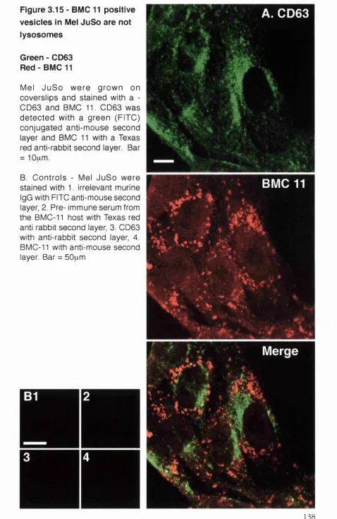
Figure 3.15 - BMC 11 positive vesicles in Mel JuSo are not lysosom es...138
Figure 3.16 - Dextran endocytosis by Mel JuSo in suspension reaches central MHC II positive a re a s ...140
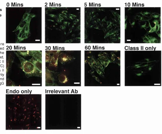
Figure 3.17 - Mel JuSo in suspension show poor definition of BMC 11 positive v esic le s... I4 l Figure 3.18 - Dextran endocytosis in situ reaches an MHC II positive vesicle after 20 m in s... 143
Figure 3.19 - Dextran endocytosis in situ does not cross a BMC 11 positive com partm ent more than 10 mins into the endocytic r o u te ... 144
Figure 3.20 - PMA activation of Mel JuSo does not alter BMC 11 distribution.... 146
Chapter 4 - C athepsin E exp ression in D endritic Cells
Figure 4.1 - Proposed relationship betw een monocytes, m acrophages and DCs derived from in vitro s tu d ie s ...151Figure 4.2 - Potential stragtegies for construction of a semi-quantitative PCR internal c o n tro l...156
Figure 4.3 - Generating an internal control for semi-quantitative PCR... 159
Figure 4.4 - Testing of the CE semi-quantitative PCR system ...160
Figure 4.5 - Phenotype of peripheral blood derived m o n o cy tes... 163
List o f Figures
Figure 4.8 - Peripheral blood derived DCs, but not peripheral blood monocytes, express the BMC 11 e p ito p e ...l67 Figure 4.9 - LPS treatm ent of DCs for 24 hours causes phenotypic changes
consistent with DC m atu ra tio n ...171 Figure 4.10 - LPS treatment of DCs for 24 hours causes phenotypic changes
consistent with DC m atu ra tio n ... 172 Figure 4.11 - LPS treatment of DCs has no effect on cathepsin E expression .... 173 Figure 4.12 - Staining of DCs with C E l.l reveals apparent cytoskeletal elements ... ...176 Figure 4.13 - Co-staining of DCs for C E l.l and the ER marker KDDD reveals a
partial o v erlap ... 177 Figure 4.14 - Dendritic cells are more efficient than lymphocytes at taking up
high m olecular weight d ex tran ... 179 Figure 4.15 - Labelled dextran bound to the dendritic cell surface at 0"C is
progressively internalised on w arm ing to 37°C... 180 Figure 4.16 - Labelled dextran moves into the dendritic cell as a c o h o r t 181 Figure 4.17 - Labelled dextran passes through a transferrin receptor positive
endosom e after 5 mins c h a s e ...184 Figure 4.18 - Labelled dextran accumulates in a CD63 positive lysosome from 30
m inutes of chase o n w a rd s... 185 Figure 4.19 - Labelled dextran passes through early and late MHC class II
containing com partm ents... 186 Figure 4.20 - Labelled dextran does not colocalise with C E l.l staining beyond 2
minutes c h a s e ... 189 Figure 4.21 - C E l.l staining overlaps dextran endocytosis at very early time
p o in ts ...190 Figure 4.22 - BMC 11 staining is diffuse and co-localises with dextran endocytosis at several time p o in ts ... 191 Figure 4.23 - BMC 11 staining shows no extensive colocalisation with C E l.l in
D C s... 192 Figure 4.24 - In LPS treated DCs transferrin receptor shows an altered
d istrib u tio n ... 195
Figure 4.25 - In LPS treated DCs there is decreased dextran delivery to CD63 positive co m p artm en ts... 196 Figure 4.26 - In LPS treated DCs there is decreased lysosomal MHC class II
retention, and increased surface MHC class I I ... 197 Figure 4.27 - LPS treatm ent of DCs has no effect on CE distribution relative to the
List of Tables
Chapter 2 - Materials & M ethods
Table 2.1 - Primary antibodies used in Western blotting... 108 Table 2.2 Antibodies used for cell surface p h en o ty p in g ... 108 Table 2.3 Antibodies used for intracellular im m unofluorescence...110
Chapter 4 - Cathepsin E in D endritic Cells
Table 4.1 Expression of C E l.l by m onocytes and peripheral blood derived DCs ... ... 168 Table 4.2 Expression of BMC 11 by monocytes and peripheral blood derived DCs
168
Table 4.3 Effect of 24 hours LPS m aturation on cathepsin E expression by
peripheral blood derived D C s...174
(3-hex 2-ME aa Ab(s) Ag(s) AP(s) APC(s) ATP ATTC BCR BFA BiP bp(s) BSA CB CD
CD(followed by number) CD-M6PR CD40L cDNA CE CPU CHO CI-M6PR CIITA CIIV Class I Class II CLIP CMV cTEC(s) CS Cys DC(s) DC-CKl DEPC DGE DM p-hexosaminidase 2-p-mercaptoethanol Amino acid(s)
Antibody (antibodies) Antigen(s)
Adaptor protein(s)
Antigen presenting cell(s) Adenosine tris phosphate
American Type Culture Collection B cell receptor
Brefeldin A
Immunoglobulin binding protein Base pair(s)
Bovine serum albumin Cathepsin B
Cathepsin D
Cluster of differentiation
Cation dependent-m annose-6-phosphate receptor CD40 ligand
Complementary deoxyribonucleic acid Cathepsin E
Colony forming unit
Chinese hamster ovary (cells)
Cation independent-m annose-6-phosphate receptor Class II transactivator
Class II containing vesicle
Major histocompatibility complex class I Major histocompatibility complex class II Class II invariant chain associated peptide Cytomegalovirus
Cortical thymic epithelial cell(s) Cathepsin S
Cysteine
Dendritic cell(s)
Dendritic cell chem okine 1 Diethylpropylcarbonate
Abbreviations
DMSO Dimethyl sulphoxide
DNA Deoxyribonucleic acid
dNTP(s) Deoxynucleotide tris phosphate(s) DO Human leucocyte antigen - DO
EBV Epstein-Barr virus
EDTA Ethylene diamine tetra-acetic acid
EE(s) Early endosome(s)
ELISA Enzyme linked im m unosorbent assay EMBL European molecular biology laboratory endo H Endoglycosidase H
ER Endoplasmic reticulum
Fc Fragment com plem ent binding
FcR Fragment com plem ent binding receptor
PCS Foetal calf serum
FITC Fluorescein isothiocyanate
fMLP Formyl methionine-leupeptin-phenylalanine
G418 Geneticin sulphate
GAP-DH Glyceraldehyde-3-phosphate dehydrogenase GFP Green fluorescent protein
GM-CSF Granulocyte/m acrophage-colony stimulating factor
GTP Guanosine tris-phosphate
HBSS Hank’s buffered saline solution
HEL Hen egg lysozyme
HIV Human immunodeficiency virus
HLA Human leucocyte antigen
HPLC High performance liquid chromatography
HRP Horseradish peroxidase
ICRF Imperial cancer research fund
IFN-y Interferon-y
Ig(s) Immunoglobulin(s)
li Invariant chain
II Interleukin
He Isoleucine
kDa Kilodalton(s)
Lamp-1 Lysosomal associated m em brane protein
LB Lauria Bertoni m edium
LC(s) Langerhans cell(s)
LE(s) Late endosome(s)
Leu Leupeptin
igp-A Lysosomal glycoprotein A
LIP(s) Leupeptin induced polypeptide(s)
LPS Lipo-polysaccharide
Lys Lysine
M-CSF Macrophage colony stimulating factor m6p r M annose-6-phosphate receptor
MCS Multiple cloning site
Met Methionine
MFI(s) Mean fluorescence intensity (intensities) MHC Major histocompatibility complex
MHC class I/MHC I Major histocompatibility complex class I MHC class II/MHC II Major histocompatibility complex class II
mig Membrane im munoglobulin
MIIC MHC class II containing com partment MLR Mixed leucocyte reaction
MR Mannose receptor
Mr Molecular weight
mRNA Messenger ribonucleic acid MTOC Microtubule organising centre
MW Molecular weight
OVA Ovalbumin
PBMC(s) Peripheral blood m ononuclear cell(s) PBS Phosphate buffered saline solution PCR Polymerase chain reaction
PDI Protein disulphide isomerase
PEG Poly-ethylene glycol
PI Propidium iodide
PI3K Phosphatidyl inositol 3 kinase
PMA 12-O-tetradecanoylphorbol-l 3-acetate PNS Post nuclear supernatant
Pro Proline
RER Rough endoplasmic reticulum
RNA Ribonucleic acid
RPMI Roswell park memorial institute (medium) RT-PCR Reverse transcriptase-polymerase chain reaction rtTA Reverse tetracycline transactivator
SDS Sodium dodecyl sulphate
Abbreviations
TCA Trichloroacetic acid
TEMED N, N, N’, N’-tetramethylethylenediamine
TfR Transferrin receptor
TGN trans-Go\gi network
T hl T helper 1
Th2 T helper 2
TNF-a Tumour necrosis factor-a
TRANCE TNF related activation induced cytokine tTA Tetracycline transactivator
TV T ubulo-vesicular
UTR Untranslated region
UV Ultra violet
WT Wild type
ZPPDK Benzyloxylcarbonyl-phenylalanylalanine- diazomethyl ketone
the atom, the nub being that they haven’t the foggiest as to w hat will h appen if they do. It may be all right. O n the other hand, it may not be all right. And pretty silly a chap w ould feel, no doubt, if, having split the atom, he suddenly found the house going up in smoke and himself torn limb from limb.’
C h a p t e r • O n
%
O
3
o K n
o
3
I n t r o d u c t i o n
chapter
Introduction
Dr. Ranee - Has the theory received m uch publicity?
Dr. Prentice - I do n ’t approve of scientists w ho publicise their theories.
Dr. Ranee - I must say I agree with you. I wish m ore scientists w ould keep their ideas to themselves.
What the Butler Saw, Joe Orton
1.1 General introduction
Central to the control of the adaptive immune response is the activation of T lymphocytes. In recent years workers asking the question of how this activation is regulated have come to examine in detail the nature of antigen presentation. In order for a T cell to recognise antigen via its T cell receptor it must first encounter that antigen, not in its native form, but following degradation within a second cell - the antigen presenting cell (APC).
A ntigen p re se n ta tio n occurs via tw o p athw ays, m ed iated by eith e r m ajor histocompatibility com plex (MHC) class I molecules, or by MHC class II molecules. MHC class I molecules generally present antigen derived from proteins synthesised within the APC itself. These proteins are degraded in the cytosol and then transported into the endoplasmic reticulum (ER) w here MHC I loading takes place. This process allows T cell surveillance of normal cellular proteins (to which the T cell arm of the immune system should be tolerant). MHC I also allows the detection of abnorm al proteins, such as those viral proteins generated w hen a virus hijacks the cellular protein synthesis m achinery for its ow n ends. More controversially MHC I may allow the detection of abnorm al cellular proteins associated with the developm ent of a tumour.
Introduction
processes which can subvert the very core of any cell, and all nucleated cells (except placental trophoblasts) express MHC class I. The T cell co-receptor for MHC I is the CD8 molecule. Recognition of an MHC class I/peptide complex by a CD8+ T cell usually activates killing mechanisms by the T cell to eliminate the infected/abnormal cell.
The MHC class I pathway does not (generally) allow detection of antigenic material from pathogens dwelling extracellularly. Such antigens are presented uia MHC class II molecules. Unlike MHC I, MHC II is expressed constitutively by only a few cells; principally B cells, macrophages and dendritic cells (DCs) - this group constituting the so called professional antigen presenting cells. MHC class II expression can be induced in many other cells by the cytokine interferon-y. The source of antigen in the MHC class II pathway is usually by uptake from the extracellular milieu. Most professional antigen presenting cells possess specialised mechanisms for the capture
A ntigen p re s e n ta tio n to T cells a t th e cell s u rfa c e
A ntigen c a p tu re a t cell s u rfa c e
A ntigen p a s s e s th ro u g h th e e n d o c y tic ro u te a n d is p ro g re ssiv e ly d e g r a d e d
ST,
MHC c la s s II is tra n s p o rte d from th e ER to e n d o s o m e s
MHC c la s s II s y n th e s is in th e ER c h a p e r o n e d by th e invariant ch ain
MHC is lo a d e d with fra g m e n ts of a n tig e n a n d tra n s p o rte d to th e cell s u rfa c e
Figure 1.1 - Overview of MHC ciass II antigen processing
of exogenous antigen. Once captured the antigen passes into the endocytic route of the cell and is degraded by poorly defined proteinase enzymes. The fragments of antigen generated encounter MHC class II within the endocytic pathway and associate with that molecule in its specialised peptide binding groove. From the endocytic pathw ay MHC class II is transported to the cell surface, w here it is presented to T cell scrutiny. An overview of this process is presented in figure 1.1.
The T cell co-receptor for MHC class II is CD4, and CD4+T cells, w hen activated, generally provide ‘help’ - either in the form of direct cell-cell signals, or mediated
via soluble protein messengers - cytokines. CD4+T cell help usually acts on B cells to allow the production of specific im m unoglobulin against an antigen, or allows the activation of inflammatory killing mechanisms by cells such as macrophages, or on dendritic cells to allow them to prime cytotoxic T cells.
In addition to T cell activation in the periphery, antigen presentation also controls the positive and negative selection of T cells in the thymus during T cell development. In this process T cells are selected to be restricted to the MHC haplotype expressed by an individual, and also deleted if they recognise thymically presented (and therefore presumably self) antigen. Surface MHC is essential for both these processes. From the description above the activation of a T cell would seem to be a relatively straightforward process. An APC merely has to capture and degrade an antigen and present the resulting MHC/peptide com plex to initiate a T cell response. However this is not the case. T cell activation requires two signals, both provided by the APC. The first signal is the M HC/peptide complex, the second signal (the costimulatory signal) derives from a variety of molecules on the surface of the APC. MHC stimulation of a T cell in the absence of a second signal can result in T cell unresponsiveness or anergy.
The two signal hypothesis of T cell activation led to a lot of interest in w hat controls the expression of the second co-stimulatory molecule. It was suggested that receptors of the innate immune system, tuned to recognise com m on foreign markers, could signal the presence of danger to the APC and activate a co-stimulatory phenotype. In a similar m anner the mediators and processes of inflammation could contribute to this immune amplification. Such theories may not explain all cases of T cell activation. However they served to re-focus attention on the antigen presenting cell as a central regulator of the immune response.
Introduction
The machinery of antigen processing itself does not appear to be fixed but is regulated. The nature of the machinery, the mechanisms by which antigen is captured and engulfed, the com partments of the endocytic route involved in antigen processing, the pH of the processing environment, the proteinases which degrade the antigen and the transport of loaded MHC to the cell surface, will all have a profound effect on the nature of the antigenic epitopes eventually encountered by T cells. For a m ore in depth textbook discussion of the points described in this general introduction see Janew ay and Travers (1997).
The detailed understanding of antigen processing is beginning to reveal a highly com plex process with many levels of possible, and indeed, probable control. Understanding this process will help us to understand the most fundam ental level at w hich the adaptive immune system is controlled, and can be m anipulated. The next section outlines the cellular biology of MHC class II antigen processing and presentation. In section 1.3 the enzymes involoved in these processes are discussed.
1.2 Cellular biology of antigen processing
1.2.1 Professional antigen presenting ceiis
1.2.1.1 B lymphocytes
B lymphocytes represent the cellular origin of antibody. All B cells produce antibody (immunoglobulin - Ig) of one specificity which is displayed on their cell surface in association with the B cell receptor. B lymphocytes present antigen with greatest efficiency if it binds their m em brane immunoglobulin (mig) (Rock et a l, 1984, Lanzavecchia, 1985 - see section 1.2.3.2). Following internalisation and processing this antigen is presented to T lymphocytes and, if recognised by a specific T cell receptor, help is obtained for production of that B cell's immunoglobulin (review ed in Clark and Ledbetter, 1994). It is essential for B cells to be able to present antigen in order to obtain help for Ig production, how ever the debate as to w hether B cells can activate naive T cells in vivo continues (Mamula and Janeway, 1993, Fuchs and Matzinger, 1993).
Early experiments in B cell depleted mice found that antigen presentation was abrogated in lymph nodes but not in spleen (Janeway et a l, 1987, Kurt-Jones et a l,
1988). Other data suggested that B cells may be able to prime naive T cells if they are first activated. Liu and Jane way found that costimulatory ability could be induced on B cells by activated T cells or the microbial cell wall component lipo-polysaccharide (LPS) (Liu and Jane way, 1991). This may be due to the induction by LPS of the potent costimulatory molecule B7 (Razi-Wolf et a l, 1992).
Experiments with both mice and chicken chimeras suggested that B cells could not prime T cells and that another cell type, possibly the dendritic cell, was required (Lassila e ta l, 1988, Ronchese and Hausmann, 1993). However B cells could efficiently activate memory T cells (Ronchese and Hausmann, 1993).
Introduction
that normal T cell priming could occur in the absence of B cells (Epstein et a l,
1995). However Constant e t a l found that responses to protein immunisation were severely diminished in B cell deficient mice (Constant et a l, 1995).
Such differences may be antigen specific (Constant et a l, 1995). The Hepatitis B virus core antigen is a T independent antigen which has the capacity to partially activate B cells independently of T cell help. Administration of this antigen to mice resulted in its rapid uptake and presentation by specific splenic B cells, w hich were present at high frequency in unprim ed mice (Milich e ta l, 1997). The binding of the Ag to the B cell surface caused the expression of the B7-2 costimulatory molecule and enabled these B cells to prime naive T cells. If the B cell priming was blocked, and priming perform ed by the remaining spleen APCs (macrophages and dendritic cells), the isotype profile of the antibody produced was shifted from IgG2a/2b to IgGl (Milich e ta l, 1997). Thus, for some antigens at least, activation of the B cell by the antigen can enable it to prime a naive T cell.
The effect of B cell antigen presentation may be to modulate the T cell response to an antigen. Stockinger et a l found that dendritic cells w ere necessary for T cell priming. Dendritic cell and m acrophage mixtures produced a T hl cytokine profile (cytokines giving help to cell m ediated immunity), how ever priming by DCs and B cells resulted in a loss of interferon-y production and the production instead of interleukin (IL)-4 (Stockinger et a l, 1996). This effect was blocked by antibodies to CD40, a molecule critical in the communication of B an d T cells (Clark and Ledbetter, 1994). Thus B cell antigen presentation may principally act to modulate, rather than initiate, the T cell response.
1.2.1.2 Macrophages
The m acrophage was the cell type in which MHC class II antigen processing and presentation w ere originally described (Unanue, 1984). Macrophages have the ability to capture much m ore antigen via fluid phase pinocytosis than B cells (Steinman and Cohn, 1972b, Chesnut e ta l, 1982 - see section 1.2.3.4). Macrophages also have an extensive phagocytic capacity, which delivers particulate antigen to lysosomal compartments (Kielian and Cohn, 1980). Antigen captured in this m anner can be subsequently presented to T cells (Watts, 1997a, Harding et a l, 1991b, Harding et a l, 1991a, Pfeifer et a l, 1992).
However, m acrophages also have an extensive role in the innate im m une system. They can capture antigen via Fc receptors and receptors for com plem ent fragments
(C3b and C3bi receptors), leading to phagocytosis (as originally observed by Elie MetchnikofO and, in the case of Fc receptor engagem ent - the release of toxic oxygen species (Johnson et a l, 1976, Wright and Silverstein, 1983).
Moreover, resting m acrophages do not express levels of MHC class II as high as those of B cells, but require treatment with IFN-yto induce such expression (Gonwa
et a l, 1986). As in B cells, macrophage costimulatory molecule expression can be induced by zymosan, a microbial product recognised by m acrophages (Liu and Jane way, 1991). Resting macrophages alone may require the presence of dendritic cells before they can effectively prime T cells (Stockinger et a l, 1996). Thus, like the B cell, the role of the macrophage in priming helper T cell responses remains ambiguous.
1.2.1.3 Dendritic ceils
The primary function of B cells during an immune response is the production of immunoglobulin, and their antigen presenting capability could be regarded solely as a m eans of obtaining T cell help for this purpose. Likewise the antigen presenting function of the macrophage is probably secondary to its role in phagocytic and inflammatory processes. In contrast to these two cell types the dendritic cell (DC) is a cell w hose raison d ’être is the capture and presentation of antigen to T cells (see reviews by Steinman, 1991, Banchereau and Steinman, 1998).
The dendritic cell was originally identified as a minor population of nucleated spleen cells (1-1.6%). The cell displayed a large cytoplasm arranged in extensive dendrites or pseudopodia - hence the term dendritic cell was coined (Steinman and Cohn, 1973). The dendritic cell displayed extensive motility, with extension and retraction of pseudopodia. The cells were detected in white pulp of spleen and in lymph nodes, but could not be found in thymus, liver or intestinal tissue (Steinman and Cohn, 1973).
Introduction
Figure 1.2 - Stages of DC maturation
A. Peripheral antigen capturing DC
Low levels of cell s u rfa c e MHC a n d co stim u lato ry m o le c u le s
High internal MHO II
High Ag cap tu rin g activity
B. Migratory DC
Lym phatic or blood v e s s e l —
Intracellular antigen/M H O II co m p lex a s s e m b ly
0
0
ê
C. Mature, Ag presenting DC In secondary lym phoid tissue
B cell in te ra c tio n s
E x ten siv e d e n d rite s
Intracellular a n tig e n p ro teo ly sis
T cell clu sterin g /A g p re s e n ta tio n to T cells
Little intracellular MHC II
High lev els of cell s u rfa c e MHC II a n d co stim u lato ry m o le c u le s
mouse spleen (Steinman etal., 1975, Steinman etal., 1979). This purification process allowed detection of surface MHC II molecules on the DC, how ever the function of these cells remained unclear.
The function of dendritic cells becam e clearer w hen it was discovered that they were far more potent than macrophages or B cells at stimulating the mixed leucocyte reaction (MLR), and could also stimulate a syngeneic MLR (Steinman and Witmer, 1978, Nussenzweig and Steinman, 1980a, Nussenzweig and Steinman, 1980b, Klinkert
e t a l , 1980).
1.2.1.3 I M aturation stages o f dendritic cells
Since these early descriptions of DCs the last 25 years has seen an explosion in the field of DC biology, which has led to the elucidation of a clearly defined m odel for DC function in the induction and control of immunity (Banchereau and Steinman,
1998, Ibrahim et a l, 1995 - see figure 1.2).
DCs in the periphery (for example Langerhans cells (LCs) in the epiderm is and interstitial DCs in internal organs) can be regarded as ‘sentinels’ of the immune system (Ibrahim et a l, 1995). These cells show high levels of antigen capturing activity (discussed in section 1.2.3.3) and high levels of MHC II and invariant chain synthesis (Sallusto and Lanzavecchia, 1994, Sallusto et a l, 1995, Winzler et a l,
1997). Thus immature DCs are in a position to monitor the sites of antigen entry into the body and have the ability to capture antigen at these sites.
A num ber of stimuli have been reported to cause migration of immature DCs from the periphery to secondary lymphoid organs. Principally migration is to draining lymph nodes via lymphatic vessels, but it can also be to the spleen via blood-borne migration (Austyn, 1996). Dendritic cells arriving in spleen from the blood pass initially to the red pulp, and after 24 hours are in the T cell areas of the w hite pulp (Austyn e ta l, 1988). Langerhans cells can be induced to migrate out of the skin by placing skin explants in culture; after 3 days LCs can be observed as ‘cords’ of cells passing through the dermal lymphatics (Larsen et a l, 1990). The bacterial cell wall com ponent LPS causes massive depletion of interstitial and epithelial DCs w hen administered systematically, and this effect was dependent on tum our necrosis factor (TNF)-a production (Roake et a l, 1995). LPS also causes migration of DCs into the T cell areas of spleen (De Smedt et a l, 1996).
Introduction
to contact sensitisers (van Wilsem e ta l, 1994). Migration involves a loss of cytoskeletal organisation and an increase in general motility as an early response to m igration/ maturation stimuli (Winzler et a l, 1997).
Many of the stimuli which cause DC migration in vivo can be used in vitro to induce DC maturation. In the case of Langerhans cells, placing them into culture alone results in a dramatic reduction in their ability to process and present exogenously supplied protein antigen, but an increase in their ability to present peptide (Schuler and Steinman, 1985, Romani et a l, 1989, Pure et a l, 1990, Inaba et a l, 1990). Culture of DCs is accom panied by a loss of the high levels of class II biosynthesis seen in immature DCs and an increase in MHC class II half-life resulting in maintained high surface MHC expression (Kampgen et a l, 1991).
In model DC culture systems a similar maturation process is induced by the addition of TNF-a, LPS or CD40 ligand (CD40L), this results in an increase in surface MHC and adhesion/costim ulatory molecule expression, but a reduction in the ability to present exogenous antigen (Sallusto and Lanzavecchia, 1994, Sallusto et a l, 1995). Thus it seems that migration from the periphery is induced by inflammatory mediators or bacterial products and is accom panied by a maturation of the DC from an antigen capturing to an antigen presenting phenotype.
1.2.1.3-2 Dendritic cell/T cell interactions
DC migration has been observed in vivo; ovalbumin pulsed DCs w ere adm inistered to mice containing transgenic T cells specific for an ovalbumin epitope. DC migration to the draining lymph node was observed, and on arrival clusters form ed betw een DCs and Ag specific T cells in the paracortical areas. These DC/T cell clusters contained proliferating T cells and w ere greatest in num ber 24h after administration of pulsed DCs. After 48h there was a decrease in the num ber of DCs rem aining in the lymph node, this DC loss appeared to be m ediated by the ovalbum in specific T cells (Ingulli e ta l, 1997). This elegant demonstration has provided direct confirmation of many aspects of the m odel of DC/T cell interaction.
Clusters of DC and T cells are the sites of T cell proliferation in the in vitro MLR (Flechner e ta l, 1988). DCs, unlike other APC types, can form antigen independent clusters with T cells (Inaba and Steinman, 1986) possibly indicating that naive T cells ‘scan’ DCs for their specific M HC/peptide complex during the initiation of an immune response. Evidence in favour of such a model is that DCs secrete a chemokine (DC-CKl) that preferentially attracts CD45RA^ naive T cells (Adema et a l, 1997). Detailed morphological analysis of the DC/T cell cluster revealed extensive mem brane
extensions from the DC which spread to contact the membranes of the surrounding T cells. Staining of the surface for MHC class II with immunogold revealed clusters or lines of MHC class II, possibly representing a cytoskeletally controlled organisation of surface class II (Setum et a l, 1993).
The DC/T cell cluster can also include B cells, these clusters w ere found to be necessary for the generation of antibody secreting cells in vitro, and the B cell in question had to express the MHC haplotype to which the clustering T cell was restricted (Inaba et a l, 1984). DCs are also im portant in B cell stimulation in such clusters. In clusters of DCs, B cells and CD40L expressing fibroblasts, DCs prom oted the class switching of memory B cells to IgG and IgA. In the presence of IL-2 DCs greatly enhanced the production of IgM by naive B cells (Dubois et a l, 1997). The mature DCs found in lymph nodes express high levels of the co-stimulatory molecule B7-2 (CD86), and blockade of this molecule leads to extensive inhibition of T cell proliferation (Inaba et a l, 1994, Caux et a l, 1994). High levels of co stimulatory molecule expression make DCs potent stimulators of the T cell response. Indeed the DC may be the only cell type w hich can activate naive T cells in vivo ~
a study w here protein was administered in adjuvant show ed that DCs rather than B cells presented the injected antigen to T cells (Guery et a l, 1996). DCs are also com petent at priming CD8+ cytotoxic T cell responses (Porgador and Gilboa, 1995).
Interleukin 12 is produced by mature DCs during T cell stimulation, this cytokine leads to the generation of a T hl type response and is a hallmark of DC T cell priming. Administration of a Toxoplasma gondii antigen in mice caused migration of DCs from the periphery into the T cell areas of spleen w here they secreted IL-12 (Reis e Sousa e ta l, 1997).
DC IL-12 production can also be stimulated by CD40L (Koch et a l, 1996, Celia et a l, 1996), a molecule expressed by activated T cells. This is evidence of a tw o w ay interaction betw een DC and T cell. DC activation of T cells could lead to T cell CD40L expression, which in turn stimulates IL-12 production by DCs. The IL-12 then acts on the T cell to produce a T h l response. This process can be dow n regulated by IL-10 and IL-4 (Koch e ta l, 1996). CD40L can also induce DC maturation
in vitro (Sallusto and Lanzavecchia, 1994), and may be important in regulating the passage of loaded MHC II to the cell surface in DCs (Pierre et a l, 1997).
Introduction
DC survival by upregulation of the Bcl-X^ gene product, this enhanced survival leads to an enhanced T cell response in the mixed leucocyte reaction (Wong et a l,
1997).
1 .2 .1 3 3 Ontogeny o f dendritic cells
Despite the intensive research into DC phenotype there is, as yet, no single definitive cell surface marker of the DC. This partially contributes to the uncertain origin of the DC. There has been a great deal of w ork on the ontogeny of DCs, which will be only briefly discussed here.
DCs can be produced from culture of CD34^ bone marrow stem cells, possibly via
a separate DC colony forming unit (CFU-DC) (Young e ta l, 1995, Caux e ta l, 1996). Most DCs are assumed to derive from the myeloid lineage, due to their reliance on the myeloid cytokine granulocyte macrophage-colony stimulating factor (GM-CSF) (Inaba e ta l, 1992, Young e ta l, 1995, Caux e ta l, 1996). In the mouse GM-CSF can prom ote developm ent of DCs, m acrophages and granulocytes side by side from class II negative bone marrow cells (Inaba et a l, 1993a), possibly indicating a com m on origin of these cell types. Indeed conversion betw een these cell types can occur. In vitro preparations of DCs can be grown from hum an blood monocytes under the influence of GM-CSF and IL-4, which suppresses monocyte outgrowth (Sallusto and Lanzavecchia, 1994, Romani e ta l, 1994, Pickl e ta l, 1996). These cells are capable of interconversion with cells of a macrophage phenotype until the final stage of DC maturity, indicating their close relationship with the macrophage (Palucka
et a l, 1998). The functional importance of a monocyte derived pathw ay as the origin of DCs in vivo is unclear.
DCs in different sites, however, may derive from different lineages and DCs themselves may not represent a single cell type of one origin. In vitro Caux et al. found that CD34^ stem cells could develop via a CD14^ or a CDla"" pathway (Caux e ta l, 1996). The final cell types w ere functionally and phenotypically very similar (with neither being CD 14 positive). However, cells derived from the CD la route show ed Birbeck granules and other markers of Langerhans cell phenotype, whilst the CD14 derived DCs seem ed to more closely resemble interstitial tissue DCs. Under the influence of m acrophage colony stimulating factor (M-CSF) the CD14, but not the CDla, derived cells could develop a macrophage like phenotype (Caux e ta l, 1996). The expression of the cutaneous lymphocyte antigen by CD34^ derived DC progenitors in hum an peripheral blood may also indicate w hether a Langerhans cell phenotype will ultimately be developed (Strunk et a l, 1997)
There is also evidence for a lymphocytic origin for some DCs, which can be derived from CD4^ progenitor cells, and are not dependent on GM-CSF for survival (Saunders
et a l, 1996, G rouard et a l, 1997). These cultures w ere dependent on a mixture of cytokines, particularly IL-3 and IL-7. Lymphoid precursors may be im portant in the origin of thymic DCs (Ardavin et a l, 1993), and in DCs responsible for T cell stimulation in germinal centres (Grouard et a l, 1996).
1.2.1 .3 -4 DCs a n d Tcell selection
In vitro DCs are capable of deleting double negative thymocytes which bear a T cell receptor (TCR) against the epitope they are presenting (Zal et a l, 1994, Tanaka et a l, 1993). In mice w here only DCs expressed the murine MHC II I-E'^, I-E'^ staining in the thymus was restricted to the thymic medulla and cortico-medullary junction. These thymic DCs w ere sufficient for the negative selection of I-E'^ restricted T cells, but w ere not involved in positive selection (Brocker et a l, 1997).
The m echanism for DC negative regulation of T cells remains unclear. A lymphoid related DC expressing CD8a has been show n to mediate killing of CD4+ T cells via
the Fas/Fas ligand mechanism, and to inhibit CD8+ T cell proliferation by inhibiting T cell IL-2 production (Suss and Shortman, 1996). These may be m echanisms by w hich the DC can act to maintain peripheral T cell tolerance.
Introduction
1.2.2 Synthesis and export of MHC class II
Both MHC class I and class II are synthesised in the endoplasmic reticulum, which is also the site of peptide binding by MHC class I. For MHC class II, with a role in presentation of exogenous antigens, it is important that it does not bind antigenic peptide in the ER, but instead passes to the endocytic route w here it can complex with peptides derived from captured antigen. Protection of the MHC class II peptide binding groove and targeting to the endocytic route are both provided by an MHC class II associated protein know n as the invariant chain (li). Invariant chain also functions to chaperone MHC class II assembly in the ER (for reviews see Cresswell
e t a l , 1990, Cresswell, 1996).
1.2.2.1 Structure of MHC class II
MHC class II is a heterodimer of a and (3 chains of molecular weight 32 and 29 kDa respectively. Each chain consists of a membrane proximal and pp and a membrane distal (ttj and pp domain (Brown et a l, 1993). The two mem brane distal domains com bine to form the peptide binding groove of the molecule. The sides of the groove consist of two a-helices and the floor of eight p-pleated sheets, each chain contributes one helix and four p-sheets (Brown et a l, 1993). MHC II interacts with the T cell CD4 via its p^ dom ain (Konig et a l, 1992, Cammarota et a l, 1992). In overall structure class II closely resembles class I MHC (Brown et a l, 1993), however, unlike class I, the peptide binding site of class II is open at both ends. This leaves class II able to bind peptides of (theoretically) any length, most being 13-25 am ino acids (aa) in length (Rudensky et a l, 1991a, Chicz et a l, 1992).
The binding site of HLA-DRl contains a conserved non-polar pocket which allows peptides to bind via an anchor residue (Brown et a l, 1993). Peptides bind to class II in an extended conformation with extensive contact betw een the peptide and the groove’s conserved residues by H-bonding along the length of the peptide (Brown
et a l, 1993, Stern et a l, 1994). The peptide in the groove lies partially ‘flush’ with the MHC m olecule e.g. - in the case of the influenza peptide HA306-318, 35% is exposed to solvent. This means that the T cell receptor will see peptide and MHC residues together (Stern et a l, 1994). The groove is lined by a series of peptide binding pockets and these are the sites of the greatest polymorphism betw een class II haplotypes, indicating that these pockets provide the differing peptide specificities of different class II haplotypes (Stern et a l, 1994).
MHC class II dimers before peptide binding are sensitive to dissociation on sodium dodecyl sulphate-poly acrylamide gel electrophoresis (SDS-PAGE); peptide binding confers resistance to this dissociation in some haplotypes (Germain and Hendrix, 1991, Sadegh-Nasseri and Germain, 1992). Peptide acquisition, as m easured by SDS stability, occurs after passage through the Golgi apparatus, probably in an acidic endosomal organelle (Tulp et al., 1994, Germain and Hendrix, 1991, Amigorena et a l, 1994 - see section 1.2.4). The structure of MHC II undergoes slight changes at the acidic pH associated with such endosomal compartments and these changes correlate with a faster on and off rates for peptide binding (Sadegh Nasseri and Germain, 1991, Sadegh-Nasseri and Germain, 1992, Runnels e ta l, 1996, Boniface et a l, 1996). The enhanced binding of peptide to MHC II at acidic pH may be due to the presence of conserved residues in the P6 pocket of I-E in the m ouse and HLA- DR in the human. The protonation of these residues may facilitate peptide binding (Fremont e ta l, 1996). Such changes in structure of MHC class II after synthesis may partially limit peptide binding to the endosomal pathway of the cell (see also sections 1.2.2.4.1 and 1.3.1.3).
1.2.2.2 MHC class ll/invariant chain complex assembly
The nucleotide sequence of the invariant chain in hum ans predicts a type II transm em brane glycoprotein of 2l6 aa in length (O ’Sullivan et a l, 1987, Strubin et a l, 1986). The commonest form of the invariant chain in humans has an approximate molecular weight of 31 kDa and is know n as p31 (p33 in some publications). Use of an upstream alternate start site for translation generates a larger p33 (p35 in some publications) form. Alternative splicing can give rise to a rarer p 4 l form in humans, by the insertion of a 64 aa stretch in the lumenal domain. Use of the alternative transcription start site in the p 4 l form gives the p43 form, which shares the extra I 6 N-terminal aa’s of p33 (O ’Sullivan et a l, 1987, Strubin et a l, 1986, reviewed in Cresswell, 1994, see figure 1.3).
In cells deficient in li, MHC class II ap dimers can still assemble at decreased levels (Lotteau e ta l, 1990, Schaiff e ta l, 1991), and the same is true for APCs derived from li deficient mice (Viville et a l, 1993, Bikoff et a l, 1993). However these dimers show very poor levels of exit from the ER and often show structural differences to wild type class II molecules. In one li deficient m ouse strain it was reported that a and p chains becam e aggregated as misfolded proteins in the ER and there was no significant passage of ap dimers beyond the Golgi apparatus (Elliott e ta l, 1994). In the absence of MHC class II most li is retained in the ER as a trimer (Marks et a l,
Introduction
COOH
280
COOH
^ 2 1 6
G l y -
Gly-Gly
Gly
Lumen
120
114
CLIP; 80-104
Gly
Gly
:
p41, 64 aa insert
120
114
CLIP; 80-104
56
Membrane
56
p33
30
Cytosol
0
-16
p43
30
0
-16
NH2
p31/33
NH2
p41/43
Figure 1.3 - Invariant chain
1992), and is dependent on residues 163 to 183 of li (p31 numbering - Bijlmakers et al., 1994). Trimers can contain any combination of the li isoforms (Roche et at.,
1991). li can bind single a and p subunits and ap dimers (Teyton et a l, 1990). The li trimer forms a framework for ap dimer assembly, and following li trimer formation three ap class II dimers are sequentially added to the li trimer (Roche et at., 1991, Lamb and Cresswell, 1992), ultimately forming a nonam er of three li chains and three class II dimers (Roche et a l, 1991).
The exact details of the assembly process remains unclear, although it seems that a and p subunits bind within a very short time of each other (Nijenhuis and Neefjes,
1994). Within 10 minutes of synthesis li, ap dimer formation is already occurring. No free ap dimers are detected, suggesting that they do not form a (significant) intermediate in the process (Lamb and Cresswell, 1992). The Ii/2ap and Ii/3(%P complexes are maximal 2h after translation begins, by 4h the number of these complexes is decreasing and free, mature ap dimers are detected (Lamb and Cresswell, 1992).
Calnexin, a ubiquitous transmembrane ER protein, also appears to play a role in nonam er formation (Schreiber e ta l, 1994, Anderson and Cresswell, 1994). Calnexin associates first with the free a, p and li units and remains with the partially assembled a/p /Ii complex until the last ap dimer is added (Anderson and Cresswell, 1994). Nonamer subunits associated with calnexin remain in the ER and thus the role of calnexin may be to stabilise the assembling nonamer subunits and retain the complex in the ER until complete. Calnexin detaches on formation of the a/p/Ii nonamer and is not found associated with free a p dimers (Anderson and Cresswell, 1994). A
Invariant ch ain
C aln ex in \
\
MHC c la s s II a ch ain MHC c la s s II p ch ain
4
^
MHC ll/li n o n a m e r
V
Introduction
study on unglycosylated li, which does not associate with calnexin, show ed that calnexin was not essential for nonam er formation, but acts to retain free elements of the complex in the ER and slow degradation of li (Romagnoli and Germain, 1995). Invariant chain/class II assembly is illustrated in figure 1.4.
Although, as discussed above, MHC II dimers can still exit the ER in the absence of li, the dimers formed do show conformational alterations as detected by panels of monoclonal antibodies (Peterson and Miller, 1990, Anderson and Miller, 1992). MHC class II molecules from li negative cells show altered patterns of glycosylation following Golgi transport (Schaiff et a l, 1991, Anderson and Miller, 1992). In the absence of li, MHC subunits associate with immunoglobulin binding protein (BiP) - an ER chaperone (Nijenhuis and Neefjes, 1994). Thus li does seem to play an important chaperone role in the assembly and post-translational processing of normal MHC class II molecules, although this effect may be more important for some MHC II haplotypes than others (Viville et a l, 1993).
1.2.2.3 MHC/invariant chain export to the endocytic route
1.2.2.3 I Invariant chain deficient cells show defective presentation o f exogenous protein antigens
In the majority of reports describing cells deficient in li, presentation of intact exogenous protein antigens by these cells is abrogated, but the presentation of peptide fragments is unaffected or even enhanced (Stockinger et a l, 1989, Bikoff, 1992, Bikoff et a l, 1993, Viville et a l, 1993). Coupled with this loss of protein presentation, is an abnormal cellular localisation of MHC class II. Class II is often largely retained in the ER and Golgi in li negative cells (Lamb et a l, 1991, Bikoff et a l, 1993) whilst that which does reach the cell surface is usually unstable on SDS gel electrophoresis (Peterson and Miller, 1990, Anderson and Miller, 1992, Bikoff et a l, 1993), indicating a lack of normal peptide loading.
O n reconstitution of li expression in these cell types, protein Ag presentation was restored (Stockinger e ta l, 1989, Nadimi e ta l, 1991). This presentation was sensitive to the drug chloroquine, w hich disrupts endosomal function (Stockinger et a l,
1989). Neefjes et al. established that on leaving the Golgi apparatus MHC class II was delayed in reaching the cell surface for l-2h by traversing the endocytic route (Neefjes et a l, 1990). Thus it seem ed likely that li targets MHC class II to the endocytic route w here it can encounter and bind exogenous antigen fragments.
1.2.23-2 Invariant chain contains signals targeting MHC class II to the endocytic route
Co-transfection of the p31 isoform of li causes redistribution of MHC class II from the ER into endocytic structures, and facilitates transport of ap dimers to the cell surface (Lotteau et al., 1990, Lamb et al., 1991). The transport of mismatched ap dimers, which do not normally reach the cell surface, is also increased by li expression (Layet and Germain, 1991). Thus, whilst li may not be obligatory for class II transport out of the ER and Golgi, it is at the very least an important accessory molecule in this process. The nature of the endosom e accessed by li is discussed in detail in section 1.2.4.
li can reach endosomes alone or with class 11 (Bakke and Dobberstein, 1990, Lotteau
et al., 1990) and this ability is lost by deletion of the N-terminus cytoplasmic tail of li, which results in li expression at the cell surface (Bakke and Dobberstein, 1990, Lotteau e ta l, 1990, Roche e ta l, 1992, Anderson e ta l, 1993). Two targeting signals have been identified in the N-terminus of li; one in residues 1-11, of which Leu, and
A ntigen
D irection of e n d o s o m a l traffic
0
E n d o s o m e
A ntigen p ro teo ly sis
a p p a r a tu s
li p ro teo ly sis
MHC/li n o n a m e r