OncoTargets and Therapy
Dove
press
O r i g i n a l r e s e a r c h
open access to scientific and medical research
Open access Full Text article
Potential role of
S
-adenosylmethionine in
osteosarcoma development
hui shi1,2,*
Wei-dong Mu1,*
Bing Zhang3
Tao Meng2
shou-tao Zhang2
Dong-sheng Zhou1
1Department of Traumatic
Orthopaedics, shandong Provincial hospital affiliated to shandong University, Jinan, 2Department of
Bone and Joint surgery, 3Department
of Urology surgery, Binzhou Medical University hospital, Binzhou, shandong, People’s republic of china
*These authors contributed equally to this work
Abstract: The metastatic form of osteosarcoma is a life threatening one since it metastasizes to the lungs. The major cause of metastatic osteosarcoma is hypomethylation of numerous genes that undergo overexpression to enable the progression of the disease. In the present study,
S-adenosylmethionine (SAM), a predominant methyl donor, was administered to find out its
effects on osteosarcoma progression. As evidence of tumor suppression, the SAM-treated mouse tissue was analyzed histologically, which exemplifies the control that SAM has over abnormal cell proliferation, especially on primary osteosarcoma, but it lacks positive effects on metastatic osteosarcoma. At the molecular level, the successful inhibition of primary osteosarcoma was found to be associated with a lower expression of Sox2, a protein highly expressed in osteosarcoma stem cells, along with an upregulated expression of TCTP. The data suggest that the administration of SAM has a positive role in treating primary osteosarcoma, but it has no role in suppressing metastatic osteosarcoma. The decreased expression of Sox2 together with upregulation of TCTP following SAM administration indicates that SAM has a control over primary osteosarcoma.
Keywords: TCTP, S-adenosylmethionine, K7M2 cells, Sox2, BALB/c mouse
Introduction
Osteosarcoma is the most common aggressive form of bone tumor that occurs in children, and adults 50 years.1 It occurs due to abnormal osteoid production and
differentiation along with genomic instability. The progression of osteosarcoma is associated with numerous genes with diverse genomic alterations.2 Previously,
surgi-cal resection therapy was practiced in osteosarcoma patients, which resulted in poor prognosis. However, prominent chemotherapy was developed during the 1980s, which increased the 5-year survival rate of osteosarcoma patients to 50%–80%.3,4 Since then,
there has been no significant progress in therapeutic modalities as well as in prognosis in patients suffering with osteosarcoma.5 Especially, the patients with the metastatic
form of osteosarcoma have only a 10%–30% longer survival rate,6 and others exhibit
less response. In order to improve the survival rate of patients, a new therapeutic target should be put forward to investigate the antimetastatic activity, or an effective drug must be required to impede the progression of osteosarcoma to metastatic stage.
Besides many reasons like deletion, translocation, amplification, and other muta-tions that contribute to tumor development,7 epigenetic changes have also paved the
way for the development of many cancers, including osteosarcoma.8–10 The major
epigenetic changes consist of DNA methylation, histone modification, and chromo-some remodeling.11 S-adenosylmethionine (SAM), an important molecule in
bio-chemistry that donates its methyl group for many biochemical reactions,12 also acts
as a precursor for forming higher polyamines. The polyamine helps to condense and
correspondence: Dong-sheng Zhou Department of Traumatic Orthopaedics,
Shandong Provincial Hospital Affiliated
to shandong University, number 324, Wuweiqi road, Jinan, shandong 250021, People’s republic of china
Tel +86 531 6877 6382 Fax +86 531 8793 3120 email [email protected]
Journal name: OncoTargets and Therapy Article Designation: Original Research Year: 2016
Volume: 9
Running head verso: Shi et al
Running head recto: Role of S-adenosylmethionine in osteosarcoma development DOI: http://dx.doi.org/10.2147/OTT.S101408
OncoTargets and Therapy downloaded from https://www.dovepress.com/ by 118.70.13.36 on 25-Aug-2020
For personal use only.
Number of times this article has been viewed
This article was published in the following Dove Press journal: OncoTargets and Therapy
Dovepress
shi et al
stabilize DNA that controls the normal function of cells. It has been reported that SAM has anticancer properties and it is used as a therapeutic agent to treat cancer.13,14 The role of
methylation especially in the promoter region was thought to repress gene expression based on the specific cell type15
and in some cases methylation-mediated gene expression, but still the more precise role of methylation in gene expression remains unclear. Similarly, there is no report that associates SAM with osteosarcoma stem cells.
TCTP is an evolutionally conserved protein from yeast to human,16–19 and has a vital role in cell cycle,20,21 apoptosis,22
cytoskeleton remodeling,21 protein synthesis,23 immune
response,17 development of normal body growth and
devel-opment of the organisms,24 and cancer.22 In addition, it was
reported that TCTP expression was elevated in cancer tissues and also during the regeneration of liver;25 besides the
pro-tein has a major role in tumor reversion.22–26 Hence, TCTP
is considered as a target for cancer therapy.
In this study, we investigated the effects of exogenous SAM on cancer stem cells of the osteosarcoma mouse model. We analyzed the progression after SAM administration and we studied SAM administration counteraction using Sox2 and TCTP that are linked to osteosarcoma stem cells.
Materials and methods
experimental animals with osteosarcoma
The animal use and care, along with the protocol carried out in this study, was approved by the institutional animal ethical committee at Binzhou Medical University Hospital. For developing an osteosarcoma mouse model, aggressive osteosarcoma-forming K7M2 cells were used.27
Five-month-old male BALB/c mice were injected with K7M2 cells (106
cells/20 µL) to establish primary and metastatic forms of tumor. Following injection, one set of mice was sacrificed on the third week to obtain primary tumor and another set was allowed up to 5 weeks to obtain the metastatic form of osteo-sarcoma. The mice were subjected to digital radiography to confirm tumor progression before sacrifice. For determining the effects of SAM on tumor development, the mice were administered SAM (100 mg/kg/day) by oral gavage from the 15th day of injection of K7M2 cells.
immunohistochemistry
For immunohistochemistry, the dissected tissues were initially fixed in 10% formalin solution and processed to get paraffin-embedded tissue. The microtome was set at 6 µm, and the sections obtained were fixed on a microscopic glass slide. Following deparaffinization with xylene, they were hydrated
with water. To block the endogenous peroxidase activity, the sections were immersed in freshly prepared 10% H2O2 and 10% methanol in 1X phosphate-buffered saline (PBS) for 20 minutes. After washing with 1XPBS, the sections were treated with 0.1% trypsin in 0.1% CaCl2 at 37°C for 5 minutes. In order to facilitate binding of specific antibodies, the sections were incubated with the primary antibody overnight at 4°C followed by the addition of a suitable secondary antibody for 30 minutes at room temperature. After washing the tissue sections, the slides were developed using a DAB (diaminobenzidine) kit as a chromogen for detecting signals. For immunohistochemistry experiments, the following primary antibodies were used: anti-Sox2 antibody (ab97959) and anti-TCTP antibody (ab37506) from Abcam (Cambridge, UK). Similarly, the secondary antibody Goat Anti-Rabbit IgG H&L (HRP) (ab6721) from Abcam was used to detect the signals.
Western blot analysis
Cell lysates were prepared from normal tissue as well as from the primary and metastatic forms of osteosarcoma tissue as per the protocol mentioned in the literature.28 The
proteins were isolated and resolved in 10% SDS-PAGE gel. They were then transferred to the membrane and incubated with the primary antibody (anti-Sox2 antibody [ab97959] or anti-TCTP antibody [ab37506]) at a dilution ratio of 1:500. The unbound primary antibody was washed out and further incubated with the secondary antibody (Goat Anti-Rabbit IgG H&L [HRP] [ab6721]) at a dilution ratio of 1:5,000. Later, the signals were visualized with the DAB kit.
Results
Positive effects of saM on the
osteosarcoma mouse model
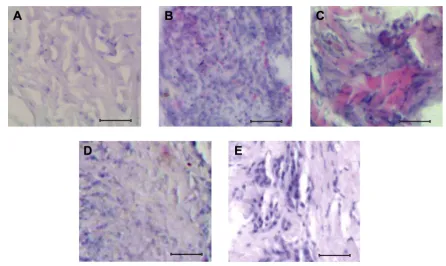
In the present study, the mouse model of osteosarcoma was successfully initiated by injecting highly tumorigenic and metastatic osteosarcoma-forming K7M2 cells. The experimen-tal mice responded well to injected K7M2 cells, which were examined through histological observation by comparing with the control mice (Figure 1A–C). Following injection, the mice were recognized with primary tumors, which were histologi-cally visualized on the third week after injection of K7M2 cells (Figure 1B). Similarly, the metastatic condition of osteosar-coma was achieved on the fifth week after injection of K7M2 cells (Figure 1C). When compared with the control, which had an even arrangement of cells (Figure 1A), the primary and metastatic osteosarcoma cells were large and deeply stained together with a disorderly arrangement of cells (Figure 1B and C). The unique feature of the metastatic form (Figure 1C)
OncoTargets and Therapy downloaded from https://www.dovepress.com/ by 118.70.13.36 on 25-Aug-2020
Dovepress role of S-adenosylmethionine in osteosarcoma development
is that it has an enlarged proliferative mass of cells with an irregular pattern of tissue arrangement compared to primary osteosarcoma (Figure 1B). Also, some of the cells were dis-placed to such an extent that the gaps between the clumps of cells were visible (Figure 1C). Upon SAM administration as described in the “Materials and methods” section, there was a remarkable improvement with reduction of proliferative cells, which is evident from the histological observation of primary osteosarcoma (Figure 1D) when compared with nontreated pri-mary osteosarcoma (Figure 1B). But no positive improvement was observed in SAM-treated metastatic osteosarcoma (Figure 1E) as was analyzed with nontreated metastatic osteosarcoma (Figure 1C). From that, it was obvious that SAM has a positive effect in the primary stage of osteosarcoma.
increased expression of sox2 in primary
and metastatic osteosarcoma
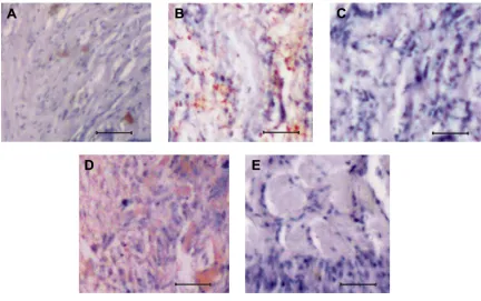
The immunohistochemistry of normal bone tissue with anti-Sox2 antibody showed a minimal expression as illustrated in Figure 2A. But in response to osteosarcoma progression, its expression got upregulated (Figure 2B and C). In the case of primary osteosarcoma, patches of cells showed sig-nals for Sox2 (Figure 2B), whereas in the metastatic form of osteosarcoma, strong signals were visualized from the
background of higher proliferative mass of cells (Figure 2C). With SAM administration, osteosarcoma tissue showed histological improvement along with the repressed expres-sion of Sox2 protein in the primary stage of osteosarcoma (Figure 2D). Following SAM treatment, the osteosarcoma development was controlled in the primary stage with a minimal expression of Sox2 along with restitution of the cellular arrangement (Figure 2D). But Sox2 expression was not restricted in the advanced stage of metastatic osteosar-coma (Figure 2E).
expression studies of TcTP in primary
and metastatic osteosarcoma
To understand the osteosarcoma progression and to cross-check the results further, the expression pattern of TCTP was studied using immunohistochemistry. The control bone tissue had limited TCTP protein expression (Figure 3A), but upon osteosarcoma development, especially in the pri-mary stages of osteosarcoma, there was an increased TCTP protein expression as shown in Figure 3B. But the advanced metastatic osteosarcoma showed only a restricted expression of TCTP with very mild signals (Figure 3C). After SAM treatment, interestingly, the primary osteosarcoma tissue showed an elevated expression of TCTP, which implies the
Figure 1 histological characteristics of normal and osteosarcoma tissues.
Notes: (A) histological section of normal bone tissue of a mouse showing uniform arrangement of cells. (B) Primary osteosarcoma tissue with abnormal mass of cells.
(C) Metastatic osteosarcoma tissue showing irregular cells that are enlarged in size when compared to normal cells. (D) Primary osteosarcoma tissue treated with saM
showing reversion in cellular pattern. (E) Metastatic osteosarcoma tissue treated with saM showing no changes in cellular pattern. scale bar: 100 µm.
Abbreviation: saM, S-adenosylmethionine.
OncoTargets and Therapy downloaded from https://www.dovepress.com/ by 118.70.13.36 on 25-Aug-2020
Dovepress
shi et al
Figure 2 analysis of sox2 expression in normal and osteosarcoma tissues.
Notes: (A) sox2 expression in normal bone tissue of a mouse. (B) sox2 expression in primary osteosarcoma tissue. (C) sox2 expression in metastatic osteosarcoma tissue.
(D) sox2 expression in saM-treated primary osteosarcoma tissue. (E) sox2 expression in saM-treated metastatic osteosarcoma tissue. scale bar: 100 µm.
Abbreviation: saM, S-adenosylmethionine.
Figure 3 analysis of TcTP expression in normal and osteosarcoma tissues.
Notes: (A) TcTP expression in normal bone tissue of a mouse. (B) TcTP expression in primary osteosarcoma tissue. (C) TcTP expression in metastatic osteosarcoma
tissue. (D) increased TcTP expression in saM-treated primary osteosarcoma tissue. (E) TcTP expression in saM-treated metastatic osteosarcoma tissue. scale bar:
100 µm.
Abbreviation: saM, S-adenosylmethionine.
OncoTargets and Therapy downloaded from https://www.dovepress.com/ by 118.70.13.36 on 25-Aug-2020
Dovepress role of S-adenosylmethionine in osteosarcoma development
fight back of cells to prevent osteosarcoma (Figure 3D), but in the case of metastatic osteosarcoma, the TCTP expression showed negative changes that resulted in the development of metastatic form.
Western blotting to analyze sox2 and
TcTP expression
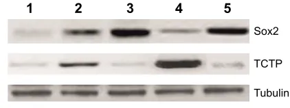
The results obtained using immunohistochemistry were further validated using Western blotting by comparing the Sox2 expression with that of TCTP. It is possible to precisely determine the osteosarcoma-regulating proteins in different tissues. The protein samples from normal bone tissue, pri-mary and metastatic osteosarcoma tissues, and SAM-treated tissues were subjected to Western blotting analysis using anti-Sox2 antibody and anti-TCTP antibody (Figure 3). Throughout the experiment, we observed that Sox2 was correlated with osteosarcoma progression, and upon treat-ment with SAM, the Sox2 level reduced along with the inhibition of tumor load in primary osteosarcoma, but SAM had no effect on metastatic osteosarcoma. The results with TCTP showed that it had an increased expression in primary osteosarcoma when compared with the normal tissue. But its expression showed some changes in metastatic osteosarcoma; on the other hand, its expression got upregulated in primary osteosarcoma after treatment with SAM (Figure 4).
Discussion
The osteosarcoma mouse model has the capability to bear a resemblance to human cancer histologically that greatly helps to understand the disease biology.29 In the present
study, the injected K7M2 cells worked effectively in the well-established BALB/c strain of mice,30 which is widely
used in the osteosarcoma mouse model to study the basic
science and for clinical investigations.31–34 In contrast,
it was reported that SAM at a concentration of 150 µM through intravenous route resulted in the development of significantly smaller skeletal lesions and at the same time marked reduction in pulmonary metastasis of osteosarcoma as compared to control groups in SCID mice.28 But in the
present experiment, osteosarcoma-developed BALB/c mice had not shown any lesions like SCID mice. It is interesting to note that SAM had different roles in the SCID than in the BALB/c mouse strain. The experimental success of develop-ing primary and metastatic osteosarcoma is visualized with an abnormal proliferative mass of cells together with deeply stained clusters of cells (Figure 1B and C). To understand the role of SAM in osteosarcoma, the mice were administered SAM. The role of SAM in inhibiting the primary osteosar-coma (Figure 1D) was effective when compared with the control (Figure 1B). But it had no effective role in inhibiting the later stages of osteosarcoma development (Figure 1E), which implies that the SAM has not much potential to stop metastatic osteosarcoma.
The molecular role of SAM was identified to have an effect on many transcription factors and genes associated with growth and apoptosis.35–37 The function of Sox2 was also well
defined in osteosarcoma that its expression is associated with triggering of the proliferative ability of normal bone tissue and the knockout of Sox2 leads to having a control over its proliferation ability.38 The present study revealed the role of
SAM in controlling the Sox2 expression. While the osteo-sarcoma progresses, the Sox2 also shows overexpression (Figure 2A and C), which implies that the development of osteosarcoma is associated with Sox2 expression. Similarly, the TCTP downregulation is also associated with osteosar-coma progression (Figures 3 and 4).
The regulation of Sox2 by SAM is more understand-able with the experiment of SAM administration into the osteosarcoma model that inhibits tumor progression along with the downregulation of Sox2 expression (Figure 2D). SAM methylate-mediated downregulation of TCTP expres-sion does not happen in primary osteosarcoma (Figure 3D), but similar effects occur in metastatic osteosarcoma (Figure 3E). On examining the results of SAM-treated metastatic osteosarcoma along with SAM-treated primary osteosarcoma, the Sox2 expression is well controlled in the primary form of osteosarcoma because SAM has a control over the initial stage of osteosarcoma but it does not hap-pen in the advanced stage of osteosarcoma. In addition, the results of immunohistochemistry with anti-TCTP antibody illustrate that TCTP has an important role in controlling
6R[ 7&73 7XEXOLQ
Figure 4 Western blotting analysis of sox2 and TcTP expression.
Notes: The protein samples were prepared from saM-treated osteosarcoma tissue
along with various controls and subjected to Western blotting analysis and then stained with anti-sox2 antibody and anti-TcTP antibody. lane 1 shows sox2 and TcTP expression in normal bone tissue samples of mice. lane 2 represents sox2 and TcTP expression in primary osteosarcoma tissue samples. lane 3 represents sox2 and TcTP expression in metastatic osteosarcoma tissue samples. lane 4 shows sox2 and TcTP expression in saM-treated primary osteosarcoma tissue samples. lane 5 represents sox2 and TcTP expression in saM-treated metastatic osteosarcoma tissue samples. Tubulin was used as a loading control.
Abbreviation: saM, S-adenosylmethionine.
OncoTargets and Therapy downloaded from https://www.dovepress.com/ by 118.70.13.36 on 25-Aug-2020
Dovepress
shi et al
the tumor. In contrast, it was already reported that normal and transformed cells show limited expression of TCTP.39
It was also reported that the TCTP levels are downregulated through activation of the tumor suppressor protein p53.22,40
Immunohistochemistry analysis shows elevated expression of TCTP, and the results coincide with the data illustrating. In addition to human osteosarcoma cells, other cell lines have also expressed the elevated levels of TCTP.41
In conclusion, the collective results show that SAM plays a major role in controlling osteosarcoma progres-sion. As a supportive effect of osteosarcoma reversion after SAM administration, we observed downregulation of Sox2 expression along with key upregulation of TCTP protein in primary osteosarcoma. The results obtained in this study are interesting, which promotes the prognosis of osteosarcoma for future investigation.
Acknowledgments
We thank the institutional review board approval committee and the ethical committee for the successful completion of this project.
Disclosure
The authors report no conflicts of interest in this work.
References
1. Picci P. Osteosarcoma (osteogenic sarcoma). Orphanet J Rare Dis. 2007;2:6.
2. Smida J, Baumhoer D, Rosemann M, et al. Genomic alterations and allelic imbalances are strong prognostic predictors in osteosarcoma.
Clin Cancer Res. 2010;16(16):4256–4267.
3. Ottaviani G, Jaffe N. The epidemiology of osteosarcoma. In: Jaffe N, Bruland O, Bielack S, editors. Pediatric and Adolescent Osteosarcoma. New York: Springer; 2009:3–13.
4. Tan ML, Choong PF, Dass CR. Osteosarcoma – conventional treatment vs gene therapy. Cancer Biol Ther. 2009;8(2):106–117.
5. Allison DC, Carney SC, Ahlmann ER, et al. A meta-analysis of osteo-sarcoma outcomes in the modern medical era. Sarcoma. 2012;2012: 704872.
6. Meyers PA. Muramyl tripeptide (mifamurtide) for the treatment of osteosarcoma. Expert Rev Anticancer Ther. 2009;9(8):1035–1049. 7. Szuhai K, Cleton-Jansen A-M, Hogendoorn PC, Bovée JV.
Molecu-lar pathology and its diagnostic use in bone tumors. Cancer Genet. 2012;205(5):193–204.
8. Esteller M. Cancer epigenomics: DNA methylomes and histone-modification maps. Nat Rev Genet. 2007;8(4):286–298.
9. Das PM, Ramachandran K, VanWert J, et al. Methylation mediated silencing of TMS1/ASC gene in prostate cancer. Mol Cancer. 2006; 5(1):28.
10. Al-Romaih K, Sadikovic B, Yoshimoto M, Wang Y, Zielenska M, Squire JA. Decitabine-induced demethylation of 5′ CpG island in GADD45A leads to apoptosis in osteosarcoma cells. Neoplasia. 2008; 10(5):471–480.
11. Cote GM, Choy E. Role of epigenetic modulation for the treatment of sarcoma. Curr Treat Options Oncol. 2013;14(3):454–464.
12. Chiang P, Gordon R, Tal J, et al. S-Adenosylmethionine and methyla-tion. FASEB J. 1996;10(4):471–480.
13. Luka Z, Mudd SH, Wagner C. Glycine N-methyltransferase and regu-lation of S-adenosylmethionine levels. J Biol Chem. 2009;284(34): 22507–22511.
14. Bale S, Ealick SE. Structural biology of S-adenosylmethionine decar-boxylase. Amino Acids. 2010;38(2):451–460.
15. Suzuki MM, Bird A. DNA methylation landscapes: provocative insights from epigenomics. Nat Rev Genet. 2008;9(6):465–476.
16. Böhm H, Benndorf R, Gaestel M, et al. The growth-related protein P23 of the Ehrlich ascites tumor: translational control, cloning and primary structure. Biochem Int. 1989;19(2):277–286.
17. MacDonald SM, Rafnar T, Langdon J, Lichtenstein LM. Molecular identification of an IgE-dependent histamine-releasing factor. Science. 1995;269(5224):688–690.
18. Thaw P, Baxter NJ, Hounslow AM, Price C, Waltho JP, Craven CJ. Structure of TCTP reveals unexpected relationship with gua-nine nucleotide-free chaperones. Nat Struct Mol Biol. 2001;8(8): 701–704.
19. Yenofsky R, Cereghini S, Krowczynska A, Brawerman G. Regula-tion of mRNA utilizaRegula-tion in mouse erythroleukemia cells induced to differentiate by exposure to dimethyl sulfoxide. Mol Cell Biol. 1983; 3(7):1197–1203.
20. Brioudes F, Thierry A-M, Chambrier P, Mollereau B, Bendahmane M. Translationally controlled tumor protein is a conserved mitotic growth integrator in animals and plants. Proc Natl Acad Sci U S A. 2010;107(37): 16384–16389.
21. Gachet Y, Tournier S, Lee M, Lazaris-Karatzas A, Poulton T, Bommer U-A. The growth-related, translationally controlled protein P23 has properties of a tubulin binding protein and associates tran-siently with microtubules during the cell cycle. J Cell Sci. 1999;112(8): 1257–1271.
22. Tuynder M, Susini L, Prieur S, et al. Biological models and genes of tumor reversion: cellular reprogramming through tpt1/TCTP and SIAH-1. Proc Natl Acad Sc U S A. 2002;99(23):14976–14981. 23. Cans C, Passer BJ, Shalak V, et al. Translationally controlled tumor
protein acts as a guanine nucleotide dissociation inhibitor on the transla-tion elongatransla-tion factor eEF1A. Proc Natl Acad Sci U S A. 2003;100(24): 13892–13897.
24. Chen SH, Wu P-S, Chou C-H, et al. A knockout mouse approach reveals that TCTP functions as an essential factor for cell proliferation and survival in a tissue- or cell type-specific manner. Mol Biol Cell. 2007;18(7): 2525–2532.
25. Zhu W-L, Cheng H-X, Han N, et al. Messenger RNA expression of translationally controlled tumor protein (TCTP) in liver regeneration and cancer. Anticancer Res. 2008;28(3A):1575–1580.
26. Arcuri F, Papa S, Carducci A, et al. Translationally controlled tumor protein (TCTP) in the human prostate and prostate cancer cells: expression, distribution, and calcium binding activity. Prostate. 2004; 60(2):130–140.
27. Khanna C, Khan J, Nguyen P, et al. Metastasis-associated differences in gene expression in a murine model of osteosarcoma. Cancer Res. 2001; 61(9):3750–3759.
28. Parashar S, Cheishvili D, Arakelian A, et al. S-Adenosylmethionine blocks osteosarcoma cells proliferation and invasion in vitro and tumor metastasis in vivo: therapeutic and diagnostic clinical applications.
Cancer Med. 2015;4(5):732–744.
29. Ek ET, Dass CR, Choong PF. Commonly used mouse models of osteo-sarcoma. Crit Rev Oncol Hematol. 2006;60(1):1–8.
30. Schmidt J, Strauß GP, Schön A, et al. Establishment and characteriza-tion of osteogenic cell lines from a spontaneous murine osteosarcoma.
Differentiation. 1988;39(3):151–160.
31. Wan X, Mendoza A, Khanna C, Helman LJ. Rapamycin inhibits ezrin-mediated metastatic behavior in a murine model of osteosarcoma.
Cancer Res. 2005;65(6):2406–2411.
32. Terabe M, Khanna C, Bose S, et al. CD1d-restricted natural killer T cells can down-regulate tumor immunosurveillance independent of inter-leukin-4 receptor-signal transducer and activator of transcription 6 or transforming growth factor-β. Cancer Res. 2006;66(7):3869–3875.
OncoTargets and Therapy downloaded from https://www.dovepress.com/ by 118.70.13.36 on 25-Aug-2020
OncoTargets and Therapy
Publish your work in this journal
Submit your manuscript here: http://www.dovepress.com/oncotargets-and-therapy-journal
OncoTargets and Therapy is an international, peer-reviewed, open access journal focusing on the pathological basis of all cancers, potential targets for therapy and treatment protocols employed to improve the management of cancer patients. The journal also focuses on the impact of management programs and new therapeutic agents and protocols on
patient perspectives such as quality of life, adherence and satisfaction. The manuscript management system is completely online and includes a very quick and fair peer-review system, which is all easy to use. Visit http://www.dovepress.com/testimonials.php to read real quotes from published authors.
Dovepress
Dove
press
role of S-adenosylmethionine in osteosarcoma development
33. Barkan D, Kleinman H, Simmons JL, et al. Inhibition of metastatic out-growth from single dormant tumor cells by targeting the cytoskeleton.
Cancer Res. 2008;68(15):6241–6250.
34. Merchant MS, Melchionda F, Sinha M, Khanna C, Helman L, Mackall CL. Immune reconstitution prevents metastatic recurrence of murine osteo-sarcoma. Cancer Immunol Immunother. 2007;56(7):1037–1046. 35. Lu SC, Mato JM. Role of methionine adenosyltransferase and
S-adenosylmethionine in alcohol-associated liver cancer. Alcohol. 2005; 35(3):227–234.
36. Chen H, Xia M, Lin M, et al. Role of methionine adenosyltransferase 2A and S-adenosylmethionine in mitogen-induced growth of human colon cancer cells. Gastroenterology. 2007;133(1):207–218.
37. Li TW, Zhang Q, Oh P, et al. S-Adenosylmethionine and methylth-ioadenosine inhibit cellular FLICE inhibitory protein expression and induce apoptosis in colon cancer cells. Mol Pharmacol. 2009;76(1): 192–200.
38. Basu-Roy U, Ambrosetti D, Favaro R, Nicolis SK, Mansukhani A, Basilico C. The transcription factor Sox2 is required for osteoblast self-renewal. Cell Death Differ. 2010;17(8):1345–1353.
39. Bommer U-A, Thiele B-J. The translationally controlled tumour protein (TCTP). Int J Biochem Cell Biol. 2004;36(3):379–385.
40. Niforou KN, Anagnostopoulos AK, Vougas K, Kittas C, Gorgoulis VG, Tsangaris GT. The proteome profile of the human osteosarcoma U2OS cell line. Cancer Genomics Proteomics. 2008;5(1):63–78.
41. Shen JH, Qu CB, Chu HK, et al. siRNA targeting TCTP suppresses osteosarcoma cell growth and induces apoptosis in vitro and in vivo.
Biotechnol Appl Biochem. 2016;63(1):5–14.
OncoTargets and Therapy downloaded from https://www.dovepress.com/ by 118.70.13.36 on 25-Aug-2020