1743
Efficient Ed3wt Method For Robust Medical
Image Watermarking
Ramanand singh ,. Piyush Shukla , Paresh Rawat
Abstract: Watermarking provides us to capability to authenticate medical imaging data. Embedding watermark within medical image adds ability to claim for copyright protection and makes image data robust against the various attacks. In this paper an efficient invisible and robust watermarking method is proposed using novel Edge Detection in Double Density discrete Wavelet Transform (ED3WT) domain method. In the proposed method the input cover image is decomposed in to wavelet coefficients using two level double densities DWT. The edge of the decomposed image s ub bands are calculated using the Sobel edge detector. Watermark is inserted to the edge of the high frequency sub-band coefficients of the decomposed image. This in turn adds the better invisibility. The benefit of using the double density DWT is its robustness against noise and filtering attacks. To add further robustness scaled morphologically dilated high frequency edge coefficient is used as key and is embedded within the image. This further improves the robustness and maintains the invisibility of the watermarking. Performance of proposed method is evaluated using medical images of different environments. The imaging data set includes skin images, MRI, Scalp images, CT scan, images. The parametric evaluation is carried out by calculating the MSE and PSNR. It is observed that method improves the invisibility and is robust to attacks like noise, mean and median filtering. Therefore method is useful for medical imaging data authentication using robust watermarking.
Index Terms: Medical Image, Watermarking, DWT, DDDWT, MRI images, Edge detection, attacks —————————— ——————————
1
I
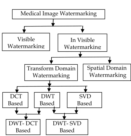
NTRODUCTIONSecurity of the medical imaging data is essential for maintaining the integrity of the patients and to avoid the surgical crime. This security of the medical images means maintaining the confidentiality and reliability of data and to make data available when needed. The reliability requires the authentication of the medical imaging data. Since medical images are shared on the intra and inter hospital and city networks therefore, there are many kinds of security risks viz. errors during transmission, lost of overwritten data on the network. The solution to these problems is medical image watermarking which provides the authentication to the medical images. The watermarked and copyrighted medical images are only allowed to use he owner or authorized persons [5]. Most of the watermarks are invisible thus observer is not able to see the same. Medical watermarking method must satisfy the requirement of the perceptual invisibility and it must be highly robust to various attacks such as noise, rotation, mean or median filtering and translation. Watermark embedding must not have many effects on the image features. It is because change in the original content of the image and may lead to wrong patient desires analysis and during the surgery it may even cost death of patients. Therefore this paper proposes a novel and efficient design for robust invisible watermarking method using the Double Density Discrete Wavelet Transform (DDDWT) which is expected to be less affected by various attacks for medical imaging data. The major reason for preferring the DDDWT as the wavelet transform is that, this method gives the better time frequency representation as compared to the conventional DWT, and more precise low and high frequency sub bands. This in turn makes the method more robust to the most of watermark attacks. Methods of the Medical image watermarking are broadly classified as shown in the Figure 1. Our main concern in this paper is to embed the watermark in DWT domain.
Figure 1 Classification of Medical Image Watermarking Techniques
2
L
ITERATURE REVIEWThe transformation based methods are widely used for medical image watermarking applications. Commonly used transformations are Discrete Cosine Transform (DCT) [13, 14], Discrete Wavelet Transformation (DWT) [3, 4], and Singular value decomposition (SVD) [5, 6]. There are certain methods which combines the features of the two transformations for adding more robustness such as DCT-DWT [7] and DWT-SVD [8]. Among all these methods the wavelet based methods are common and efficient due to its multi-resolution characteristics and simultaneous time-frequency analysis features. The edge detection based watermarking methods are widely used in transform domain to improve the robustness and invisibility of the watermarking techniques
3. Wavelet Domain Advantages
Why one have to prefer watermarking using wavelet transform domain following major benefits can be stated.
Medical Image Watermarking
Visible
Watermarking Watermarking In Visible
Transform Domain Watermarking
Spatial Domain Watermarking
DCT Based
DWT Based
SVD Based
DWT- DCT Based
1744 Application like image and video compression such as
JPEG and MPEG4 standards are all based on wavelet transform. Therefore the watermarking in wavelet domain is less sensitive to compression attacks. ‗
Wavelet bas watermarking methods uses the multi-resolution characteristics of wavelet transform thus embedded watermark is robust.
The watermarking on the high frequency sub bands using the edge detection of the image makes the watermark invisible to human eye.
Wavelet domain offers higher robustness to signal processing. Wavelet transform is compatible to the HVS and DCT much better.
However DWT has spatial frequency locality, which means if signal is embedded it will affect the image locally. Hence a wavelet transform provides both frequency and spatial description for an image.
4. Need for Medical Image Watermarking
Three main objectives are foreseen in the medical domain4.1 Imperceptible/Reversible Watermarking
Medical tradition is very strict with the quality of biomedical images. Thus the watermarking method must be reversible, in that the original pixel values must be exactly recovered (Macq and Dewey 1999). This limits significantly the capacity and the number of possible methods. An alternative way is to define regions of interest, to be left intact, and leave us with regions of insertion where a watermark could be inserted and does not interfere or disturb the radiologist.
4.2 Integrity Control
The ―secure camera‖ concept applies also to biomedical images, especially in the context of legal aspects and insurance claims. There is thus a need to prove that the images on which the diagnoses and any insurance claims are based have preserved their integrity.
4.3 Authentication
A critical requirement in patient records is to authenticate the different parts of the electronic patient record, in particular the images. More often an attached file or a header, which carries all the needed information, identifies an image. However, keeping the meta-data of the image in a separate header file is prone to forgeries or clumsy practices. An alternative would be to embed all such information into the image data itself.
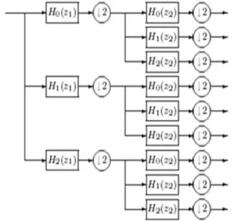
5 DOUBLE-DENSITY
DWT
The double-density Discrete Wavelet Transformation (DDDWT) is basically represented by one scaling function and two wavelets function and . Using the double density discrete wavelet transform improves the multi-resolution characteristic. Since, this transformation uses three filters (one low pass and two high pass filters) for generating multi-resolution framework as shown in the Figure 3. The scaling and wavelet functions are given as;
Where are defined as filters. The process is given in the Figure 2.
Figure 2 DDDWT implementation
DDDWT was analyzed and concluded that transform performs better than the standard DWT in terms of denoising; however, there is room for improvement because not all of the wavelets are directional. That is why, although the double-density DWT utilizes more wavelets, some lack a dominant spatial orientation, which prevents them from being able to isolate those directions. The down sampling process of DDDWT is given in the Figure 3.
Figure 3 implementation of 2D discrete wavelet transformation
6 PROPOSED
DOUBLE
DENSITY
DWT
BASED
WATERMARKING
1745
Figure 4 Proposed DWT based Watermarking method
Initially, the input image is decomposed into two levels by a DWT, resulting in an approximation sub band with low frequency components and L1 detail sub bands with high frequency components. The proposed algorithm finds the edge of HH sub-band using either sobel edge detector. Morphological dilation operations are not required and are removed. Another coefficients are the edge coefficients
Finally, Gaussian noise template is added as watermark and is distributed to edged groups with a variable standard deviations strength. The receiver detects the watermark data by correlating the watermarked image with the watermark sequence. In this present work watermark is embedded to the HH coefficients instead of LL coefficients. The double density discrete wavelet transform is implemented for medical images and the second level decomposition coefficients are shown in the Figure 5.
Figure 6 Flow chart of watermark insertion with DWT
The Gaussian noise template is used as a watermark and is inserted to the selected high frequency (HH) sub-band coefficients as rule mentioned in equation (3). The sub-band with minimum entropy is chosen for watermark insertion usually having diagonal edges. Because adding watermarking to these coefficients add the invisibility to watermark. A simplified watermark insertion rule is defined which removes the need of using image dilation. Watermark is scaled using according to the group that coefficients and image ut belong to. The watermarked image is generated using the inverse wavelet transform. The modified new watermark insertion rule is defined as;
Where is the edge detected coefficients of HH wavelet sub-band
is the zero mean noisy template
Where is defined as the clipping parameter and for better invisibility the value of should be smaller. This watermark is added to the HH decomposition DDDWT sub-band as,
In this paper the value of is set to 0.1 for better invisibility
7 EXPERIMENTAL
RESULTS
AND
DISCUSSION
In this paper efficient edge detection based DDDWT decomposition coefficients are used to generate watermarked images with Gaussian noise as watermark. This section presents some experimental results of the proposed watermarking method for medical images. The Sobel edge detections is implemented to have less visual artefacts. The DDDWT
IDDDWT
Sobel / Canny edge detector
Noise tamplate
Watermark Insertion Rule X(i,j)
Input
Image
W(i,j) Output
Image
XLx,y
YLx,y
W(x,y)
W(x,y)
Figure 5 second level DDDWT decompositions for CT image 1
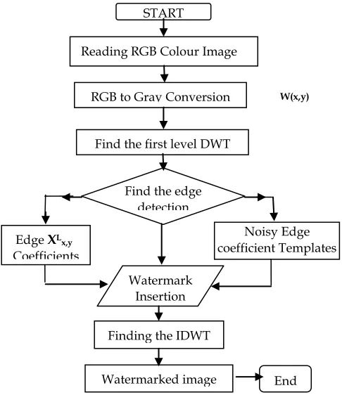
START
Reading RGB Colour Image
RGB to Gray Conversion
Find the edge detection Coefficients
Edge XLx,y
Coefficients
Noisy Edge coefficient Templates
Watermark Insertion
Rule Finding the IDWT
Watermarked image
out End
Find the first level DWT Coefficients
1746 sequential results of the work are presented in the Figure 7.
The sequential results comparisons of watermark embedding process are presented in section 7.2 Then finally in section 7.2 performance evaluation of results are carried out
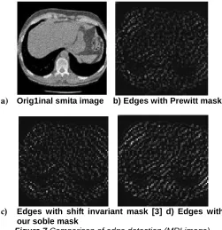
7.1 Results of Edge Detection
Results of edge detection with different masks for image1_smita a medical image is given in Figure 7. It can be observed that Sobel edge detector performs better compared to other edge masks. It is sharper and brighter with more features. Therefore in this paper it is proposed to use Soble mask for edge detection. The edge detection is calculated on the decomposed DDDWT coefficients having minimum entropy. Also our method with DDDWT transform performs robust and better than existing edge masks.
a) Orig1inal smita image b) Edges with Prewitt mask
c) Edges with shift invariant mask [3] d) Edges with
our soble mask
Figure 7 Comparison of edge detection (MRI image)
7.2 Sequential Results
In Figure 8 comparison of the sequential results with various processing steps are presented.
First original image and the watermark images are taken.
Then edge detection is applied on the image
The watermark is aided with the edge component.
Scaled watermark is replaced by coefficient of DDDWT. For embedding.
Then IDDDWT is calculated to find the watermarked image
Noisy attacked is applied.
The image is retrieved from it.
a) Original image b) Watermark image c) edge detected CT image d) Summation of Watermark
e) Scaled Watermark f) IDDDWT watermarked image
g) Noisy attacked watermark h) Retrieved Image
Figure 8 Sequential Results of the proposed watermarking method
7.3 Results of Watermarking
1747
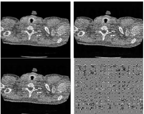
a) Original Image2_smita b) Watermarked image c) Reconstructed attacked image d) the difference a-c
Figure 9 Watermarked and reconstructed results for Image1_smita
b) Original Image2_smita b) Watermarked image c) Reconstructed attacked image d) the difference a-c
Figure 10 Watermarked and reconstructed results for Image2_smita
8 Conclusion
In this paper a robust watermarking is implemented using edge detector in 2-D double density discrete wavelet transform (DDDWT) domain. Results of classical edge detectors are compared for different medical images. The work presented in this dissertations are presented for watermarking using DDDWT method. Edge detectors are Implemented with two level DDDWT and the comparative results are presented. It is found that wavelet based results with multi resolution are better than the conventional edge based methods and are more robust to watermark attacks. The soble edge detectors perform equally for all kind of medical images. The proposed method removes the need of the image dilation and the watermark is selected as Gaussian noise tamplates. In order to add the robustness the watermark is embedded to the wavelet coefficients having minimum features. In the proposed method the Gaussian noise template is added as watermark and is added to the high frequency component instead of low frequency component. Watermarking with to high frequency component increases the invisibility of the watermark. in order to improve the performance watermark is scaled using scaling parameter which gives the better invisibility.
A
CKNOWLEDGMENTAuthors are highly grateful to each and every individual associated to complete this research and also acknowledges to each authors referred here
R
EFERENCES[1] Rahul K. Sarawale , S. R. Chougule, ― Noise removal using double-density dual-tree complex DWT‖ , IEEE Second International Conference on mage Information Processing (ICIIP), 2013
[2] Ivan W. Selesnick.―The Double-Density Dual-Tree Discrete Wavelet Transform‖ o IEEE Transactions on Signal Processing. 2001.
[3] J. N. Ellinas, D. E. Manolakis, ―A robust watermarking scheme based on edge detection and contrast sensitivity function,‖ in VISAPP Proc. Int. Conf. Computer Vision Theory and Applications, Barcelona, 2007.
[4] Narong Mettripun, ―Robust Medical image watermarking based on DWT for Patient Identification‖, in the IEEE 13th international conference on electrical engineering/electronics, computer, telecommunications and information technology (ECTI-CON) July 2016.
[5] Nilesh Rathi, Ganga Holi ―Securing Medical Images by Watermarking Using DWT-DCT-SVD‖, International Journal of Computer Trends and Technology (IJCTT) – volume X Issue Y–Month 2014
[6] Md Saiful Islam and Ui Pil Chong ―A Digital Image Watermarking Algorithm Based on DWT DCT and SVD ―,International Journal of Computer and Communication Engineering, Vol. 3, No. 5, September 2014
[7] Apeksha Tiwari, Virendra Singh ―Digital Image Watermarking Using DWT and Shift Invariant Edge Detection ―International Journal of Computer Technology and Electronics Engineering (IJCTEE) Volume 3, Issue 6, December 2013
[8] Nai-Kuei Chen, Chung-Yen Su, Che-Yang Shih, Yu-Tang Chen ―Reversible Watermarking for Medical Images Using Histogram Shifting with Location Map Reduction‖, IEEE international Conference on Industrial Technology (ICIT) 2016,
[9] S. C. Liew and J. M. Zain, ―Reversible medical image watermarking for tamper detection and recovery‖, 3 rd IEEE International Conference on Computer Science and Information Technology (ICCSIT), pp. 417-420, 2010.
[10]Smita Agrawal, Manoj Kumar, ―‖Reversible Data Hiding for Medical Images using Integer-to-Integer Wavelet Transform‖, In IEEE Students‘ Conference on Electrical, Electronics and Computer Science, (SCEECS) 2016.
[11]R. Dugad, K. Ratakonda, and N. Ahuja, ―A new wavelet-based scheme for watermarking images,‖ in IEEE Proc. Int. Conf. Image Processing, USA, pp. 419-423, 1998.
[12]J. R. Kim, and Y. S. Moon, ―A robust wavelet-based digital watermarking using level-adaptive thresholding,‖ in IEEE Proc. Int. Conf. Image Processing, Japan, pp. 226-230. 1999,
1748 Random Mid-band Coefficient Exchange Scheme for
Gray Scale Images ―, International Journal of Computer Applications (0975 – 8887) Volume 100– No.8, August 2014
[14]P.Tejaswini, K.Manjunath, A Mahendran, ―Digital Watermarking Using DWT-SVD‖, International Journal of Scientific & Engineering Research Volume 3, Issue 8, August-2012
[15]R. J. Beattie, 1984, ―Edge detection for semantically based early visual processing, ―dissertation, Univ. Edinburgh, Edinburgh, U.K.
[16]Vikas Tyagi ―Data Hiding in Image using least significant bit with cryptography‖, International Journal of Advanced Research in Computer Science and Software Engineering, Vol. 2, Issue 4, April 2012. [17]S. Mallat, S. Zhong, , ―Characterization of signals