Volume 18 Number 6 pp. 647–661 C The Author(s) 2015 doi:10.1017/thg.2015.82
Hormone Replacement Therapy Associated White
Blood Cell DNA Methylation and Gene Expression
are Associated With Within-Pair Differences of
Body Adiposity and Bone Mass
Aileen Bahl,1,∗Eija P ¨oll ¨anen,2,∗Khadeeja Ismail,1Sarianna Sipil ¨a,2Tuija M. Mikkola,2Eva Berglund,3
Carl M ˚arten Lindqvist,3Ann-Christine Syv ¨anen,3Taina Rantanen,2Jaakko Kaprio,1,4,5Vuokko Kovanen,2
and Miina Ollikainen1
1Department of Public Health, University of Helsinki, Helsinki, Finland
2Gerontology Research Center, Department of Health Sciences, University of Jyv ¨askyl ¨a, Jyv ¨askyl ¨a, Finland 3Department of Medical Sciences, Molecular Medicine and Science for Life Laboratory, Uppsala University, Uppsala,
Sweden
4Institute for Molecular Medicine FIMM, University of Helsinki, Helsinki, Finland 5National Institute for Health and Welfare, Helsinki, Finland
The loss of estrogen during menopause causes changes in the female body, with wide-ranging effects on health. Estrogen-containing hormone replacement therapy (HRT) leads to a relief of typical menopausal symptoms, benefits bone and muscle health, and is associated with tissue-specific gene expression profiles. As gene expression is controlled by epigenetic factors (including DNA methylation), many of which are environmentally sensitive, it is plausible that at least part of the HRT-associated gene expression is due to changes in DNA methylation profile. We investigated genome-wide DNA methylation and gene expression patterns of white blood cells (WBCs) and their associations with body composition, including muscle and bone measures of monozygotic (MZ) female twin pairs discordant for HRT. We identified 7,855 nominally significant differentially methylated regions (DMRs) associated with 4,044 genes. Of the genes with DMRs, five (ACBA1, CCL5, FASLG, PPP2R2B, and UHRF1) were also differentially expressed. All have been pre-viously associated with HRT or estrogenic regulation, but not with HRT-associated DNA methylation. All five genes were associated with bone mineral content (BMC), andABCA1, FASLG, and UHRF1 were also associated with body adiposity. Our study is the first to show that HRT associates with genome-wide DNA methylation alterations in WBCs. Moreover, we show that five differentially expressed genes with DMRs associate with clinical measures, including body fat percentage, lean body mass, bone mass, and blood lipids. Our results indicate that at least part of the known beneficial HRT effects on body composition and bone mass may be regulated by DNA methylation associated alterations in gene expression in circulating WBCs.
Keywords:hormone replacement therapy, HRT, menopause, discordant monozygotic twin pair design, gene expression, DNA methylation, epigenetic regulation, body composition, skeletal muscle composition, bone mineral content
Menopause leads to constantly low circulating 17 -estradiol (E2) levels compared with pre-menopausal cyclic monthly fluctuation of high to low systemic E2. The most commonly experienced adverse consequences are vasomo-tor symptoms such as hot flushes and night sweats, which are effectively treated with E2-based HRT (Sassarini & Lumsden,2015). HRT has also been shown to be associated with better muscle and bone health (Mikkola et al.,2011; Ronkainen et al.,2009). Aging is typically associated with
deterioration of bone properties, reduced skeletal muscle mass, and increased ectopic fat accumulation, which affect
RECEIVED10 September 2015;ACCEPTED10 October 2015.
ADDRESS FOR CORRESPONDENCE: Miina Ollikainen, PhD,
Depart-ment of Public Health, PO BOX 41, FI-00014, University of
Helsinki, Finland. [email protected]
muscle quality and function. E2deprivation has been shown to result in gain of fat mass and loss of fat free mass (Shea et al.,2015) as well as in increased risk for osteoporotic changes (Riggs et al.,2002; Szulc et al.,2006), while post-menopausal HRT partially counteracts aging related mus-culoskeletal deteriorations (Cheng et al.,2002; Komulainen et al.,1999; Sipil¨a et al.,2001; Taaffe et al.,2005). However, there is considerable variation in response to HRT between individuals, which can be partly explained by genetic factors (Langdahl,2009).
Studying HRT-discordant post-menopausal MZ twins eliminates differences in the DNA sequence between HRT users and their non-user twin sisters. This enables investiga-tion of potential epigenetic influences of HRT. The purpose of this study was to investigate the association of long-term HRT on WBC DNA methylation and thereby gene expression as a potential mechanism associated with dif-ferences in total body, muscle, and bone characteristics in post-menopausal MZ twin pairs discordant for E2-based HRT use. We hypothesized that HRT-associated differences in DNA methylation could underlie gene expression dif-ferences that in turn would manifest as difdif-ferences at the phenotype level. To test this hypothesis, we investigated 20 HRT-discordant MZ twin pairs for genome-wide DNA methylation and determined DMRs within the discordant twin pairs. Next, we examined if genes with DMRs are also differentially expressed in WBCs in a subgroup of nine HRT-discordant twin pairs. Finally, the within-pair expression differences were correlated with within-pair dif-ferences of the phenotype characteristics to further link the identified DMR and gene expression profiles with body adi-posity, muscle characteristics, and bone mass of the HRT-discordant post-menopausal MZ twin pairs.
Methods
Study Design and Data Collection
The participants of the current study originate from two separate twin studies, the Finnish Twin Study on Aging (FITSA) and the Sarcopenia — Skeletal MuscleAdaptation to Post-menopausal Hypogonadism and Effects of HRT and Physical Activity in Older Women: a Genetic and Molecu-lar Biological Study on Estrogen-related Pathways (SAWEs), for which the subjects were recruited from the Older Finnish Twin Cohort (Kaprio & Koskenvuo, 2002; Kaprio et al., 1978), consisting of 13,888 twin pairs of known zygosity. The FITSA study investigates the genetic and environmen-tal effects on the disablement process in older female twins (Tiainen et al., 2004). The SAWEs study investigates the associations of HRT on muscle properties at molecular level in a genetically controlled design (Ronkainen et al., 2009). Zygosity of all twins was confirmed by genotyping multiple polymorphic markers. Study participants visited our laboratory on two consecutive days. The evening of the first day consisted of the computed tomography
assess-ments for muscle composition. The second day consisted of blood sampling and measurements of body and bone composition.
MZ HRT-discordant twin pairs (N=20) were studied. The HRT-using co-twins were on E2-based HRT with the preparation containing either E2only (n=12) with 1–2 mg of estrogenic agent or E2in combination with progesterone (n=8) with estrogenic (1–2 mg) and progestogenic com-pounds. The other co-twin had never used HRT. The age of the participants ranged from 54 to 72 years (mean age 62.4 ±6.0 years). The mean duration of HRT use was 8.8±5.7 years (range 1–25 years). Physical activity was assessed with the scale of Grimby (1986), with slight modifications and categorized as sedentary (no other activities, but at the most light walking two times/week), moderately active (walking or other light exercise at least three times/week, but no other more intensive activities), and active (moderate or vigorous exercise at least three times/week; Ronkainen et al.,2009).
The subjects provided written informed consent. The protocol was designed and performed according to the principles of the Helsinki Declaration and approved by the Ethics Committee of the Hospital District of Central Finland.
Total Body and Muscle Composition and Bone Mineral Content
Total body fat percentage and lean body mass were as-sessed by bioelectrical impedance analysis (Spectrum II, RJL Systems, Detroit, MI, USA). Computed tomography scans (Siemens Somatom motion scanner, Siemens, Erlan-gen, Germany) were obtained from the midpoint between the greater trochanter and the lateral joint line of the knee and analyzed with the Geanie 2.1 software (Commit, Es-poo, Finland) to separate lean and fat tissue on the basis of the given radiological density limits. Total cross-sectional area (CSA) of thigh muscle compartment was determined by manually outlining the muscle compartment along the fascial plane to exclude subcutaneous fat. Within this area, lean muscle CSA was further separated from inter- and intra-muscular (IM) fat tissue infiltrated into the muscle compartment. As an indicator of muscle quality, the mean attenuation coefficient in Hounsfield Units (HU; i.e., mean density value within lean muscle CSA) was defined. Low HU values are associated with high amount of lipids infil-trating into muscle tissue, therefore indicating poor muscle quality (Goodpaster et al.,2000).
density threshold of 130 mg/cm3was used to separate bone from the surrounding soft tissue.
Blood Sampling and Assessment of
Systemic Hormones, Lipids, and CRP
Blood samples were taken after overnight fasting and sera were stored at −80°C until further analyses. Serum hor-mone assessments were performed as previously described (Ronkainen et al.,2009). Serum E2was determined using an extraction radioimmunoassay (Ankarberg-Lindgren & Norjavaara,2008). Serum concentrations of sex hormone-binding globulin (SHBG) and high sensitivity C-reactive protein (CRP) were measured using solid-phase, chemi-luminescent immunometric assays (Immulite 1000; Di-agnostic Products, Los Angeles, CA, USA). Free E2 lev-els were calculated from E2 and SHBG levels accord-ing to (Bjornerem et al., 2004). Serum total cholesterol, low-density lipoprotein-cholesterol (LDL), high-density lipoprotein-cholesterol (HDL), and triglycerides were mea-sured by enzymatic colorimetric assays using a KoneLab20 Clinical Chemistry Analyzer (Thermo Scientific, Vantaa, Finland).
DNA Extraction and Methylation Analysis
High molecular weight WBC DNA was extracted using QI-Aamp DNA Mini kit (QIAGEN Nordic, Sollentuna, Swe-den). Bisulfite conversion of DNA was completed using EZ-96 DNA Methylation-Gold Kit (Zymo Research, Irvine, CA, USA) according to the manufacturer’s instructions, and the co-twins were always converted on the same plate to minimize potential batch effects.
Genome-wide DNA methylation of 40 twin samples (20 pairs) was measured using the Illumina Infinium Hu-manMethylation450 BeadChip (450k array) according to the manufacturer’s instructions (Illumina, San Diego, CA, USA). Data pre-processing was carried out using R soft-ware R-3.02 (http://www.R-project.org) and Bioconduc-tor 2.13 (http://www.bioconductor.org), as described previ-ously (Ollikainen et al.,2015). First, we filtered out cytosine-guanine (CpG) sites that did not fulfill certain quality mea-sures. We discarded: (1) probes with a detectionpvalue> .001 in any sample (n=2653); (2) ‘rs’ (n=65) and ‘ch’ (n
=3076) probes reflecting single nucleotide polymorphism (SNP) genotyping probes that are only included for DNA tracing and non-CpG probes (Nordlund et al.,2013; Price et al.,2013), respectively, on the 450k array; (3) unreliable probes (n= 103421), according to Naeem et al. (2014); as well as (4) probes on sex chromosomes (n=8100), as differingvalues of the probes might be the result of ran-domXinactivation by DNA methylation. As a final filtering step, we removed uninformative probes, that is the 50% of probes with lowest variance (n=184131) among the studied samples.
We used R Bioconductor package lumi for background correction and quantile normalization of the 450k data. Color normalization was performed using the All Sample Mean Normalization (ASMN) algorithm. Additionally, we used Beta Mixture Quantile (BMIQ) from the wateRmelon package to account for differences in the distributions of the two different probe types. For removing batch effects, we used ComBat function from the sva package. As neighbor-ing CpG sites are known to have highly similar methylation values, we averaged over thevalues of CpG sites located in the same CpG island (Figure S1). We identified the CpGs belonging to the same CpG island and computed the mean values of the betas for each individual. These CpGs thus define a region with a potential of becoming a DMR in downstream analyses. All probes not belonging to any CpG islands remain as singletons in the analyses. For uniformity, these probes will also be referred to as DMRs (instead of differentially methylated probes, DMPs) in case they show differential methylation in further analyses. The CpG is-lands were defined as in Price et al. (2013) and Figure S1. Briefly, high-density CpG islands (HC) have CG content of >55%, the observed/expected ratio is>0.75, and the length of the island is>500 bp. Intermediate-density CpG islands (IC) have CpG content of>50%, the observed/expected ratio>0.48, and the length of the island is>200 bp. CpGs not belonging to CpG islands are classified as LC CpGs (low-density CpGs). The final dataset consisted of 1,04976 regions that were used in the analyses.
RNA Extraction and Gene Expression
Analysis
Statistics
All statistical analyses were performed in R. Differential within-pair analysis of genome-wide DNA methylation and candidate gene expression was performed in limma (http://bioconductor.org/packages/release/bioc/manuals/ limma/man/limma.pdf). Linear regression models were applied to all methylation probes in the final dataset to de-termine significant DMRs within twin-pairs discordant for HRT. In the same way, limma was used to assess differential expression of only those genes with DMRs. Methylation and expression probes were matched based on Entrez IDs. In both analyses, the data was adjusted for smoking discordance (ever vs. never smoker) within the twin pairs. Under- or overrepresentation of DMRs in specific genomic regions or in relation to CpG density in comparison to the CpG distributions in the whole dataset was assessed using Fisher’s test. Global HRT-associated methylation patterns were computed by categorizing each DMR as hypo- or hypermethylated in HRT users. This categorization was performed on within-pair methylation differences over all pairs. Significance of the global overrepresentation of differential methylation in one direction (global hypo-or hypermethylation at a higher rate than expected) was assessed using Binomial test. Pearson correlations between significantly differentially expressed genes with DMRs and clinical measures were computed on within-pair differences of expression values and within-pair differences of trait values. All p values were adjusted for multiple testing using Benjamini–Hochberg procedure. Adjusted
pvalues (FDR) .05 were considered significant, if not specified otherwise.
Gene Ontology Analysis
Gene ontology (GO) analysis of within-pair differen-tial methylation was carried out using the gene set analysis (GSA) package in R (http://statweb.stanford. edu/tibs/ftp/GSA.pdf). Methylation probes were assigned to probe-sets representing GO terms. In order to avoid con-sidering too small or too large and thus irrelevant GO terms, we restricted the analysis to sets with a minimum of 15 and a maximum of 500 genes. GSA was applied on within-pair methylation differences with 1000 permutations. GO terms with FDR<0.05 were considered as significant. GSA does not take into account the number of CpG sites on individ-ual genes in a GO term; however, there was no obvious bias related to number of probes per gene (Figure S2).
Results
Phenotypic Characteristics
General properties and metabolic measures of the HRT users and non-users belonging to the discordant twin pairs are presented inTable 1. Their mean age was 62.4± 6.0 years. As expected, HRT users had higher mean circulat-ing levels of total and free E2compared to their non-user
co-twins (202.8±205.8 vs. 27.1±22.0 pmol/L,p= 9.6 ×10⁻⁵; 3.5±2.9 vs. 0.6± 0.5 pmol/L,p=9.5×10−6, respectively). HRT users also had higher serum SHBG con-centration than their non-using co-twins (86.2±41.3 vs. 55.3±27.1,p=3.6×10⁻⁵). There was no evidence for differences in the concentrations of blood lipids and CRP between HRT users and non-users (p>.05 for both). No evidence for the differences between twins in the level of physical activity assessed by a three-scale self-assessment score was observed (p>.05). In addition, we did not ob-serve any significant differences in WBC count estimates (Houseman et al.,2012) between HRT users versus non-users (p>.05 for all WBC estimates), which is an impor-tant point considering the downstream DNA methylation analyses that could otherwise be confounded by differences in cell-type distributions.
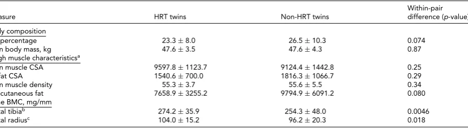
Table 2presents the phenotypic characteristics of HRT users and non-users. There was no evidence of differences in the body composition between the HRT users and non-users (p>.05). The more precise analysis of thigh muscle composition, which we had available from nine twin pairs, revealed no differences in the lean muscle area of thigh, the amount of subcutaneous fat surrounding thigh, IM fat infiltration within thigh muscle compartment, nor in the amount of IM fat assessed as the lean muscle density of thigh (p>.05). HRT users had higher BMC than their non-user co-twins, both in tibia (274.2±35.9 vs. 254.3±48.0,p=
.0046) and radius (104.0±15.2 vs. 96.2±20.3,p=.018).
Genome-Wide DNA Methylation Analysis
Genome-wide DNA methylation analysis within the 20 HRT-discordant MZ twin pairs did not result in any sig-nificant DMRs after multiple testing correction. However, 7,855 DMRs with nominalpvalue.05 (Table S1) were observed. DMRs were enriched at intergenic regions and de-pleted at gene promoters (FDR<0.001,Figure 1a). When investigating the location of the DMRs in relation to CpG density, the majority were at non-island (low density, LC) CpG sites. Consistent with this, high density CpG (HC) is-lands were significantly under-represented (FDR<3.3× 10⁻¹⁶), whereas LC CpG sites were over-represented (FDR
<3.3×10⁻¹⁶) among the DMRs when compared to the whole array distribution of the CpG sites (Figure 1b). Fi-nally, the DMRs were more frequently hypomethylated than hypermethylated (p<2.2×10⁻¹⁶), suggesting that HRT associates with global hypomethylation.
TABLE 1
General Properties and Metabolic Measures of the HRT-Discordant MZ Twin Pairs in the Study
Within-pair Measure HRT usersN=20 Non-usersN=20 difference (p value)
General properties
Time of HRT use (in years) 8.8±5.7 Never
E2treatment (# of users) 12 None
Combined treatment (# of users) 8 None Mean physical activity score 2.55±0.6 2.55±0.6 (# of active/moderately active/sedentary) (12/7/1) (12/7/1) Smoking status
Smoker while co-twin is non-smoker 3 2
Both non-smokers 11
Both smokers 4
Blood cell count estimates
CD8 T cells -8.7×10⁻²⁰±3.9×10⁻¹⁹ 0.001±0.002 0.18
CD4 T cells 0.141±0.007 0.139±0.007 0.23
NK cells 0.261±0.007 0.261±0.006 0.96
B cells 0.210±0.004 0.210±0.006 0.76
Monocytes 0.072±0.006 0.073±0.004 0.35
Granulocytes 0.211±0.006 0.211±0.004 0.78
Serum hormones
E2, pmol/L 202.8±205.8 27.1±22.0 9.6e-05
Free E2, pmol/L 3.5±2.9 0.6±0.5 9.5e-06
SHBG, nmol/L 86.2±41.3 55.3±27.1 3.6e-05
Blood lipids and CRPa
Total cholesterol, mmol/L 5.3±0.5 5.3±0.4 0.82 LDL cholesterol, mmol/L 3.1±0.8 3.2±0.4 0.91 HDL cholesterol, mmol/L 1.6±0.5 1.5±0.4 0.23 Triglycerides, mmol/L 1.2±1.0 1.1±0.5 0.48 High sensitivity CRP, mg/L 1.2±1.0 1.2±0.9 0.91
Note:aVariables of blood lipids and CRP were available from nine twin pairs. E2=17-estradiol, CRP=C-reactive protein, LDL=low-density lipoprotein, HDL=high-density lipoprotein.
TABLE 2
Phenotype Characteristics of the MZ Twin Pairs in the Study
Within-pair Measure HRT twins Non-HRT twins difference (p-value)
Body composition
Fat percentage 23.3±8.0 26.5±10.3 0.074
Lean body mass, kg 47.6±3.5 47.6±4.3 0.87
Thigh muscle characteristicsa
Lean muscle CSA 9597.8±1123.7 9124.4±1442.8 0.25 IM fat CSA 1540.6±700.0 1816.3±1066.7 0.29 Lean muscle density 55.3±3.7 55.6±5.5 0.34 Subcutaneous fat 7658.9±3255.2 9794.9±6091.2 0.080 Bone BMC, mg/mm
Distal tibiab 274.2±35.9 254.3±48.0 0.0046
Distal radiusc 104.0±15.2 96.2±20.3 0.018
Note:aVariables of thigh muscle characteristics were available from nine twin pairs. bDistal tibia was measured from eight twin pairs.
cDistal radius was measured from nine twin pairs. CSA=cross-sectional area, IM=intra-/inter-muscular, BMC=bone mineral content.
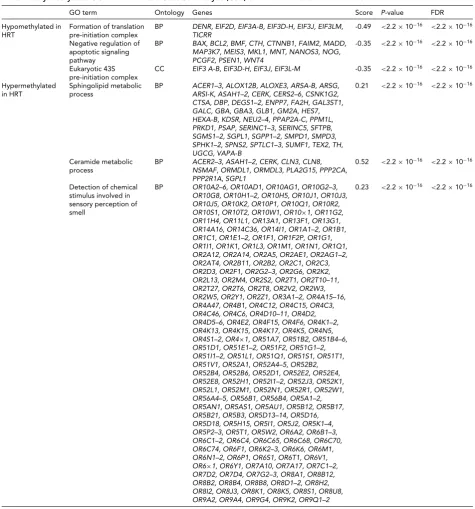
and five hypermethylated GO terms (FDR<2.2×10⁻¹⁶, Table 3). The significant biological processes were related to regulation of translation and apoptosis, and to chemical reactions and pathways involving sphingolipids. The molec-ular functions were associated to signal transduction.
Differential Expression Analysis of Genes
Associated With DMRs
To investigate the potential biological relevance of the iden-tified within-pair methylation differences, we applied a hypothesis-based approach and tested whether the genes
FIGURE 1
Genomic distribution of the DMRs in relation to genes and CpG density. Note: Bar plots show the proportions of the DMRs at genes and intergenic regions (a), and at CpG islands and non-island CpGs (b).P values denote which of the CpG categories are over- or under-represented among the DMRs (n=7855) in the HRT-discordant twin pairs. Fisher’s exact test was used to determine under- or overrepresentation of the CpG categories among the DMRs. Promoters include CpGs at 5UTRs, CpGs<1500 bp from transcription start site, and at first exon. HC, CpGs at high density CpG island, IC; CpGs at intermediate density CpG island; LC, non-CpG island CpG sites;∗FDR<0.01;∗∗FDR<3.3×10⁻¹⁶.
(TNF superfamily member 6,FASLG), and they function in apoptosis and cell survival (FASLG; Zhang et al.,2009; andPPP2R2B; Paluszczak et al.,2014), transcription and epigenetic regulation (UHRF1; Berkyurek et al.,2014), im-munoregulation (CCL5; Viola & Luster,2008), and in cel-lular cholesterol removal and assembly of HDL (ABCA1; Schmitz & Langmann,2001).
Correlation of the Expression of Genes
With DMRs With Clinical Measures of the
HRT-Discordant Twin Pairs
In an attempt to understand how the differentially expressed genes with DMRs (ABCA1, CCL5, UHRF1, PP2R2B,and
FASLG) link to the clinical outcomes, we correlated the pair gene expression differences with the
within-pair differences of the relevant clinical measures. The ob-served strong correlations between the within-pair differ-ences in serum concentration of both total and free E2with the within-pair difference in gene expression of each of the identified five genes supported the hypothesis that these genes are targets for HRT-mediated regulation (Figure 3 and Table S2). Negative association was found forABCA1
(r=-0.92, FDR= 8.63×10⁻⁴) andUHRF1(r=-0.90, FDR=1.18×10⁻³) and positive association withFASLG
TABLE 3
Differentially Methylated GO Terms from the Gene Set Analysis (GSA) of HRT-Discordant MZ Twin Pairs
GO term Ontology Genes Score P-value FDR
Hypomethylated in HRT
Formation of translation pre-initiation complex
BP DENR, EIF2D, EIF3A-B, EIF3D-H, EIF3J, EIF3LM, TICRR
-0.49 <2.2×10⁻¹⁶ <2.2×10⁻¹⁶
Negative regulation of apoptotic signaling pathway
BP BAX, BCL2, BMF, CTH, CTNNB1, FAIM2, MADD, MAP3K7, MEIS3, MKL1, MNT, NANOS3, NOG, PCGF2, PSEN1, WNT4
-0.35 <2.2×10⁻¹⁶ <2.2×10⁻¹⁶
Eukaryotic 43S pre-initiation complex
CC EIF3 A-B, EIF3D-H, EIF3J, EIF3L-M -0.35 <2.2×10⁻¹⁶ <2.2×10⁻¹⁶
Hypermethylated in HRT
Sphingolipid metabolic process
BP ACER1–3, ALOX12B, ALOXE3, ARSA-B, ARSG, ARSI-K, ASAH1–2, CERK, CERS2–6, CSNK1G2, CTSA, DBP, DEGS1–2, ENPP7, FA2H, GAL3ST1, GALC, GBA, GBA3, GLB1, GM2A, HES7, HEXA-B, KDSR, NEU2–4, PPAP2A-C, PPM1L, PRKD1, PSAP, SERINC1–3, SERINC5, SFTPB, SGMS1–2, SGPL1, SGPP1–2, SMPD1, SMPD3, SPHK1–2, SPNS2, SPTLC1–3, SUMF1, TEX2, TH, UGCG, VAPA-B
0.21 <2.2×10⁻¹⁶ <2.2×10⁻¹⁶
Ceramide metabolic process
BP ACER2–3, ASAH1–2, CERK, CLN3, CLN8, NSMAF, ORMDL1, ORMDL3, PLA2G15, PPP2CA, PPP2R1A, SGPL1
0.52 <2.2×10⁻¹⁶ <2.2×10⁻¹⁶
Detection of chemical stimulus involved in sensory perception of smell
BP OR10A2–6, OR10AD1, OR10AG1, OR10G2–3, OR10G8, OR10H1–2, OR10H5, OR10J1, OR10J3, OR10J5, OR10K2, OR10P1, OR10Q1, OR10R2, OR10S1, OR10T2, OR10W1, OR10×1, OR11G2, OR11H4, OR11L1, OR13A1, OR13F1, OR13G1, OR14A16, OR14C36, OR14I1, OR1A1–2, OR1B1, OR1C1, OR1E1–2, OR1F1, OR1F2P, OR1G1, OR1I1, OR1K1, OR1L3, OR1M1, OR1N1, OR1Q1, OR2A12, OR2A14, OR2A5, OR2AE1, OR2AG1–2, OR2AT4, OR2B11, OR2B2, OR2C1, OR2C3, OR2D3, OR2F1, OR2G2–3, OR2G6, OR2K2, OR2L13, OR2M4, OR2S2, OR2T1, OR2T10–11, OR2T27, OR2T6, OR2T8, OR2V2, OR2W3, OR2W5, OR2Y1, OR2Z1, OR3A1–2, OR4A15–16, OR4A47, OR4B1, OR4C12, OR4C15, OR4C3, OR4C46, OR4C6, OR4D10–11, OR4D2, OR4D5–6, OR4E2, OR4F15, OR4F6, OR4K1–2, OR4K13, OR4K15, OR4K17, OR4K5, OR4N5, OR4S1–2, OR4×1, OR51A7, OR51B2, OR51B4–6, OR51D1, OR51E1–2, OR51F2, OR51G1–2, OR51I1–2, OR51L1, OR51Q1, OR51S1, OR51T1, OR51V1, OR52A1, OR52A4–5, OR52B2, OR52B4, OR52B6, OR52D1, OR52E2, OR52E4, OR52E8, OR52H1, OR52I1–2, OR52J3, OR52K1, OR52L1, OR52M1, OR52N1, OR52R1, OR52W1, OR56A4–5, OR56B1, OR56B4, OR5A1–2, OR5AN1, OR5AS1, OR5AU1, OR5B12, OR5B17, OR5B21, OR5B3, OR5D13–14, OR5D16, OR5D18, OR5H15, OR5I1, OR5J2, OR5K1–4, OR5P2–3, OR5T1, OR5W2, OR6A2, OR6B1–3, OR6C1–2, OR6C4, OR6C65, OR6C68, OR6C70, OR6C74, OR6F1, OR6K2–3, OR6K6, OR6M1, OR6N1–2, OR6P1, OR6S1, OR6T1, OR6V1, OR6×1, OR6Y1, OR7A10, OR7A17, OR7C1–2, OR7D2, OR7D4, OR7G2–3, OR8A1, OR8B12, OR8B2, OR8B4, OR8B8, OR8D1–2, OR8H2, OR8I2, OR8J3, OR8K1, OR8K5, OR8S1, OR8U8, OR9A2, OR9A4, OR9G4, OR9K2, OR9Q1–2
0.23 <2.2×10⁻¹⁶ <2.2×10⁻¹⁶
expression, and body composition (Figure 3and Table S2).
ABCA1was negatively andFASLGandPPP2R2Bpositively correlated with lean body mass, and ABCA1andUHRF1
were negatively andFASLGpositively correlated with body fat percentage, indicating that these genes associate with to-tal body composition in HRT/E2related manner. In order to determine more precisely the nature of this association between gene expression and lean versus fat mass, we did correlation analysis with the available variables of lean mass
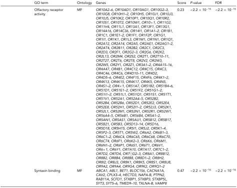
TABLE 3 Continued
GO term Ontology Genes Score P-value FDR
Olfactory receptor activity
MF OR10A2–6, OR10AD1, OR10AG1, OR10G2–3, OR10G8, OR10H1–2, OR10H5, OR10J1, OR10J3, OR10J5, OR10K2, OR10P1, OR10Q1, OR10R2, OR10S1, OR10T2, OR10W1, OR10×1, OR11G2, OR11H4, OR11L1, OR13A1, OR13F1, OR13G1, OR14A16, OR14C36, OR14I1, OR1A1–2, OR1B1, OR1C1, OR1E1–2, OR1F1, OR1F2P, OR1G1, OR1I1, OR1K1, OR1L3, OR1M1, OR1N1, OR1Q1, OR2A12, OR2A14, OR2A5, OR2AE1, OR2AG1–2, OR2AT4, OR2B11, OR2B2, OR2C1, OR2C3, OR2D3, OR2F1, OR2G2–3, OR2G6, OR2K2, OR2L13, OR2M4, OR2S2, OR2T1, OR2T10–11, OR2T27, OR2T6, OR2T8, OR2V2, OR2W3, OR2W5, OR2Y1, OR2Z1, OR3A1–2, OR4A15–16, OR4A47, OR4B1, OR4C12, OR4C15, OR4C3, OR4C46, OR4C6, OR4D10–11, OR4D2, OR4D5–6, OR4E2, OR4F15, OR4F6, OR4K1–2, OR4K13, OR4K15, OR4K17, OR4K5, OR4N5, OR4S1–2, OR4×1, OR51A7, OR51B2, OR51B4–6, OR51D1, OR51E1–2, OR51F2, OR51G1–2, OR51I1–2, OR51L1, OR51Q1, OR51S1, OR51T1, OR51V1, OR52A1, OR52A4–5, OR52B2, OR52B4, OR52B6, OR52D1, OR52E2, OR52E4, OR52E8, OR52H1, OR52I1–2, OR52J3, OR52K1, OR52L1, OR52M1, OR52N1, OR52R1, OR52W1, OR56A4–5, OR56B1, OR56B4, OR5A1–2, OR5AN1, OR5AS1, OR5AU1, OR5B12, OR5B17, OR5B21, OR5B3, OR5D13–14, OR5D16, OR5D18, OR5H15, OR5I1, OR5J2, OR5K1–4, OR5P2–3, OR5T1, OR5W2, OR6A2, OR6B1–3, OR6C1–2, OR6C4, OR6C65, OR6C68, OR6C70, OR6C74, OR6F1, OR6K2–3, OR6K6, OR6M1, OR6N1–2, OR6P1, OR6S1, OR6T1, OR6V1, OR6×1, OR6Y1, OR7A10, OR7A17, OR7C1–2, OR7D2, OR7D4, OR7░G2–3, OR8A1, OR8B12, OR8B2, OR8B4, OR8B8, OR8D1–2, OR8H2, OR8I2, OR8J3, OR8K1, OR8K5, OR8S1, OR8U8, OR9A2, OR9A4, OR9G4, OR9K2, OR9Q1–2
0.23 <2.2×10⁻¹⁶ <2.2×10⁻¹⁶
Syntaxin binding MF ABCA1, ABL1, BET1, BLOC1S6, CACNA1A, CAV2, CPLX3–4, HECTD3, NAPA-B, PTPN2, RAB11A, SCFD1, STXBP1, STXBP3, STXBP5L, SYT3, SYT5–6, TMED9–10, TXLNA-B, VAMP8
0.47 <2.2×10⁻¹⁶ <2.2×10⁻¹⁶
TABLE 4
Differentially Expressed Genes With DMRs Within HRT-Discordant Twin Pairs
Expression probe DMR Gene symbol Expression Log FCa p value FDR
ILMN_1766054 cg13430450 ABCA1 -0.281 2.4×10−07 0.0016 ILMN_1773352 IC_cg10315334 and cg12455187 CCL5 0.144 1.1×10−06 0.0026 ILMN_1786065 IC_cg14759209/cg23867903 UHRF1 -0.135 6.7×10−06 0.0060 ILMN_1660732 cg19494588 PPP2R2B 0.138 2.8×10−05 0.020 ILMN_1781824 cg10161121 FASLG 0.145 6.3×10−05 0.036
Note:aNegative log fold change denotes downregulation and positive log fold change upregulation in the HRT using co-twins compared with their non-using co-twins. Log fold changes as well as associatedp-values and FDRs result from the candidate gene expression analysis in which only genes with DMRs were considered. DMR, differentially methylated region; FC, fold change.
CCL5,PPP2R2B,andFASLG,Table S2). However, lower leg and arm BMC (distal tibia and distal radius, respectively) was highly significantly associated with expression of all five genes (Figure 3and Table S2).
Discussion
In the current study, we report associations of post-menopausal HRT with genome-wide DNA methylation and
gene expression in WBCs by investigating HRT-discordant MZ twin pairs. Through within-pair genome-wide DNA methylation analysis, we identified a number of genes with HRT-associated DMRs, of which five were differentially ex-pressed.ABCA1andUHRF1were down-regulated whereas
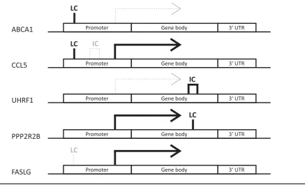
FIGURE 2
DMR patterns were unique for each of the identified differentially expressed genes with DMRs. Note: Genes are split up into three genomic locations: promoter, gene body and 3UTR. The position of the indicated DMR shows the genomic location in which it appears in the given gene, the tag above gives its location in relation to CpG density. Bold solid black line under the tag indicates hypermethylation and thin gray dotted line hypomethylation. LC refers to low density and IC intermediate density CpG island. No differentially expressed genes with high density CpG island was found. The differential expression is indicated by arrows. Solid black arrows indicate higher and thin dotted gray arrows lower expression in HRT users compared to non-users, respectively.
composition, bone mass, and blood lipids strongly associate with the differentially expressed genes with DMRs. These data indicate that at least part of the known beneficial HRT effects on the clinical outcomes may be regulated by DNA methylation in circulating WBCs.
Alterations in DNA methylation, both at global and individual gene level, have long been shown to associate with many human diseases, such as cancer (Lin & Wag-ner,2015; Stefansson et al.,2015), type 2 diabetes (Nilsson et al.,2014), BMI (Dick et al.,2014), and obesity-associated metabolic disturbances (Ollikainen et al.,2015), as well as aging (Marttila et al., 2015; Yuan et al.,2015). However, to our knowledge, there are no genome-wide methylation studies on the effects of HRT. One previous study has shown that conjugated equine estrogen therapy is associated with reduced plasma homocysteine and increment of total level of genomic methylation in peripheral mononuclear cells (Friso et al., 2007). We, however, identified 7,855 DMRs with nominal significance and global DNA hypomethy-lation in WBCs associated with HRT. This potential dis-crepancy may be partially explained by differences in HRT specimen (equine estrogen vs. estradiol). In our study, the genomic distribution of the DMRs in relation to genes and CpG density was highly similar to what has been previously reported for complex diseases; gene promoters and HC is-lands were under-represented and intragenic regions and
LC CpGs over-represented among DMRs compared to the whole array distribution of the CpG sites (Irizarry et al., 2009; Nilsson et al.,2014; Ollikainen et al.,2015; Yang et al., 2015). Hence, HRT seems to affect DNA methylation in similar genomic regions as reported for common multi-factorial diseases. As most of the complex diseases are age related, it is possible that at least part of these commonly ob-served associations between DNA methylation and different diseases are due to aging-associated methylation changes, including global hypomethylation (Heyn et al.,2012; Jo-hansson et al.,2013; Yuan et al.,2015). As our study was based on within-pair comparisons of HRT-discordant MZ twin pairs, the effect of age could potentially only mani-fest as increased methylation variation by age, as reported previously for unrelated individuals (Tserel et al., 2015). Also, the age range that we studied was relatively limited, so we cannot draw conclusions about very old women or pre-menopausal women.
FIGURE 3
(Colour online) Heat map of correlations between significantly differentially expressed genes with DMRs and clinical measures. Note: The heat map shows genes on thex-axis and clinical measures on the y-axis. Colors of squares in the heat map indicate correlation values (see color key). Dark red squares stand for high negative correlations between gene and trait, dark blue squares indicate strong positive correlations. Stars in the squares indicate significantp-values (∗) and FDRs (∗∗) with 0.05 as cut-off, respectively. The dendrogram on the left-hand side shows clustering of the clinical measures, the one above the heatmap shows clustering of the genes according to the similarity of their correlation profiles.
apoptosis (Fang et al.,2015), and to increase cognitive abil-ities and other brain functions (Comasco et al.,2014; Mah-mutyazicioglu et al.,2014). Dysfunction of the olfactory system is an early sign of neurodegeneration, and HRT has a positive effect on olfactory function (Doty et al.,2015), explaining our GO findings related to sensory perception of smell and olfactory receptor activity. Unfortunately, in the current study we did not assess cognition or other neural functions. Therefore, further studies are needed to see if the HRT-associated methylation and gene expression indeed have a role in neural function.
In order to go beyond only reporting associations on genome-wide DNA methylation, we investigated the poten-tial biological relevance of the identified within-pair methy-lation differences by exploring whether any of the genes with DMRs are differentially expressed. This analysis re-vealed that five genes with DMRs (ABCA1,UHRF1,FASLG,
PPP2R2B, and CCL5) were also differentially expressed within pairs, and showed that the relationships between
gene body or promoter methylation and gene expression are widespread, as previously reported (Wagner et al.,2014).
ABCA1,CCL5,andFASLGhave been previously shown to associate with HRT and menopause (Christodoulakos et al., 2007; Darabi et al.,2011b; Kangas et al.,2014), and both
UHRF1andPPP2R2Bhave been associated to estrogen re-lated breast cancer by epigenetic mechanisms (Jin et al., 2010; Klajic et al.,2013; Muggerud et al.,2010). In addition,
PPP2R2Bis a member of a PP2-family, whose expression levels have been reported to be maintained by estrogen (Yi & Simpkins,2008). Moreover, this family is directly involved in synaptic plasticity and memory formation. In addition,
FASLGmay also play a role in neuroprotection (Knight et al., 2010). This goes nicely hand in hand with our GO findings, and thus increased expression and differential methylation ofPPP2R2BandFASLGin HRT users in our study further support the potential neuroprotective role of HRT.
correlated the expression of the DMR-containing genes with clinical measures obtained from the HRT-discordant twin pairs (Figure 3, Table S2). This analysis revealed three dis-tinct clusters. First, both measures of total body size, body fat percentage, and lean and body mass were in the same cluster with serum E2, sharing the direction of the association with gene expression.ABCA1andUHRF1were negatively and
FASLG,PPP2R2B,andCCL5positively associated with E2 and total body size. Second, triglycerides shared the associ-ation pattern with BMC measures.ABCA1andFASLG ex-pression was negatively, whileUHRF1,PPP2R2B,andCCL5
expression was positively associated with triglycerides and BMC. The third cluster was formed by the cholesterol mea-sures, of which only LDL showed significant association with gene expression.ABCA1andFASLGexpression was positively whilePPP2R2BandCCL5negatively associated with LDL concentration.
Our findings support the known influence of HRT on whole body lipid levels. Individuals on HRT generally show higher HDL and apoA levels while LDL cholesterol and apoB levels are usually decreased (Darabi et al., 2011b).
ABCA1, which we found to be differentially expressed and methylated, is a specialized transmembrane receptor of pe-ripheral cells involved in the reverse cholesterol process mediating cholesterol efflux to HDL particles. Some stud-ies have shown HRT to increaseABCA1 gene expression (Darabi et al.,2011a; 2011b), while others, including our current study, have observed decreasedABCA1levels due to HRT (Cerda et al.,2013). We did not observe differences in blood lipids within HRT user and non-user twin pairs, possibly due to the fact that genetic regulation is a stronger determinant of blood lipid concentrations than the effects of HRT. However, within-pair difference inABCA1 expres-sion was strongly positively correlated with within-pair dif-ference in LDL and negatively with within-pair difdif-ference in triglycerides.ABCA1mediated reverse cholesterol trans-port has also been linked to inflammation and apoptosis of WBCs (Mineo & Shaul,2012).
As another indicator of the association of HRT with inflammation, we found two inflammatory cytokinesCCL5
andFASLGto be differentially expressed and methylated.
FASLGalso removes monocytes after the immune response by inducing their apoptosis (Mor et al.,2003; Perlman et al., 2001). Thus, higherFASLGexpression may participate in prevention of inflammation.
Estrogen-mediated upregulation ofFASLGhas also been shown to be important in maintaining bone re-modeling balance (Shao et al., 2015; Wang et al., 2015). Although DNA methylation has been previously proposed as a reg-ulatory mechanism ofFASLGexpression (Castellano et al., 2006), HRT has not been suggested to be inductor of its dif-ferential methylation before. Our study shows thatFASLG
has LC type DMR in the gene promoter area, is differentially expressed, and moreover, its expression is significantly as-sociated with distal tibia and radius BMC. Thus, these data
support the role of estrogen as a regulator ofFASLG ex-pression and further suggest methylation as a mediating mechanism of the regulation.
Finally, our results support the role of HRT in maintain-ing healthier body composition. The main contributors to lean body mass are lean muscle mass and bone mass. Of these, we assessed muscle mass as thigh lean muscle CSA and bone mass as the BMC of tibia and radius, and found bone variables to negatively associate with the expression ofABCA1andFASLGand positively with the expression ofUHRF1, PPP2R2B,andCCL5. Another important body composition measure is fat percentage that is a measure of total body adiposity, including both abdominal subcu-taneous and visceral adipose tissue as well as peripheral gluteofemoral and intra-muscular adipose tissue. Since ab-dominal and gluteofemoral adipose tissue has been shown to possess opposite roles in metabolic health (Manolopou-los et al.,2010), we examined the associations between gene expression and thigh subcutaneous and IM fat, as rough es-timates of gluteofemoral fat, but found no significant corre-lations. However, total body fat percentage correlated nega-tively withABCA1andUHRF1, and positively withFASLG
expression. This observation may indicate that these associ-ations largely come from abdominal fat depots. Therefore, we can conclude that the identified differentially expressed and methylated genes are involved in HRT-associated regu-lation of body composition; however, without further stud-ies it is difficult to determine how clinically relevant these associations are.
No previous DNA methylation studies in relation to menopausal symptoms or factors influencing the decision to start using HRT exist. Although precaution in interpret-ing our results is warranted, we are confident that our obser-vations are due to HRT-discordance associated differences in circulating estradiol concentrations rather than due to other factors related to HRT.
To conclude, our study is the first attempt to asso-ciate post-menopausal HRT use with genome-wide DNA methylation and associated differences in gene expression of WBCs. Our results suggest that HRT-associated methyla-tion associated with differential gene expression may be one of the biological mechanisms driving beneficial HRT effects on body adiposity and bone mass. However, further studies are needed to evaluate and confirm the clinical relevance of our findings.
Acknowledgments
We thank the participants for their invaluable contribu-tions to the study. The technical staffs at the Finnish Twin Cohort Study and at the Department of Health Sciences and Gerontology Research Center at University of Jyv¨askyl¨a are acknowledged for their help in collecting the data. The Gerontology Research Center is a joint effort between the University of Jyv¨askyl¨a and the University of Tampere. The 450k DNA methylation analysis was performed by the SNP&SEQ Technology Platform in Uppsala, which is part of the National Genomics Infrastructure hosted by Science for Life Laboratory.
This study was supported by the Academy of Finland (M. O., grant number 251316 V. K., grant number 114310, J. K., grant numbers 265240 and 263278, T. R., grant number 69818), Finnish Ministry of Culture and Edu-cation (V. K., grant number 89/627/2008, T. R., grant number 120/722/2003), The Sigrid Juselius Foundation (M. O.); EPITRAIN - Innovative techniques and models to understand epigenetic regulation in the pathogenesis of common diseases (EPITRAIN - FP7-PEOPLE-2012-ITN, grant number 316758); ENGAGE,- European Network for Genetic and Genomic Epidemiology, (FP7-HEALTH-F4– 2007, grant number 201413); the Swedish Research Council (A-C.S., grant number E0226301, C0524801); the Swedish Cancer Society (A-C. S., grant number 140581) the Swedish Childhood Cancer Foundation (A-C. S., grant number PR2014–0100), the Swedish Foundation for Strategic Re-search (A-C. S., grant number RBc08–008); the Erik, Karin and G¨osta Selander’s Stiftelse (E. B.).
Ethical Standards
The authors assert that all procedures contributing to this work comply with the ethical standards of the relevant na-tional and instituna-tional committees on human experimen-tation and with the Helsinki Declaration of 1975, as revised in 2008.
Supplementary Material
To view supplementary material for this article, please visit http://dx.doi.org/10.1017/thg.2015.82.
References
Ankarberg-Lindgren, C., & Norjavaara, E. (2008). A purifi-cation step prior to commercial sensitive immunoassay is necessary to achieve clinical usefulness when quantifying
serum 17 beta-estradiol in prepubertal children.European
Journal of Endocrinology/European Federation of Endocrine Societies,158, 117–124.
Berkyurek, A. C., Suetake, I., Arita, K., Takeshita, K., Nakagawa, A., Shirakawa, M., & Tajima, S. (2014). The DNA methyltransferase Dnmt1 directly interacts with the SET and RING finger-associated (SRA) domain of the mul-tifunctional protein Uhrf1 to facilitate accession of the
cat-alytic center to hemi-methylated DNA.The Journal of
Bio-logical Chemistry,289, 379–386.
Bjornerem, A., Straume, B., Midtby, M., Fonnebo, V., Sundsfjord, J., Svartberg, J., . . . Berntsen, G. K. (2004). Endogenous sex hormones in relation to age, sex, lifestyle factors, and chronic diseases in a general population: The
tromso study. The Journal of Clinical Endocrinology and
Metabolism,89, 6039–6047.
Castellano, R., Vire, B., Pion, M., Quivy, V., Olive, D., Hirsch, I., . . . Collette, Y. (2006). Active transcription of the human FASL/CD95 L/TNFSF6 promoter region in T lymphocytes involves chromatin remodeling: Role of DNA methylation and protein acetylation suggest distinct mechanisms of
transcriptional repression.The Journal of Biological
Chem-istry,281, 14719–14728.
Cerda, A., Issa, M. H., Genvigir, F. D., Rohde, C. B., Cavalli, S. A., Bertolami, M. C., . . . Hirata, R. D. (2013). Atorvas-tatin and hormone therapy influence expression of ABCA1, APOA1 and SCARB1 in mononuclear cells from
hyper-cholesterolemic postmenopausal women. The Journal of
Steroid Biochemistry and Molecular Biology,138, 403–409. Cheng, S., Sipil¨a, S., Taaffe, D. R., Puolakka, J., & Suominen,
H. (2002). Change in bone mass distribution induced by hormone replacement therapy and high-impact physical
exercise in post-menopausal women.Bone,31, 126–135.
Christodoulakos, G. E., Lambrinoudaki, I. V., Economou, E. V., Papadias, C., Vitoratos, N., Panoulis, C. P., . . . Creatsas, G. C. (2007). Circulating chemoattractants RANTES, negatively related to endogenous androgens, and MCP-1 are differentially suppressed by hormone therapy
and raloxifene.Atherosclerosis,193, 142–150.
Comasco, E., Frokjaer, V. G., & Sundstrom-Poromaa, I. (2014). Functional and molecular neuroimaging of menopause and
hormone replacement therapy.Frontiers in Neuroscience,8,
388.
Darabi, M., Ani, M., Panjehpour, M., Rabbani, M., Movahedian, A., & Zarean, E. (2011a). Effect of estro-gen receptor beta A1730G polymorphism on ABCA1 estro-gene expression response to postmenopausal hormone
replace-ment therapy.Genetic Testing and Molecular Biomarkers,15,
Darabi, M., Rabbani, M., Ani, M., Zarean, E., Panjehpour, M., & Movahedian, A. (2011b). Increased leukocyte ABCA1 gene expression in post-menopausal women on hormone
replacement therapy.Gynecological Endocrinology,27, 701–
705.
Dick, K. J., Nelson, C. P., Tsaprouni, L., Sandling, J. K., Aissi, D., Wahl, S., . . . Samani, N. J. (2014). DNA methylation
and body-mass index: A genome-wide analysis. Lancet,
383(9933), 1990–1998.
Doty, R. L., Tourbier, I., Ng, V., Neff, J., Armstrong, D., Battistini, M., . . . Sondheimer, S. J. (2015). Influences of hormone replacement therapy on olfactory and cognitive
function in postmenopausal women.Neurobiology of Aging,
36, 2053–2059.
Fang, D., Yang, H., Lin, J., Teng, Y., Jiang, Y., Chen, J., & Li, Y. (2015). 17beta-estradiol regulates cell proliferation, colony formation, migration, invasion and promotes apop-tosis by upregulating miR-9 and thus degrades MALAT-1 in osteosarcoma cell MG-63 in an estrogen
receptor-independent manner.Biochemical and Biophysical Research
Communications,457, 500–506.
Friso, S., Lamon-Fava, S., Jang, H., Schaefer, E. J., Corrocher, R., & Choi, S. W. (2007). Oestrogen replacement therapy reduces total plasma homocysteine and enhances genomic
DNA methylation in postmenopausal women.The British
Journal of Nutrition,97, 617–621.
Goodpaster, B. H., Kelley, D. E., Thaete, F. L., He, J., & Ross, R. (2000). Skeletal muscle attenuation determined by com-puted tomography is associated with skeletal muscle lipid
content.Journal of Applied Physiology,89, 104–110.
Grimby, G. (1986). Physical activity and muscle training in
the elderly.Acta Medica Scandinavica. Supplementum,711,
233–237.
Heyn, H., Li, N., Ferreira, H. J., Moran, S., Pisano, D. G., Gomez, A., . . . Esteller, M. (2012). Distinct DNA
methy-lomes of newborns and centenarians.Proceedings of the
Na-tional Academy of Sciences of the United States of America, 109, 10522–10527.
Houseman, E. A., Accomando, W. P., Koestler, D. C.,
Christensen, B. C., Marsit, C. J., Nelson, H. H., . . . Kelsey, K. T. (2012). DNA methylation arrays as surrogate
mea-sures of cell mixture distribution.BMC Bioinformatics,13,
86.
Irizarry, R. A., Ladd-Acosta, C., Wen, B., Wu, Z., Montano, C., Onyango, P., . . . Feinberg, A. P. (2009). The human colon cancer methylome shows similar hypo- and
hypermethyla-tion at conserved tissue-specific CpG island shores.Nature
Genetics,41, 178–186.
Jin, W., Chen, L., Chen, Y., Xu, S. G., Di, G. H., Yin, W. J., . . . Shao, Z. M. (2010). UHRF1 is associated with epigenetic
silencing of BRCA1 in sporadic breast cancer.Breast Cancer
Research and Treatment,123, 359–373.
Johansson, A., Enroth, S., & Gyllensten, U. (2013). Continu-ous aging of the human DNA methylome throughout the
human lifespan.PloS One,8, e67378.
Kangas, R., P¨oll¨anen, E., Rippo, M. R., Lanzarini, C., Prattichizzo, F., Niskala, P., . . . Kovanen, V. (2014). Cir-culating miR-21, miR-146 a and fas ligand respond to
post-menopausal estrogen-based hormone replacement therapy
— A study with monozygotic twin pairs.Mechanisms of
Ageing and Development,143–144, 1–8.
Kaprio, J., & Koskenvuo, M. (2002). Genetic and environ-mental factors in complex diseases: The older finnish twin
cohort.Twin Research,5, 358–365.
Kaprio, J., Sarna, S., Koskenvuo, M., & Rantasalo, I. (1978). The finnish twin registry: Formation and compilation, ques-tionnaire study, zygosity determination procedures, and
re-search program.Progress in Clinical and Biological Research,
24 Part B, 179–184.
Klajic, J., Fleischer, T., Dejeux, E., Edvardsen, H., Warnberg, F., Bukholm, I., . . . Kristensen, V. N. (2013). Quantitative DNA methylation analyses reveal stage dependent DNA methylation and association to clinico-pathological factors
in breast tumors.BMC Cancer,13, 456.
Knight, J. C., Scharf, E. L., & Mao-Draayer, Y. (2010). Fas
ac-tivation increases neural progenitor cell survival.Journal of
Neuroscience Research,88, 746–757.
Komulainen, M., Kroger, H., Tuppurainen, M. T., Heikkinen, A. M., Alhava, E., Honkanen, R., . . . Saarikoski, S. (1999). Prevention of femoral and lumbar bone loss with hor-mone replacement therapy and vitamin D3 in early postmenopausal women: A population-based 5-year
ran-domized trial.The Journal of Clinical Endocrinology and
Metabolism,84, 546–552.
Langdahl, B. L. (2009). The genetics of response to estrogen
treatment.Clinical Cases in Mineral and Bone Metabolism,
6, 44–49.
Lin, Q., & Wagner, W. (2015). Epigenetic aging signatures are
coherently modified in cancer.PLoS Genetics,11, e1005334.
Mahmutyazicioglu, K., Besir, F. H., Bardakci, M., Tanriverdi, H. A., & Ankarali, H. (2014). Hormone replacement therapy-related changes in the early postmenopausal pe-riod (critical window): An in vivo brain proton magnetic
resonance spectroscopy study.Turkish Journal of Medical
Sciences,44, 853–861.
Manolopoulos, K. N., Karpe, F., & Frayn, K. N. (2010). Glu-teofemoral body fat as a determinant of metabolic health. International Journal of Obesity,34, 949–959.
Marttila, S., Kananen, L., Hayrynen, S., Jylhava, J., Nevalainen, T., Hervonen, A., . . . Hurme, M. (2015). Ageing-associated changes in the human DNA methylome: Genomic
loca-tions and effects on gene expression.BMC Genomics,16,
179.
Mikkola, T. M., Heinonen, A., Kovanen, V., Cheng, S., Kujala, U. M., Suominen, H., . . . Sipil¨a, S. (2011). Influence of long-term postmenopausal hormone-replacement therapy on estimated structural bone strength: A study in discordant
monozygotic twins.Journal of Bone and Mineral Research,
26, 546–552.
Mineo, C., & Shaul, P. W. (2012). Novel biological functions of
high-density lipoprotein cholesterol.Circulation Research,
111, 1079–1090.
Mor, G., Sapi, E., Abrahams, V. M., Rutherford, T., Song, J., Hao, X. Y., . . . Kohen, F. (2003). Interaction of the es-trogen receptors with the fas ligand promoter in human
Muggerud, A. A., Ronneberg, J. A., Warnberg, F., Botling, J., Busato, F., Jovanovic, J., . . . Tost, J. (2010). Frequent aber-rant DNA methylation of ABCB1, FOXC1, PPP2R2B and PTEN in ductal carcinoma in situ and early invasive breast
cancer.Breast Cancer Research,12, R3.
Naeem, H., Wong, N. C., Chatterton, Z., Hong, M. K., Pedersen, J. S., Corcoran, N. M., . . . Macintyre, G. (2014). Reducing the risk of false discovery enabling identification of biologically significant genome-wide methylation status
using the HumanMethylation450 array.BMC Genomics,15,
51.
Nilsson, E., Jansson, P. A., Perfilyev, A., Volkov, P., Pedersen, M., Svensson, M. K., . . . Ling, C. (2014). Altered DNA methylation and differential expression of genes influenc-ing metabolism and inflammation in adipose tissue from
subjects with type 2 diabetes.Diabetes,63, 2962–2976.
Nordlund, J., Backlin, C. L., Wahlberg, P., Busche, S., Berglund, E. C., Eloranta, M. L., . . . Syvanen, A. C. (2013). Genome-wide signatures of differential DNA methylation in
pedi-atric acute lymphoblastic leukemia. Genome Biology, 14,
105.
Ollikainen, M., Ismail, K., Gervin, K., Kyllonen, A., Hakkarainen, A., Lundbom, J., . . . Kaprio, J. (2015). Genome-wide blood DNA methylation alterations at reg-ulatory elements and heterochromatic regions in
monozy-gotic twins discordant for obesity and liver fat. Clinical
Epigenetics,7, 39.
Paluszczak, J., Hemmerling, D., Kostrzewska-Poczekaj, M., Jarmuz-Szymczak, M., Grenman, R., Wierzbicka, M., & Baer-Dubowska, W. (2014). Frequent hypermethylation of WNT pathway genes in laryngeal squamous cell
car-cinomas.Journal of Oral Pathology & Medicine,43, 652–
657.
Perlman, H., Pagliari, L. J., Nguyen, N., Bradley, K., Liu, H., & Pope, R. M. (2001). The fas-FasL death receptor and PI3K pathways independently regulate monocyte homeostasis. European Journal of Immunology,31, 2421–2430.
Price, M. E., Cotton, A. M., Lam, L. L., Farre, P., Emberly, E., Brown, C. J., . . . Kobor, M. S. (2013). Additional anno-tation enhances potential for biologically-relevant analysis of the illumina infinium HumanMethylation450 BeadChip
array.Epigenetics & Chromatin,6, 4.
Riggs, B. L., Khosla, S., & Melton, L. J., 3rd. (2002). Sex steroids and the construction and conservation of the adult skeleton. Endocrine Reviews,23, 279–302.
Ronkainen, P. H., Kovanen, V., Alen, M., P¨oll¨anen, E., Palonen, E. M., Ankarberg-Lindgren, C., . . . Sipil¨a, S. (2009). Post-menopausal hormone replacement therapy modifies skele-tal muscle composition and function: A study with
monozy-gotic twin pairs.Journal of Applied Physiology,107, 25–33.
Ronkainen, P. H., P¨oll¨anen, E., Alen, M., Pitkanen, R., Puolakka, J., Kujala, U. M., . . . Kovanen, V. (2010). Global gene expression profiles in skeletal muscle of monozygotic female twins discordant for hormone replacement therapy. Aging Cell,9, 1098–1110.
Sassarini, J., & Lumsden, M. A. (2015). Oestrogen replacement
in postmenopausal women.Age and Ageing,44, 551–558.
Schmitz, G., & Langmann, T. (2001). Structure, function and
regulation of the ABC1 gene product.Current Opinion in
Lipidology,12, 129–140.
Shao, B., Liao, L., Yu, Y., Shuai, Y., Su, X., Jing, H., . . . Jin, Y. (2015). Estrogen preserves fas ligand levels by inhibiting microRNA-181 a in bone marrow-derived mesenchymal
stem cells to maintain bone remodeling balance. FASEB
Journal,29, 3935–3944.
Shea, K. L., Gavin, K. M., Melanson, E. L., Gibbons, E., Stavros, A., Wolfe, P., . . . Kohrt, W. M. (2015). Body composition and bone mineral density after ovarian hormone
suppres-sion with or without estradiol treatment.Menopause,22,
1045–1052.
Sipil¨a, S., Taaffe, D. R., Cheng, S., Puolakka, J., Toivanen, J., & Suominen, H. (2001). Effects of hormone re-placement therapy and high-impact physical exercise on skeletal muscle in post-menopausal women: A
random-ized placebo-controlled study.Clinical Science,101, 147–
157.
Stefansson, O. A., Moran, S., Gomez, A., Sayols, S., Arribas-Jorba, C., Sandoval, J., . . . Esteller, M. (2015). A DNA methylation-based definition of biologically distinct breast
cancer subtypes.Molecular Oncology,9, 555–568.
Szulc, P., Seeman, E., Duboeuf, F., Sornay-Rendu, E., & Delmas, P. D. (2006). Bone fragility: Failure of periosteal apposi-tion to compensate for increased endocortical resorpapposi-tion
in postmenopausal women.Journal of Bone and Mineral
Research,21, 1856–1863.
Taaffe, D. R., Sipil¨a, S., Cheng, S., Puolakka, J., Toivanen, J., & Suominen, H. (2005). The effect of hormone replacement therapy and/or exercise on skeletal muscle attenuation in
postmenopausal women: A yearlong intervention.Clinical
Physiology and Functional Imaging,25, 297–304.
Tiainen, K., Sipil¨a, S., Alen, M., Heikkinen, E., Kaprio, J., Koskenvuo, M., . . . Rantanen, T. (2004). Heritability of maximal isometric muscle strength in older female twins. Journal of Applied Physiology,96, 173–180.
Tserel, L., Kolde, R., Limbach, M., Tretyakov, K., Kasela, S., Kisand, K., . . . Peterson, P. (2015). Age-related profiling
of DNA methylation in CD8+T cells reveals changes in
immune response and transcriptional regulator genes.
Sci-entific Reports,5, 13107.
Viola, A., & Luster, A. D. (2008). Chemokines and their
recep-tors: Drug targets in immunity and inflammation.Annual
Review of Pharmacology and Toxicology,48, 171–197. Wagner, J. R., Busche, S., Ge, B., Kwan, T., Pastinen, T., &
Blanchette, M. (2014). The relationship between DNA methylation, genetic and expression inter-individual
varia-tion in untransformed human fibroblasts.Genome Biology,
15, R37.
Wang, L., Liu, S., Zhao, Y., Liu, D., Liu, Y., Chen, C., . . . Jin, Y. (2015). Osteoblast-induced osteoclast apoptosis by fas ligand/FAS pathway is required for maintenance
of bone mass. Cell Death and Differentiation, 22, 1654–
1664.
and childhood asthma in the inner city. The Journal of Allergy and Clinical Immunology,136, 69–80.
Yi, K. D., & Simpkins, J. W. (2008). Protein phosphatase 1, pro-tein phosphatase 2A, and calcineurin play a role in
estrogen-mediated neuroprotection.Endocrinology,149, 5235–5243.
Yuan, T., Jiao, Y., de Jong, S., Ophoff, R. A., Beck, S., & Teschendorff, A. E. (2015). An integrative multi-scale
anal-ysis of the dynamic DNA methylation landscape in aging. PLoS Genetics,11, e1004996.
Zhang, J., Chen, X., Zhang, S., Zhou, G., Xia, X., & Lu, L. (2009). Effects of transdermal estrogen therapy on expres-sions of estrogen receptors and T-lymphocyte apoptosis in
surgically menopausal women.Cellular & Molecular