Available Online: http://journal-ecobiotechnology.com/
Selective isolation and antimicrobial activity of rare actinomycetes from
mangrove sediment of Karwar
Sateesh V. Naikpatil* and J.L.Rathod
Department of Marine Biology, Karnatak University P.G and Research Centre, Kodibag, Karwar 581303, India
Abstract
The mangrove ecosystem is a largely unexplored source for actinomycetes. Various pretreatment procedures and selective media were applied to assess the optimal conditions for the isolation of rare actinomycetes from Mangrove sediment.
Pretreatment of wet-heating for 15 min at 70oC and phenol treatment of soil suspension were the most effective methods for
the isolation of those microorganisms. Hair hydrolysate vitamin agar (HHVA) was the most suitable medium for the recovery of rare actinomycetes. Fifty-three rare actinomycete strains were chosen using selective isolation approaches, then morphological and chemical properties of the isolates were determined. The isolates belonged to one of the following genus, Micromonospora, Microbispora, Actinoplanes, and Actinomadura. Later Micromonospora and Actinomadura were selected for antimicrobial activity. Minimum bactericidal concentration (MBC) of ethyl acetate extract against Staphylococcus aureus were 1.20 mg/ml for Micromonospora species and 5mg/ml for Actinomadura species. Thin layer chromatography (TLC) of the ethyl acetate extracts were carried out in duplicate using Chloroform: methanol (4:1) as solvent system and Tetracycline as
reference antibiotic. Under UV light they gave greenish yellow spots with Rf value 0.85 for the antimicrobial from
Actinomadura species and 0.88 for that from Micromonospora species. In bioautography (using Staphylococcus auras as test organism) inhibition zones were obtained and they were associated with the yellowish green spots of the chromatogram as detected under UV light. This may indicate the same compounds were responsible for the antibacterial activity of those actinomycetes isolates.
Keywords: Actinomadura, Actinoplanes, HHVA, Micromonospora, Pretreatment, Rare actinomycetes.
INTRODUCTION
The actinomycetes are Gram positive bacteria having high G+C (>55%) content in their DNA. The name ‘Actinomycetes’ was derived from Greek ‘aktis’ (a ray) and ‘mykes’ (fungus) and given to these organisms form initial observation of their morphology. Actinomycetes were originally considered to be an intermediate group between bacteria and fungi but now are recognized as prokaryotic organisms.
Actinomycetes are widely distributed in natural and man-made environments, and play an important role in the degradation of organic matter. They are also well known as a rich source of antibiotics and bioactive molecules, and are of considerable importance in industry. When conventional isolation techniques were applied, most of the isolates recovered on agar plates have been
identified as genus Streptomyces, which are the dominant
actinomycetes in soil. Several factors must be considered for the purpose of screening novel bioactive molecules: choice of screening source, pretreatment, selective medium, culture condition, and recognition of candidate colonies on a primary isolation plate. The
role of rare actinomycetes as bioactive molecule sources became apparent as these organisms provided about 25% of the antibiotics of actinomycete origin reported during 1975 to 1980.Rare actinomycetes have usually been regarded as strains of actinomycetes whose isolation frequency by conventional methods is much lower than that of streptomycete strains.Subsequently, employing pretreatments of soil by drying and heating stimulated the isolation of rare actinomycetes.An alternative approach was to make the isolation procedure more selective by adding chemicals such as phenol to the soil suspension. Many actinomycetes have shown multiple resistances to wide ranges of antibiotics. Several antibiotic molecules were used in selective medium to inhibit the competing bacteria including fast-growing actinomycetes. Macromolecules such as casein, chitin, hair hydrolysate, and humic acid were chosen as carbon and nitrogen sources of rare actinomycetes. Diagnosis of isolates on a primary isolation plate and recognition as a novel taxa was very important both for practical and taxonomical purposes. Until recently, bacterial systematic was based on the morphological and behavioral properties of microorganisms. Chemical information can be used at all taxonomic levels and it is likely that chemical properties will become an important part of minimal descriptions of many genera and species of actinomycetes. In this study, we estimated the efficiency of pretreatment methods and selective isolation medium for rare actinomycete genera such as Actinoplanes, Micromonospora, Microbispora and Actinomadura. The isolates were characterized with chemical techniques such as fatty acid profile, major menaquinone composition, cell wall diaminopimelic acid pattern, and whole cell sugars pattern.
The actinomycetes are noteworthy as antibiotic producers,
Received: Aug 25, 2011; Revised: Oct 16, 2011; Accepted: Nov 24, 2011. *Corresponding Author
Sateesh V. Naikpatil
Department of Marine Biology, Karnatak University P.G and Research Centre, Kodibag, Karwar 581303, India Tel: +919902669585
making three quarters of all known products; the Streptomyces are especially prolific and can produce a great many antibiotics and other class of biologically active secondary metabolites. They cover around 80% of total antibiotic product, with other genera trailing numerically; Micromonospora is the runner up with less than one-tenth as many as Streptomyces. If we include secondary metabolites with biological activities other than antimicrobial, actinomycetes are still out in front, over 60%; Streptomyces spp. accounting for 80% of these (Hopwood, et al., 2000).
MATERIALS AND METHODS Sampling and pretreatment of soil
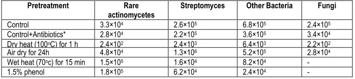
Soil samples were collected from the mangroves forest of Karwar. Four different pretreatment methods as described in Table 1 were carried out in the first 24 h after sampling.
Selective isolation of rare actinomycetes
Serially diluted soil suspensions were spread onto selective
isolation medium, and incubated for 4 weeks at 25oC. Starch casein
nitrate agar (SCA), humic acid vitamin agar (HVA), hair hydrolysate vitamin agar (HHVA), and Bennet's agar (BA) were used for the selective isolation of rare actinomycetes. Preliminary designation of rare actinomycete colonies were done by microscopic observation with a long working distance microscope. The criteria for classification between streptomycete and rare actinomycete strains were taken from previous works. Single colonies were successively transferred onto glucose yeast extract (GYE) agar and incubated until pure isolates were obtained. Spore mass and mycelium
fragments of the pure isolates were stored at -20oC as glycerol (20%,
v/v) suspension.
Morphology
Production of spore mass and its color, substrate mycelium color and production of diffusible pigment were detected on the 21-day old cultures on oatmeal agar.
Amino acid analysis
Chemical composition of the isolates were determined. Biomasses were obtained from liquid cultures in GYE broth medium
at 28oC for 7 days, freeze dried, and kept refrigerated for further
analysis. Diaminopimelic acid isomers were determined as described in Seong et al. Acid hydrolysate of the biomass was applied on TLC plates and developed for 4 h in a solvent system containing methanol-water-10 N HCl-pyridine (80 : 26.25 : 3.75 : 10, v/v). Dry plates were sprayed with a solution of ninhydrine in acetone (0.2%, w/v).
Fatty acid analysis
Extraction of fatty acids as their methyl esters were performed by alkaline methanolysis ). Fatty acid methyl esters (FAMEs) were
separated in a HP-1 capillary column in a gas chromatograph
equipped with a flame ionization detector. The temperature was
programmed to hold at 170oC for 1 min, then to rise by 5oC/min.
Injector temperature was held at 250oC, and the detector at 300oC.
Whole cell sugar analysis
Whole cell sugars were extracted as alditol acetates, and were analyzed using a gas chromatograph fitted with a flame ionization detector. Separation was achieved using a fused silica
capillary column. The temperature was programmed to hold at 160oC
for 2 min, then to rise by 5oC/min. Injector temperature was held at
250oC, and the detector at 300oC.
Quinone analysis
For the extraction of menaquinone, 50 mg of dried biomass was treated with chloroform/methanol (2:1, v/v) by shaking overnight. The extracts were concentrated using an electric aspirator, and cell debris was removed by centrifugation. The solvents were evaporated completely, and the remnants were resuspended in hexane. High performance liquid chromatographic separation of the quinones was
done with an ODS Hypersil column and acetonitrile/tetrahydrofuran
(70:30, v/v) as the mobile phase. The flow rate was 1 ml/min at 37oC,
and the detector was a UV detector operated at 254 nm.
Screening of actinomycetes for antimicrobial activity
The screening method consists of two steps; Primary screening and secondary screening. In primary screening the antimicrobial activity of pure isolates were determined by perpendicular streak method (Egorov, 1985) on Nutrient agar (NA).
The test organisms used were; Bacillus subtilis, Staphylococcus
aureus, Enterobacter aerogens, Escherichia coli, Klebsiella species, Proteus species, Pseudomonas species, Salmonella typhi and Shigella species. Secondary screening was performed by agar well
method against the standard test organisms Escherichia coli,
Staphylococcus aureus, Salmonella typhi, Bacillus subtilis, and Proteus spp.
Characterization of actinomycetes
The potent actinomycetes selected from secondary screening were characterized by morphological and biochemical methods. Morphological methods consist of macroscopic and microscopic methods. The microscopic characterization was done by cover slip culture method (Kawato and Sinobu, 1979).The mycelium structure, color and arrangement of conidiospore and arthrospore on the mycelium was observed through the oil immersion (1000X). The observed structure was compared with Bergey’s manual of Determinative Bacteriology, Ninth edition (2000) and the organism was identified. Various biochemical tests performed for the identification of the potent isolates are as follows: Casein hydrolysis, Starch hydrolysis, Tween 20 hydrolysis, Urea hydrolysis, Esculin hydrolysis, Acid production from sugar, NaCl resistance, Temperature tolerance.
Identification of the isolates was also carried out by chemical properties. Discrimination between actinoplanetes and Actinomadura
was primarily done bysugar pattern. Diagnostic sugar of the latter is
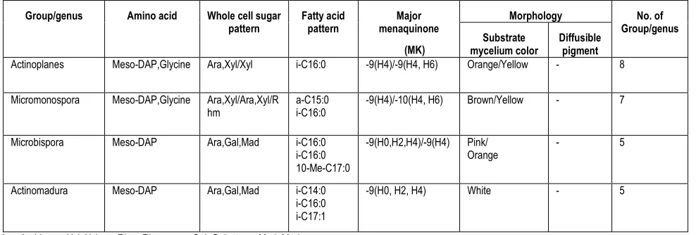
madurose, while the former is xylose (Table 2).
Fermentation process
Isolation of antibacterial metabolites
Antibacterial compound was recovered from the filtrate by solvent extraction method following the process described by
Westley et.al, 1979.and Liu et.al, 1986. Ethyl acetate was added to
the filtrate in the ratio of 1:1(v/v) and shaken vigorously for 1 hour for complete extraction. The ethyl acetate phase that contains antibiotic was separated from the aqueous phase. It was evaporated to dryness in water bath at 80°- 90°C and the residue obtained was weighed. Thus obtained compound was used to determine antimicrobial activity, minimum inhibitory concentration and to perform bioautography.
Determination of the antimicrobial activity
The antimicrobial activity was determined by agar well method
(Sen. et al., 1995). The partially purified extract obtained by the
evaporation of the ethyl acetate extract was dissolved in 1 ml 0.2M phosphate buffer (pH 7.0). Then 100µl of it was loaded into well bored and test organism swabbed Muller Hinton agar plates. The plates were incubated at 37°C for 18-24 hrs and examined. The diameter of the zones of complete inhibition was measured to the nearest whole millimeter.
Determination of minimum inhibitory concentration
It was determined by the serial dilution of the antimicrobial in nutrient broth, two fold dilutions at each time, against Staphylococcus aureus.
Thin layer chromatography
Silica gel plates, 10X20 cm, 1mm thick, were prepared. They were activated at 150°C for half an hour. Ten microliters of the ethyl acetate fractions and reference antibiotics were applied on the plates and the chromatogram was developed using chloroform: methanol (4:1) as solvent system. The plates were run in duplicate; one set was used as the reference chromatogram. The spots in the chromatogram were visualized in the iodine vapour chamber and UV chamber.
RESULTS
Isolation medium for rare actinomycetes
The dominance of other bacteria and fungal contamination inhibited the colonization of actinomycetes on isolation medium. When antifungal agents such as nystatin (50µg/ml) and nalidixic acid (20µg/ml) were supplemented into the isolation medium, the number of fungi decreased. Thus, the isolation medium was supplemented with those antibiotics in succeeding experiments. BA and SCA
supported the growth of actinomycetes including Streptomyces as
well as fungi and yeast. The brown color of HVA made it difficult to discriminate the morphology of colonies. However, the number of Streptomyces, other bacteria and fungi decreased, allowing rare actinomycete colonies to dominate on HVA. Rare actinomycetes as
well as Streptomyces grew well on HHVA. Although the growth rate
of actinomycetes is low, discrimination of typical morphology of colonies was easy on HHVA. Thus, for the isolation of rare
actinomycetes, HHVA is recommended (Fig. 1).
Fig 1. Screening of actinomycetes for antimicrobial activity
Effect of pretreatment of soil
When the mangrove soils were cultured without pretreatment, the number of colonies recovered was in the order of other bacteria, Streptomyces, fungi and non-streptomycete actinomycetes. When the soil was air-dried, other bacterial numbers decreased, and streptomycete colonies increased. All kinds of microorganisms including rare actinomycetes decreased when the soil was dried at
100oC for 1 h. Heating the soil suspension at 70oC for 15 min
inhibited the fungal and bacterial colonies, thus the recovery of actinomycetes, specifically, rare actinomycetes, increased up to 50% of the total microorganisms. Phenol treatment of soil suspension lowered the number of fungi and other bacteria, but the actinomycetes were less affected, thus 65% of the colonies belonged to rare actinomycetes (Fig. 2; Table 1). Rare actinomycetes were preliminarily selected from HHVA by morphological examination. Categorization of the colonies as non-streptomycetes was done by naked eye observation and microscope, and the criteria for classification between streptomycete or non-streptomycete strains were derived from previous works. From this preliminary selection, 53 strains were isolated and pure cultured. Morphological and chemical properties of the isolates were determined.
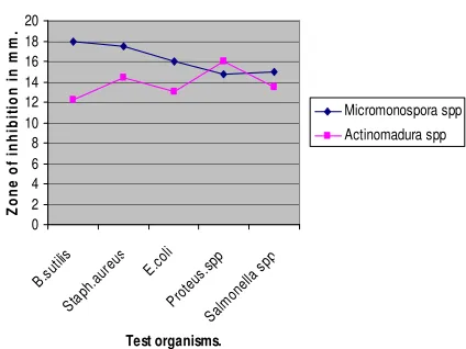
Fig 2. Antagonastic activity of putative isolates
0 2 4 6 8 10 12 Bacil lus su
btilis Pseu dom onas spp Enter obac
ter a erog
enosaE.c oli Kleb siella spp Prot eus spp Salm onel
la sp p
Shige lla sp
p
Stap h. A
ureu s Test Microorganisms N o . o f P u ti ti v e is o la te s Primary screening Secondary screening 0 2 4 6 8 10 12 14 16 18 20 B.su tilis Stap h.au
reus E.co li Prot eus. spp Salm onel
Table 1. Recovery of microorganisms (CFU/g Sediment sample) on hair hydrolysate agar (HHVA) from sediment.
*Nystatin (50µg/ml) +Nalidixic acid (20µg/ml)
Morphology
Only two strains did not produce aerial spore masses on oatmeal agar. Twenty-three strains produced the specific color of substrate mycelium. Only four strains produced diffusible pigments. Strain KW01 produced green pigment on oatmeal agar. Distinguished white color of mycelium was found in five strains including strain KR01. . Strain KR01 produced branched mycelium and globose sporangia on aerial mycelium. Sporangiospores are formed by septation of hypha within sporangium. Strain KW02 produced branched mycelium and spores in characteristic longitudinal pairs on aerial mycelium. Strain KN02 produced no fragmenting branched mycelium. Subspherical spores are produced within spherical sporangia. Strain KS03 had scanty aerial mycelium and its spores are borne singly.
Chemical properties
Among 53 isolates which were defined as non-streptomycete
actinomycetes with preliminary selection, 46 strains had meso-DAP,
and it was found that preliminary selection on agar plate show 87% of efficiency for the discrimination as rare actinomycetes. Aspartic acid, lysine, and glycine were the main amino acids of 46 strains.
Whole cell sugar patterns of actinomycetes containing meso-DAP
were defined on the basis of previous work. Twenty strains had xylose either alone or with arabinose or rhamnose, which belongs to pattern D (Table 2). Eleven strains had madurose, and their sugar pattern belongs to B. Four strains containing L-DAP had no characteristic sugars (pattern C). Whole organism methanolysates of the isolates contained fatty acids having 15 to 18 carbon chains, which are commonly found in prokaryotes.. While Streptomyces had
saturated iso-fatty acid of 15, 16 and 17 carbon numbers, and
antiiso-fatty acids of 15 and 17 carbon numbers, the isolates had n-heaxadecanoic acid (n-C16:0) as well as 14-methylpentadecanoic acid (i-C16:0),
Methyltetradecanoic acid (i-C15:0) and
12-methyltetradecanoic acid (a-C15:0) as the major species (Table 2).
Also, cis-15-methylhexadecenoic acid isomers (i-C17:1),
14-methylhexadecanoic acid (a-C17:0), and 10-methyl hexadecanoic acid (10-Me-C17:0) were the attendant fatty acids of the strains. Branched fatty acids were dominant in all cases, constituting 63.6% of the total fatty acids on average, and linear ones, 15.1%. The unsaturated fatty acids, mostly hexadecenoic acid isomers (C16:1) and heptadecenoic acid isomers (C17:1), accounted for 16.3% of the total. Most of the test strains had menaquinones of 9 isoprene units, designated as 9 with 1 to 4 double bonds saturated, MK-9(H2)~MK-9(H8). MK-9(H4) or MK-9(H6) was dominant in all cases.
Two strains had MK-10(H4) and MK-10(H6).
Antimicrobial Activity
Out of 53 actinomycetes subjected for primary screening process, only 13 isolates showed the activity against test organisms. Of the 13 isolates, 02 were active against only gram negative organism, 04 against gram positive organisms and 07 against both gram positive and gram negative organisms. Among them, 11 of the
isolates were active against Bacillus subtilis, 09 against
Staphylococcus aureus, 07 against Escherichia coli, 05 against Salmonella typhi and 06 against Proteus species.
Out of the 13 isolates that were subjected for the secondary
screening, 09 isolates were active against Bacillus subtilis, 06
against Staph. aureus, 07 against E. coli, 03 against Proteus species and 01 against Salmonella typhi.
Minimum inhibitory concentration
The minimum inhibitory concentration for the extract from Actinomadura spp. was 5mg/ml and that from Micromonospora spp was 1.20mg/ml.
Identification of isolates
Identification of the isolates was carried out using morphological and chemical properties. Discrimination between actinoplanetes and maduromycetes was primarily done by sugar pattern. Diagnostic sugar of the latter is madurose, while the former is xylose (Table 2). Main menaquinone profile was the useful criteria
in actinoplanetes. Dactylosporangium whose manaquinone is
MK-9(H6) and MK-9(H8) was distinguished from Actinoplanes and
Micromonospora. Discrimination between Actinoplanes and Micromonospora whose sugar pattern and menaquinone profile are
similar was carried out with fatty acid profile and spore
ornamentation. Actinoplanes had I C16:0 as main fatty acid more
than 50%. In contrast, Micromonospora had i-C15:0, i- C16:0, and
a-C15:0 as major ones. Because of similar sugar and main
menaquinone profile of maduromycetes, discrimination between
Microbispora and Actinomadura was carried out with fatty acid profile
and spore ornamentation. The former had 10-Me- C17:0 as
attendant fatty acid about 14% in addition to major fatty acid i-C16:0.
The latter had i-C14:0, i-C16:0 and i-C17:1 as main fatty acids in a
similar ratio of 20%. Characteristic longitudinal spore pairs are found
in aerial mycelium of Microbispora. Sporangiospores are formed by
septation of hypha within sporangium of Actinomadura.
Pretreatment Rare
actinomycetes
Streptomyces Other Bacteria Fungi
Control 3.3×10⁴ 2.6×10⁵ 6.8×10⁵ 2.4×10⁵
Control+Antibiotics* 2.8×10⁴ 2.2×10⁵ 3.6×10⁵ 3.4×10⁴
Dry heat (100oC) for 1 h 2.4×10² 2.4×10³ 6.4×10³ 2.2×10²
Air dry for 24h 4.8×10⁴ 1.3×10⁶ 5.2×10⁵ 2.8×10⁴
Wet heat (70oc) for 15 min 1.5×10⁵ 1.6×10⁴ 8.2×10⁴ -
Table 2. Identification of isolates with morphological and chemical properties.
Group/genus Amino acid Whole cell sugar
pattern
Fatty acid pattern
Major menaquinone
(MK)
Morphology No. of
Group/genus Substrate
mycelium color
Diffusible pigment Actinoplanes Meso-DAP,Glycine Ara,Xyl/Xyl i-C16:0 -9(H4)/-9(H4, H6) Orange/Yellow - 8
Micromonospora Meso-DAP,Glycine Ara,Xyl/Ara,Xyl/R hm
a-C15:0 i-C16:0
-9(H4)/-10(H4, H6) Brown/Yellow - 7
Microbispora Meso-DAP Ara,Gal,Mad i-C16:0 i-C16:0 10-Me-C17:0
-9(H0,H2,H4)/-9(H4) Pink/ Orange
- 5
Actinomadura Meso-DAP Ara,Gal,Mad i-C14:0 i-C16:0 i-C17:1
-9(H0, H2, H4) White - 5
Ara, Arabinose; Xyl, Xylose; Rhm, Rhamnose; Gal, Galactose; Mad, Madurose. i-C16:0, 14-methylpentadecanoic acid; i-C15:0, 13-methyltetradecanoic acid; a-C15:0, 12-methyltetradecanoic acid; 10-Me-C17:0, 10-methylhexadecenoic acid; i-C14:0, 12-methyltriadecanoic acid; and i-C17:1, cis, 15-methylhexadecenoic acid
Thin layer chromatography
The spot given by the extract of Actinomadura spp was a
circular with Rf value 0.88 and that of Micromonospora spp was an
extended spot with Rf value 0.85. The fluorescence colours of the spots were greenish yellow. The reference antibiotic, Tetracycline, didn’t move with the solvent system. .
DISCUSSION
Various pretreatment procedures and selective media were applied to assess the optimal conditions for the isolation of rare actinomycetes from soil. Pretreatment of wet-heating for 15 min at
70oC and phenol treatment of soil suspension were the most
effective methods for the isolation of those microorganisms. Hair hydrolysate vitamin agar (HHVA) was the most suitable medium for the recovery of rare actinomycetes. Isolation medium was supplemented with those antibiotics in succeeding experiments. The dominance of other bacteria and fungal contamination inhibited the colonization of actinomycetes on isolation medium. When antifungal agents such as nystatin (50 µg/ml) and nalidixic acid (20 µg/ml) were supplemented into the isolation medium, the number of fungi decreased.
For antimicrobial activity the putative isolates of primary screening when subjected to secondary screening, showed different activity from that of primary screening; some of the active isolates didn’t show the activity in the secondary screening while some showed little activity and some showed improved activity. According to Bushell (1993), during the screening of the novel secondary metabolite, actinomycetes isolates are often encountered which show antibiotic activity on agar but not in liquid culture.
The result of primary and secondary screening revels that most of the active isolates were active against gram positive bacteria
(Bacillus subtilis and Staph. aureus) than gram negative bacteria.
The reason for different sensitivity between gram positive and gram negative bacteria could be ascribed to the morphological differences between these microorganisms, gram negative bacteria having an
outer polysaccharide membrane carrying the structural
lipopolysaccharide components. This makes the cell wall
impermeable to lipophilic solutes, The gram positive should more susceptible having only an outer peptidoglycan layer which is not an effective permeability barrier (Scherrer & Gerhardt, 1971).
Although various biochemical tests were performed, it was unable to identify the actinomycetes up to species level due to the lack of other tests. According to Kutzner (1972) for proper identification of genera and species of actinomycetes, besides morphological and physiological properties, various other biochemical properties such as cell wall chemo type, whole-cell sugar pattern, peptidoglycan type, phospholipids type and G+C % of DNA should be determined.
The minimum inhibitory concentration (MIC) for the
antimicrobial extracted from Streptomyces spp was 5mg/ml and that
from Micromonosporaso spp was 1.25mg/ml. This show that the
antimicrobial from Micromonospora spp was more active than from
Streptomyces spp but there are various factors affecting the activity. The Streptomyces spp can be a poor fermenter than the later one or the solvent used for extraction may not be suitable for it or the compound may not be properly extracted by the solvent.
The MIC is not a constant for a given agent, because it is affected by the nature of the test organism used, the inoculum size, and the composition of the culture medium, the incubation time, and aeration.
For complete characterization of an antibiotic it should be isolated in pure form as a single component but this is impractical in a screening programme like this. However, a little effort was made in this approach. According to the TLC separation, the two extracts
yielded components with Rf values may be the antibacterial
compounds. (By referring to bioautogram). This may mean that the same compounds are responsible for antibacterial activity of those isolates. Although the antimicrobial agents obtained in this study can’t be declared as new antibiotics, there is the probability of finding new antibiotics in Karwar because of its wide biodiversity.
REFERENCES
[1] Berdy, J, 1967. Recent developments of Antibiotic Research and
[2] Bergey’s manual of determinative bacteriology, 2000. Actinomycetales. 9thedition.
[3] Englyst, H.N. and J.H. Cummings. 1984. Simplified method for
the measurement of total non-starch polysaccharides by gas-chromatography of constituent sugars as alditol acetates. Analyst. 101, 937-942.
[4] Goodfellow, M. 1989. Suprageneric classification of
actinomycetes. p. 2333-2339. In S.T. Williams, M.E. Sharpe,
and J.G. Holt (eds.), Bergey's manual of systematic
bacteriology. Williams and Wilkins, London.
[5] Hayakawa, M. and H. Nonomura. 1987a. Efficacy of artificial Vol.
39, No. 1 Selective Isolation of Rare Actinomycetes from Forest Soil 23 humic acid as a selective nutrient in HV agar
used for the isolation of soil actinomycetes. J. Ferment.
Technol. 65, 609-616.
[6] Hayakawa, M. and H. Nonomura. 1987b. Humic acid-vitamin
agar, a new medium for the selective isolation of soil actinomycetes. J. Ferment. Technol. 65, 501-509.
[7] Iwai, H. and Y. Takahashi. 1992. Selection of microbial sources
of bioactive compounds. p. 281-302, In S.Oumra (ed.), The
Search for bioactive compounds from microorganisms. Springer-Verlag, New York.
[8] Kim, C.J., K.H. Lee, A. Shimazu, O.S. Kwon, and D.J. Park.
1995. Isolation of rare actinomycetes on various types of soil. K. J. Appl. Microbiol. Biotechnol. 23, 36-42.
[9] Kim, C.J., K.H. Lee, O.S. Kwon, A. Shimazu, and I.D. Yoo. 1994.
Selective isolation of actinomycetes by physical pretreatment of soil sample. K. J. Appl. Microbiol. Biotechnol. 22, 222-225.
[10]Kawato, M., Shinobu, R., 1959. A simple technique for the
microscopical observation, memoirs of the Osaka University
Liberal Arts and Education, 114.
[11]Lechevalier, M.P., A.C. Horan and H.A. Lechevalier. 1971. Lipid
composition in the classification of nocardiae and mycobacteria. J. Bacteriol. 105, 313-318.
[12]Minnikin, D.E. and M. Goodfellow. 1980. Lipid composition in the
classification and identification of acid-fast bacteria, p. 189-
255. In M. Goodfellow and R.G. Board (eds.), Microbiological
classification and identification, Academic Press, London
[13]Minnikin, D.E., A.G. O'Donnell, M. Goodfellow, G. Alderson, M.
Athalye, A. Schaal, and J.H. Parlett. 1984. An integrated procedure for the extraction of bacterial isoprenoid quinines and polar lipids. J. Microbiol. Methods 2, 233-241.
[14]Nolan, R.D. and T. Cross. 1988. Isolation and screening of
actinomycetes, p. 2-8. In M. Goodfellow, S.T. Williams, and M.
Mordarski (eds.), Actinomycetes in biotechnology. Academic
Press, London.
[15]O'Donnell, A.G. 1985. Numerical analysis of chemotaxanomic
data, p. 403-414. In M. Goodfellow, D. Jones and F.G. Priest
(eds.), Computer assisted bacterial systematics, Academic Press, London.
[16]Okami, Y. and K. Hotta. 1988. Search and discovery of new
antibiotics, p. 39. In M. Goodfellow, S.T. Williams, and M.
Mordarski (eds.), Actinomycetes in biotechnology. Academic
Press, London.
[17]Okazaki, T. 1987. Rare actinomycetes -new breed of
actinomycetes. J.Microorganism 3, 453-461.
[18]Seong, C.N., Y.S. Kim, K.S. Baik, S.D. Lee, Y.C. Hah, S.B. Kim,
and M. Goodfellow. 1999. Mycolic acid-containing
actinomycetes associated with activated sludge foam. J.
Microbiol. 37, 66-72.
[19]Waksman, S.A., 1968. Actinomycetes; Nature, Formation and
Activities, Applied Microbiology.5, 235-293.
[20]Watve, M.G. et al., How many antibiotics are produced by the
genus Streptomyces. Arch. Microbiol. 2001, 176, 386-390.
[21]Bérdy, J. Bioactive microbial metabolites. J. Antibiot. 2005, 58,
1-26.
[22]Lazzarini, A.; Cavaletti, L.; Toppo, G.; Marinelli, F. Rare genera
of actinomycetes as potential producers of new antibiotics. Antonie van Leeuwenhoek 2000, 78, 388-405.
[23]Ara, I.; Kudo, T.; Matsumoto, A.; Takahashi, Y.; Omura, S. 2007.
Nonomuraea maheshkhaliensis sp.nov., a novel actinomycete
isolated from mangrove rhizosphere mud. J. Gen. Appl.
Microbiol. 53, 159-166.
[24]Yi, H.; Schumann, P.; Sohn, K.; Chun, J. 2004. Serinicoccus
marinus gen. nov., sp. nov., a novel actinomycete with
L-ornithine and L-serine in the peptidoglycan. Int. J. Syst. Evol.
Microbiol. 54, 1585-1589.
[25]Fenical, W.; Jensen, P.R.2006. Developing a new resource for
drug discovery: marine actinomycete bacteria. Nature Chem.
Biol. 2, 666-673.
[26]Gao, H.; Hong, K.; Zhang, X.; Liu, H.W.; Wang, N. L.; Zhang, L.;
Yao, X.S. 2007.Polyhydroxylated sterols and new sterol fatty
esters from the mangrove fungus Aspergillus awamori
exhibiting potent cytotoxic activity. Helv. Chim. Acta 90, 1165-1178.
[27]Pisano, A. M; Sommer, J.M.; Lopez, M.M. 1986. Application of
pretreatment for the isolation of bioactive actinomycetes from marine sediments. Appl. Microbiol. Biotechnol. 25, 285-288.
[28]Eccleston, G. P.; Brooks, P. R.; Kurtböke, D. I. 2008.The
occurrence of bioactive micromonosporae in aquatic habitats of the Sunshine Coast in Australia. Mar. Drugs . 6, 243-261.
[29]solvent extraction method following the process described by
Westley et.al, 1979.and Liu et.al, 1986
[30]Williams, P.G.; Buchanan, G.O.; Feling, R.H.; Kauffman, C.A.;
Jensen, P.R.; Fenical, W. 2005.New cytotoxic
salinosporamides from marine actinomycete Salinispora
tropica. J. Org. Chem.70, 6196-6203.
[31]Bredholt, H.; Fjærvik, E.; Johnsen, G.; Zotchev, S.B.