Prehospital: Emergency Care
Eleventh Edition
Chapter 31
Head Trauma
Slides in this presentation contain hyperlinks. JAWS users should be able to
Learning Readiness
• EMS Education Standards, text p. 927.
• Chapter Objectives, text p. 927.
• Key Terms, text p. 927.
Setting the Stage
• Overview of Lesson Topics
– Anatomy of the Skull and Brain
Case Study Introduction
EMTs Matt Brooks and Luis Garcia have arrived on the scene of a motorcycle collision, involving a single patient who was ejected from his bike when he struck the side of a car. The patient, a male in his 30s, was not wearing a
Case Study
(1 of 3)• What are the first steps the EMTs must take?
• What findings would lead the EMTs to suspect traumatic brain injury?
Introduction
• Head injuries can be life threatening, and can lead to permanent disability.
• The patient’s mental status can make assessment difficult.
Anatomy of the Skull and Brain
(1 of 5)• The Skull
– The cranial skull surrounds the brain.
– The face is made up of 14 bones.
– The basilar skull is the weakest portion.
– Some of the basilar skull bones are thin and
Anatomy of the Skull and Brain
(2 of 5)• The Brain
– Cushioned by cerebrospinal fluid (CSF)
▪ CSF leaking from the nose or ears is an indication of basilar skull fracture.
– The Meninges
▪ Three layers surround the brain.
– Outermost is the dura mater
– The next layer is the arachnoid
Anatomy of the Skull and Brain
(3 of 5)• The Brain
– The Meninges
▪ Bleeding that occurs between the dura mater and the skull is called epidural.
▪ Subdural bleeding occurs beneath the dura and is usually venous.
▪ Bleeding that occurs between the arachnoid
Anatomy of the Skull and Brain
(4 of 5)• The Brain
– Parts of the Brain
▪ Cerebrum
– Two hemispheres.
– Each hemisphere has four lobes.
– Responsible for conscious and sensory functions, emotion, personality.
▪ Cerebellum
Anatomy of the Skull and Brain
(5 of 5)• The Brain
– Parts of the Brain
▪ Brainstem
– Made up of the pons, midbrain, and medulla oblongata.
– Controls automatic functions, including cardiac, respiratory, and vasomotor function.
Head Injury
(1 of 33)• Scalp Injuries
– Soft tissue injuries.
– Blood vessels do not constrict well; bleeding can be profuse.
Head Injury
(2 of 33)• Skull Injuries
– Deformity requires extreme trauma.
– Most skull fractures are linear.
– Depressed skull fracture occurs when the bone ends are pushed in toward the brain.
– Skull fractures can be open or closed.
– A basilar skull fracture involves the floor of the cranium.
Head Injury
(3 of 33)• Brain Injuries
– Swelling and bleeding within the skull can increase the pressure within the skull, which decreases brain tissue perfusion.
Head Injury
(4 of 33)• Pathophysiology of Traumatic Brain Injury
– Traumatic brain injuries can occur from penetrating trauma, blunt trauma, or secondary injury.
Head Injury
(5 of 33)• Pathophysiology of Traumatic Brain Injury
– Primary Brain Injury
▪ The primary brain injury is the result of trauma to the brain that occurs at the time of insult from
Head Injury
(6 of 33)• Pathophysiology of Traumatic Brain Injury
– Secondary Brain Injury
▪ Traumatic brain injury will be worsened by:
– Hypoxemia
– Hypercarbia
– Hypoglycemia
– Hyperglycemia
– Hyperthermia
Head Injury
(7 of 33)• Pathophysiology of Traumatic Brain Injury
– Secondary Brain Injury
▪ To minimize secondary injury maintain:
– A patient airway
– Adequate ventilation
– SpO2 ≥ 94%
– Systolic BP > 90 mmHg
– Normal body temperature
Head Injury
(8 of 33)• Pathophysiology of Traumatic Brain Injury
– Secondary Brain Injury
▪ Stop seizures as quickly as possible.
Head Injury
(9 of 33)• Pathophysiology of Traumatic Brain Injury
– Brain Herniation
▪ Increased intracranial pressure from bleeding or swelling forces the brain out of its normal position.
▪ Compression of the brain causes dysfunction.
Head Injury
(10 of 33)• Pathophysiology of Traumatic Brain Injury
– Brain Herniation
▪ Brain herniation signs and symptoms:
– Dilated or sluggish pupil on one side
– Weakness or paralysis
– Severe alteration in consciousness
– Abnormal posturing
– Abnormal breathing pattern
Head Injury
(11 of 33)• Types of Head and Brain Injuries
– Closed and Open Head Injuries
▪ An open wound to the head does not signify a more severe brain injury.
▪ The lack of a wound to the head does not indicate a lesser brain injury.
▪ Closed head injury- The scalp can be lacerated but the skull remains intact.
Head Injury
(12 of 33)• Types of Head and Brain Injuries
– Brain injury from shearing, tearing, and stretching nerve fibers is known as a diffuse axonal injury
(DAI).
– DAI interferes with transmission of nerve impulses.
– DAI is related to severe acceleration and deceleration forces.
Head Injury
(13 of 33)• Types of Head and Brain Injuries
– Concussion
▪ Mild DAI.
▪ Presentation ranges from momentary confusion to brief complete loss of responsiveness.
▪ Usually results in headache.
Head Injury
(14 of 33)• Types of Head and Brain Injuries
– Contusion
▪ Bruising and swelling of the brain
▪ Can accompany concussion
▪ May or may not cause increased intracranial pressure
▪ Caused by coup/contrecoup or
Head Injury
(15 of 33)• Types of Head and Brain Injuries
– Subdural Hematoma
▪ Collection of blood between dura mater and arachnoid.
▪ Results from damage to bridging veins in skull.
Head Injury
(16 of 33)• Types of Head and Brain Injuries
– Subdural Hematoma
▪ Acute: signs and symptoms begin immediately.
▪ Occult: signs and symptoms are delayed for days to weeks.
Head Injury
(17 of 33)• Types of Head and Brain Injuries
– Epidural Hematoma
▪ More rare, but extremely emergent.
▪ Often associated with fracture of the temporal skull.
▪ Bleeding is rapid and severe, causing a rapid increase in intracranial pressure.
Head Injury
(18 of 33)• Types of Head and Brain Injuries
– Laceration
▪ May occur with closed or open injury.
▪ Results in bleeding and nervous system disruption.
Case Study
(2 of 3)Matt immediately performs in-line spinal stabilization and uses a jaw-thrust maneuver to open the airway, as Luis prepares to suction the airway.
The patient has bleeding from his nose, a hematoma over the frontal area of his head, and many abrasions to his
face.
His breathing is deep and rapid; his radial pulse is strong at about 60 per minute.
Case Study
(3 of 3)• What immediate indications are there of a traumatic brain injury?
• What additional information do the EMTs need to obtain through their assessment?
Click on the Sign That Is Most Consistent
with a Concussion
A. Unequal pupils
B. Paralysis on one side of the body
C. Nonpurposeful response to pain
Head Injury
(19 of 33)• Assessment-Based Approach: Head Injury
– Scene size-up
▪ Unresponsiveness or altered mental status suggest head injury.
▪ Signs of head injury may be apparent.
▪ Never assume that mental status changes in a trauma patient are due to drug or alcohol
Head Injury
(20 of 33)• Assessment-Based Approach: Head Injury
– Primary assessment
▪ Manual in-line stabilization of the spine.
▪ If needed, establish an airway with a jaw-thrust maneuver.
▪ Maintain an SpO2 of 94% or above.
Head Injury
(21 of 33)• Assessment-Based Approach: Head Injury
– Primary assessment
▪ Determine the mental status with AVPU.
– Alert
– Verbal
– Painful
Head Injury
(22 of 33)• Assessment-Based Approach: Head Injury
– Primary assessment
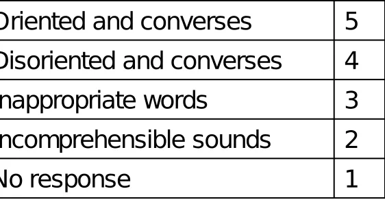
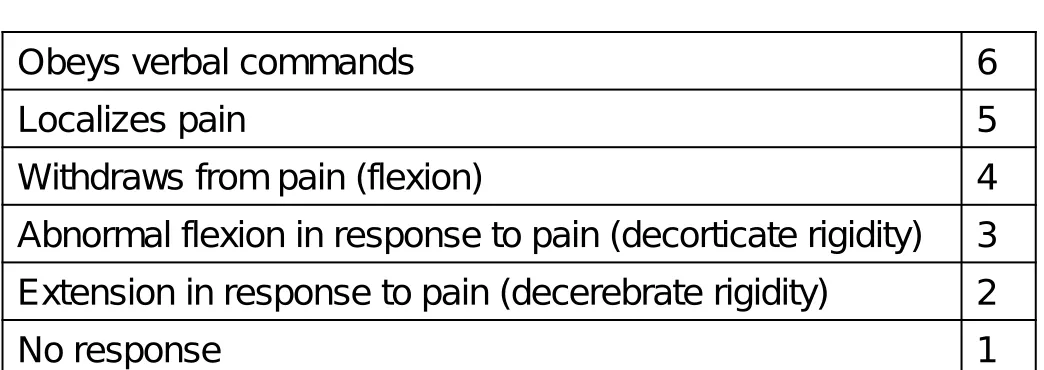
▪ The Glasgow Coma Scale is a more precise way to determine the mental status.
▪ The GCS is a measure of the patient’s eye
Table 31-1 Glasgow Coma Scale
(1 of 2)Eye Opening
Spontaneous 4
To verbal command 3
To pain 2
No response 1
Verbal Response
Oriented and converses 5
Disoriented and converses 4
Table 31-1 Glasgow Coma Scale
(2 of 2)Motor Response
Obeys verbal commands 6
Localizes pain 5
Withdraws from pain (flexion) 4
Abnormal flexion in response to pain (decorticate rigidity) 3
Extension in response to pain (decerebrate rigidity) 2
Head Injury
(23 of 33)• Assessment-Based Approach: Head Injury
– Secondary assessment
▪ Perform a physical exam.
▪ Anticipate altered pain response.
▪ Check vital signs.
Examine the Head for Deformities,
This Patient was Riding on a B
M
X Track, Doing
Flips on a Bicycle without a Helmet or Other
Protective Gear
Head Injury
(24 of 33)• Assessment-Based Approach: Head Injury
– Secondary assessment
▪ Vital signs
– Record every five minutes.
– Blood pressure
• High systolic BP indicates increasing intracranial pressure.
Head Injury
(25 of 33)• Assessment-Based Approach: Head Injury
– Secondary assessment
▪ Vital signs
– Pulse
• Increased rate indicates another source of bleeding.
• Decreased rate indicates increasing
Head Injury
(26 of 33)• Assessment-Based Approach: Head Injury
– Secondary assessment
▪ Vital signs
– Respirations
• Assess the rate, depth, and pattern.
Head Injury
(27 of 33)• Assessment-Based Approach: Head Injury
– Secondary assessment
▪ If the following signs are present, consider hyperventilation at rate of 20/minute.
– Unequal pupils
– Fixed pupil
– Cushing reflex
– Hemiplegia or hemiparesis
Head Injury
(28 of 33)• Assessment-Based Approach: Head Injury
– Secondary assessment
▪ History
– Ask questions relevant to head injury.
– Sometimes an injury to the head, days or
Head Injury
(29 of 33)• Assessment-Based Approach: Head Injury
– Secondary assessment
▪ Look for pertinent signs and symptoms
▪ Retrograde amnesia—the patient is unable to remember circumstances leading up to the incident
Head Injury
(30 of 33)• Assessment-Based Approach: Head Injury
– Emergency Medical Care
▪ Standard Precautions.
▪ Manual in-line spinal stabilization.
▪ Maintain the airway, breathing, and oxygenation.
Head Injury
(31 of 33)• Assessment-Based Approach: Head Injury
– Emergency Medical Care
▪ Monitor the airway, breathing, pulse, and mental status for deterioration.
▪ Control bleeding.
– Do not apply pressure to an open or depressed skull injury.
Head Injury
(32 of 33)• Assessment-Based Approach: Head Injury
– Emergency Medical Care
▪ Be prepared for seizures.
▪ Continuously monitor mental status.
Head Injury
(33 of 33)• Assessment-Based Approach: Head Injury
– Reassessment
▪ Every five minutes.
Case Study Conclusion
(1 of 3)Matt and Luis recognize the indications of severe traumatic brain injury. Their priorities are continuing to manage the airway, breathing, and oxygenation while immobilizing the spine in preparation for transport.
Case Study Conclusion
(2 of 3)At the hospital, the patient is found to have a severe traumatic brain injury with a subdural hematoma and
increased intracranial pressure. He is intubated and there is immediate intervention to relieve the intracranial
Case Study Conclusion
(3 of 3)The patient is admitted to critical care. Despite rapid and appropriate care by all involved, his prognosis for recovery is poor, as it is for many patients with devastating head
Lesson Summary
• Brain injuries can be devastating, leading to death or disability.
• Increasing ICP worsens damage to the brain and can lead to herniation.
• Treatment is aimed at protecting the spine and managing airway, breathing, oxygenation and circulation.
Correct!
A concussion is characterized by an initial period of unresponsiveness or confusion followed by improving mental status.
Incorrect
(1 of 3)Unequal pupils are an indication of severe brain injury with increasing intracranial pressure.
Incorrect
(2 of 3)Paralysis on one side of the body is an indication of severe brain injury with increasing intracranial pressure.
Incorrect
(3 of 3)Nonpurposeful response to pain is an indication of severe brain injury with increasing intracranial pressure.