DEVELOPMENT REPORT
Development of an implicit method
for directing weight shifting to the affected side
in patients with stroke: a proof of concept study
Kazuhiro Yasuda
1*†, Kenta Saichi
2†, Yu Kitaji
3, Hiroaki Harashima
3and Hiroyasu Iwata
2Abstract
Weight-shift training during stroke rehabilitation requires patient effort, potentially causing leg spasticity and anxiety, which disturb motor learning. The purpose of this study was (1) to devise an implicit guidance method for weight bearing that uses vibratory cues (and is therefore low exertion) and (2) to determine if the implicit guidance method is feasible. The first experiment included 12 healthy subjects. We conducted an experiment to produce a Weber’s fraction capable of calculating a just-noticeable difference during a weight-shifting task. We then applied this Weber’s fraction to a weight-shifting task in a patient with stroke. Using the implicit guidance method, the patient did not perceive an increase in weight bearing while weight shifting. Furthermore, the implicit guidance method appeared to reduce anxiety during training. This implicit guidance system warrants further investigation.
Keywords: Stroke, Rehabilitation, Postural control, Weight shift, Fear of falling, Human–machine interaction, Wearable device, Just-noticeable difference, Perception
© The Author(s) 2017. This article is distributed under the terms of the Creative Commons Attribution 4.0 International License (http://creativecommons.org/licenses/by/4.0/), which permits unrestricted use, distribution, and reproduction in any medium, provided you give appropriate credit to the original author(s) and the source, provide a link to the Creative Commons license, and indicate if changes were made.
Background
Reduced postural control, caused by sensory and motor impairments after stroke, impacts activities of daily life, and affects independent walking [1, 2]. Improv-ing postural control in patients after stroke is extremely important and may help them lead independent lives, participate in society, and maintain optimum levels of health. Asymmetrical weight bearing and weight-shifting ability correlate with gait [3]. Stroke-related limitations in weight shifting manifest in the anterior–posterior (AP) and medio–lateral (ML) directions [4, 5], with marked limitations typically noted on the affected side [6, 7]. Additional limitations include reduced weight-shifting speed and accuracy [8, 9], reduced single leg support ability on the affected side [10, 11], and reduced floor reaction force on the affected side [12, 13]. Over time, patients who demonstrate these limitations form motor
patterns that use only part of their support base as means of compensating for reduced postural control.
In clinical settings, these issues are often addressed through targeted weight-shifting training. Patients with stroke must expend considerable effort to weight shift, which often induces associated reactions (AR), and spas-ticity [14, 15]. One example of AR is where muscular strain in the paretic upper limb temporarily increases, upon exertion of the lower limb. Spasticity involves increased muscular strain caused by motor paralysis, and is exacerbated by muscle extension and exertion [14, 15].
Weight-shifting tasks can cause anxiety. Previous research into the relationships between emotional changes and postural control found that maintaining a standing position on an elevated surface causes “stiffness behavior” where the leg muscles display excessive ten-sion [16, 17]. This phenomenon helps maintain postural stability during standing [18], whereas in complex, eve-ryday environments it impedes trouble-free movement, increasing the risk of falls [19]. In patients with a history of stroke and hemiplegia, increased leg muscle tension exacerbates spasticity in the paretic limb, thereby imped-ing left–right symmetrical gait.
Open Access
*Correspondence: [email protected]
†Kazuhiro Yasuda and Kenta Saichi contributed equally to this paper 1 Research Institute for Science and Engineering, Waseda University, 3-4-1
Okubo, Shinjuku-ku, Tokyo 169-8555, Japan
One means of engineering support for limited weight-shifting is through the use of visual or auditory bio-feedback (BF). Previous studies examined foot center of pressure movement in a visual display [20–23], the use of visual cues for recognizing asymmetry [24], or visual and auditory feedback [23, 25, 26]. These methods enable the perception of foot pressure and weight asymmetry, thereby allowing the conscious modification of move-ment and weight shifting. Mostly, visual BF is used in clinical practice; however, patients are highly dependent on vision in the early period following the initial onset of stroke [27, 28]. In detail, Marigold et al. reported that patients with stroke tend to more heavily rely on visual input to maintain frontal plane (mediolateral) sway [29]; thus, sensory supplementation, particularly in the form of tactile BF, may help avoid inadequate sensory inte-gration. Furthermore, review articles concluded that visual or auditory BF does not improve functional bal-ance ability [30, 31]. Several cases have used visual BF up until now during balance training; however, this training method has not been sufficiently effective and could also reinforce visual reliance.
In a clinical trial, vibrotactile BF improved body sway during quiet standing in patients with vestibular dis-orders and Parkinson disease [32–36], and we recently found that the immediate beneficial effects on postural stability in patients with stroke [37]. In addition, several studies used haptic feedback as a navigation for weight-shifting [38, 39]. A study of Parkinson disease reported that weight induction by stimulus of visual and haptic feedback combined led to immediate increases in the anteroposterior and left–right limits of stability (LOS)
[39]. However, no studies have applied haptic BF to weight shifting of the paretic leg in stroke patients. Fur-ther, no system has been devised to reduce the aforemen-tioned AR, and the anxiety experienced, which are typical in stroke patients.
Thus, the purpose of this study was to devise a method of implicit weight-bearing guidance, using haptic stimu-lation. A secondary objective was to examine the feasibil-ity of implicit guidance in patients with stroke, and verify the validity of the technique in advance of future, large-scale investigations.
Methods
Overview of the haptic‑based weight‑shift system
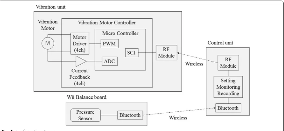
The system consists of a Wii balance board (Wiiboard, Nintendo, Japan) for capturing center of foot pres-sure (CoP) data. The subject was fitted with a vibration unit, with four vibrators worn around the pelvic girdle, which displayed CoP motion, coupled with a personal computer (PC) (Fig. 1). We measured CoP using a Wii balance board (WBB), with signals relayed to a PC by Bluetooth. CoP measurements (sampling) were taken every 20 ms, and the transmission period was 20 ms. The software screen displayed CoP and target weight bear-ing in real time (Fig. 2). The target weight-shift length can be set at any position from 0 to 1vibration motors (Z7AL2B169208200% of body weight by inputting the subject’s weight. Vibrations were transmitted to a vibra-tion unit (i.e., pelvic belt) using a USB connecvibra-tion wire-less module (nRF24LE1-F16Q32, Nordic, Norway). The communication cycle is 20 ms. The frequency band-width of the module ranges from 2400 to 2525 MHz.
The vibration unit includes four built-in , KOTL, China). Vibration motors are mapped on a bony ridge (i.e., the bilateral anterior and posterior superior iliac spines) to efficiently convey vibrotactile stimulation (Fig. 3b). The rate of the vibration motor speed is 12,000 ± 2500 rpm. The intensity of vibrations can be changed by PC accord-ing to the degree of patient paralysis. Until the CoP posi-tion reaches the target weight-shift length, the device will continue to vibrate. Vibrations cease once the subject reaches the target weight-shift length.
Implicit weight‑shift length‑extension algorithm
Weight-shifting length is automatically determined by an implicit weight-shift length-extension. To reduce exces-sive voluntary movements and fear of falling, we applied Weber’s Law to the sense of weight, increasing the length of weight shifting [40]. This law enables calculation of the just-noticeable difference (JND), which is the mini-mum amount of change that can be perceived. In the perceptual literature, JND represents the smallest detect-able change by which a subject can reliably discrimi-nate between a comparator and an original stimulus. For example, in a size-discrimination task a participant might be asked to verbally report whether the length of a visually presented line (i.e., the comparator) differs from the length of a reference line. In this context, JNDs are defined statistically with the appropriate detection of a magnitude change based on an experimentally deter-mined criterion [41]. Equation (1) represents Weber’s Law, where W is the current amount of weight bearing, K is the Weber ratio, and ΔW is the JND
Experiment to derive the Weber ratio for weight‑shift task
Because the Weber ratio for sense of weight bearing has not been used previously, we conducted an experiment with 12 young, healthy subjects to derive the Weber ratio. Subjects included 12 healthy adults who provided written
(1) W = K × W
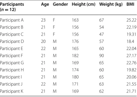
consent for participation (mean age 21.25 years, SD 0.72, male 9 participants, female 3 participants). Body mass index (BMI) for each participant is shown in Table 1. The present study was performed with approval of the Ethical Review Board of Waseda University.
Subjects stood with their feet 30 cm apart, and the experimental task was repeated weight-shift task to both left and right leg (Fig. 3a). First, we instructed subjects to shift 60% of their body weight to the left side by using a vibratory cue on the left side waist (i.e., L1/2; Fig. 2). When the weight shift reached 60% of the body weight, vibrations for the left side stopped, and the vibrator acti-vated on the right side; this process was repeated. After weight shifting once to the left and once to the right, we increased the percentage of weight shifting, based on the set Weber ratio (initial value 0.05), without communi-cating this change to the participants (Fig. 3c). The ini-tial value of 0.05 was based on preliminary experiments that found a somatic sensory Weber ratio of 0.03–0.3 [42]. This procedure was repeated at 90-second intervals, and after the experiment subjects reported if they felt an increase in the amount of weight shifting, according to two conditions (“Yes” = it increased; “No” = unchanged). After performing the weight-shift task using the ini-tial Weber ratio, the Weber ratio was either increased or decreased according to participants’ responses (decreased when the participant felt an increase in the amount of weight shifting; increased if the participant did not). Once we established the point where a change of 0.01 in the Weber ratio caused a change in response, decreases were made in 0.002 increments from the noticeable value to find the Weber ratio where increases for weight shifting were unnoticeable for each participant (Fig. 4).
Fig. 2 Screen shot of the software. On the software screen, the CoP and target weight bearing could be displayed in real time
Table 1 Characteristic of the participants
BMI body mass index
Participants
(n = 12) Age Gender Height (cm) Weight (kg) BMI
Participant A 23 F 163 67 25.22
Participant B 21 F 156 54 22.19
Participant C 21 F 156 47 19.31
Participant D 20 M 176 57 18.4
Participant E 22 M 165 60 22.04
Participant F 21 M 182 90 27.17
Participant G 21 M 169 65 22.76
Participant H 21 M 174 60 19.82
Participant I 21 M 180 65 20.06
Participant J 22 M 171 63 21.55
Results
Table 2 shows the participants’ Weber ratios. The mean Weber ratio was 0.045, SD 0.0078, maximum 0.054, and minimum 0.024, with a 95% CI of 0.040–0.050.
Examination of the test validity in patients
As a preliminary step toward a large-scale clinical trial, we applied the device to one patient with stroke to exam-ine method feasibility. In this examination, we conducted simulated weight-shift training using the derived Weber ratio, and examined the following variables: (1) percep-tion of vibratory stimuli: whether or not vibrapercep-tion cues could be perceived on the paretic side, (2) perception of increased weight bearing: whether or not the patient could perceive increases in weight bearing, (3) anxiety: anxiety during the task, and referent data on the influ-ence of training, (4) weight-bearing dose on the paretic side was measured before and after intervention, and comparisons made.
Table 3 shows patient characteristics. Our patient was a 73-year-old man with left hemiplegia (Brunstrom’s recov-ery stage; see Appendix [43]), caused by right cerebral infarction. The patient was able to walk independently, and had no cognitive or mental disorders (i.e., Mini-Men-tal State Examination score above 23 [44]; no dementia).
We familiarized the patient with the weight-bearing guidance by vibration device. (At this point, the patient was not informed that the amount of weight bearing would increase.) For the weight-shift task, the patient assumed a left–right symmetrical standing position, with feet 30 cm apart on the WBB, with eyes open. In these conditions, the patient focused on a marker placed approximately 2 m in front of him. In the pretest,
Fig. 4 Derivation process of the Weber ratio (WR). After the repeated weight-shift task, participants responded whether or not they felt an increase in the amount of weight shifting according to two condi-tions (response opcondi-tions “Yes; increased”, “No; unchanged”)
Table 2 Weber ratio in 60% of the weight-bearing
n number of participants SD standard deviation CI confidence interval
Par‑ ticipants (n = 12)
Weber
ratio SD Max value Min value 95%CI
0.045 ± 0.0078 0.054 0.024 0.040–0.050
Table 3 Characteristic of the patient
Brs Brunstrom’s recovery stage, LE Lower extremity, MMSE Mini mental state examination
Patient: F.M (n = 1)
Age 73
Gender Male
Paralyzed side Left
Days from onset 90
Superficial sensation Moderate
Pain sensation Normal
Vibratory sensation Moderate
Proprioception Moderate
Motor impairment Brs; LE/VI
MMSE 28/30
the patient performed the task of weight shifting to the paretic limb five times in order to evaluate baseline weight-shift ability. After the pre-testing, a continu-ous weight-shift tasks of both left and right sides was repeated over 70 s × 4 sessions with one-min rest inter-vals. During the weight-shift task, when the vibrations for one side stopped, he shifted his weight to the other side. The target weight-bearing dose was set at pre-test value (baseline) × 0.8. In doing so, the amount of weight bear-ing was increased for each session based on the Weber ratio. Lastly, we performed a post-test of the weight-shift task identical to the pre-test (i.e., weight shifting to the paretic limb five times to evaluate weight-shift ability).
The patient was questioned about his perception of vibratory stimuli applied from the left and right, and his perception of increased weight bearing in each ses-sion after completion of the post-test. We used a visual analog scale (VAS) to measure anxiety after the pre-test, and after the weight-shift training [45]. The VAS is fre-quently used to measure pain intensity. The pain VAS is a continuous scale comprised of a horizontal (HVAS) or vertical (VVAS) line, usually 10 cm (100 mm) in length, anchored by two verbal descriptors, one for each symp-tom extreme. VAS is a valid measure of fall anxiety, according to a recent study [46]. We asked the patient to indicate his fear of falling, on a scale from 1 to 10. The amount of weight bearing in the pre- and post-test was calculated using the data obtained from the WBB. For weight bearing, variables were compared pre-and post-testing. Statistical analyses were performed using a Wil-coxon signed-rank test as a non-parametric test because of the small sample size [47]. Significance level was set at
p < 0.05.
Results
The patient perceived the vibratory stimulation, applied from the left and right, on both the paretic and non-paretic side. Perception could therefore be used as a signal for weight shifting. The patient did not notice the increase in weight-shift between sessions. The VAS for anxiety was 5 (moderate) in the pre-test weight-shifting task, but 0 during the weight-shifting task. Compared with the pre-test, the amount of weight bearing was sig-nificantly increased from 67.34 (66.92–67.36) kg to 68.45 (68.44–69.24) kg in the post-test (p = 0.0431) with a medium effect size (r = 0.30) [48].
Discussion
In the present study, we applied a method of implicit weight-bearing guidance using vibratory cues. We cal-culated the Weber ratio during a weight-shift task, per-formed by young healthy individuals. In the range of 60% weight bearing, the Weber ratio was 0.045 (SD 0.0076).
We applied a repeated weight-shift task with vibratory cues, using the calculated Weber ratio, to a patient with stroke. The patient did not notice any increase in weight bearing during the task. This investigation had no con-trol conditions, therefore due care should be exercised during interpretation. The patient’s perceived level of anxiety was less during this task, compared with previ-ous weight-shift tasks. Use of a weight-bearing guidance system, using vibratory stimulation, was feasible for use in a patient with stroke. Additional studies are needed to apply this system to larger cohorts of patients.
This was the first study to examine an implicit weight-bearing guidance system, using haptic-based feedback. It is also the first study to examine the feasibility of applying this system to patients with stroke. During the weight-shift task, the participant did not perceive sub-tle increases in the amount of weight bearing. Although the underlying mechanisms are unclear, we can postu-late that sensory information is collected and maintained in the prefrontal cortex and in primary sensory corti-ces. This information is later collated with new sensory information. By applying the Weber ratio to the amount of weight bearing, subliminal sensory information may input into memory centers, enabling implicit weight-bearing guidance.
From a clinical perspective, providing visual infor-mation is a common training strategy for patients with stroke. Traditionally, this training involves checking postural swaying and the state of left–right leg weight bearing [20]. However visual BF appears ineffective for weight-bearing training [30, 31]. Furthermore, during high effort tasks, AR and spasticity temporarily increase, often causing anxiety, potentially reducing motivation to train [16, 17]. Future application of this system for ena-bling implicit weight shifting, through haptic-based feed-back, may assist patients with stroke.
Furthermore, the positive effect described in this study may represent a reliable basis for future applications (e.g., turning the device from a training equipment into something that can be worn in daily life, using an insole-type pressure sensor). Our device does not interfere with other sensory modalities (i.e., visual or auditory); hence, we expect that patients and physical therapists alike will appreciate the potential of the device use in clinical set-ting or activities of daily life.
studies should compare the implicit weight-bearing guid-ance system with conventional weight-shifting methods, in a patient population.
Conclusions
In the present study, we derived a Weber ratio during a weight-shifting task in young healthy subjects. Based on the derived Weber ratio, we applied a method of implicit weight-bearing training using vibratory cues in a stroke patient, to examine the feasibility of the method. The patient used vibratory cues to shift weight bearing to the left or right. The patient did not notice increases in weight bearing during training. Furthermore, this implicit method reduced anxiety during the weight-shift-ing task. Future studies should examine the effectiveness of this method, from different perspectives, in a cohort of patients with stroke in clinical settings.
Appendix
Brunnstrom’s recovery stages [43]. Lower extremity function.
Stage I Flaccidity.
Stage II Minimal voluntary movements.
Stage III Hip flexion, knee flexion, and ankle dorsiflex-ion performed as a combined motdorsiflex-ion in sit-ting and standing.
Stage IV In sitting: knee flexion beyond 90°; ankle dor-siflexion with the heel on the floor.
Stage V In standing: isolated knee flexion with hip extended; isolated ankle dorsiflexion with knee extended.
Stage V Almost normal movement but declining
movement speed.
Abbreviations
BF: biofeedback; PC: personal computer; CoP: center of foot pressure; AR: associated reaction; WBB: Wii balance board; VAS: visual analog scale.
Authors’ contributions
KY and HI constructed the study concept and design. KS and YK collected and analyzed data. KY prepared the draft manuscript. All members verified the content of their contributions. All authors read and approved the final manuscript.
Author details
1 Research Institute for Science and Engineering, Waseda University, 3-4-1
Okubo, Shinjuku-ku, Tokyo 169-8555, Japan. 2 Graduate School of Creative
Science and Engineering, Waseda University, 3-4-1 Okubo, Shinjuku-ku, Tokyo 169-8555, Japan. 3 Department of Rehabilitation, Tokyo General
Hospi-tal, 3-15-2 Ekota, Nakano-ku, Tokyo 165-0022, Japan.
Acknowledgements
Authors would like to thank Zenyu Ogawa for his support in designing hard-ware. Authors would like to thank Shuntarou Horikawa for help with software programming.
Competing interests
The authors declare that they have no competing interests.
Consent for publication
Written informed consent for publication of their clinical details and/or clinical images was obtained from the patient.
Ethics approval and consent to participate
Ethical approval was granted by the Ethics Committee of Waseda University.
Funding
This study was supported by the Japan Society for the Promotion of Science (JSPS), Grant-in-Aid for Young Scientists (B) No. 15K21446 and supported by the Research Institute for Science and Engineering, Waseda University, Grant-in-Aid for Junior Researchers, and Global Robot Academia Institute, Waseda University.
Publisher’s Note
Springer Nature remains neutral with regard to jurisdictional claims in pub-lished maps and institutional affiliations.
Received: 15 June 2017 Accepted: 19 October 2017
References
1. Keenan MA, Perry J, Jordan C (1984) Factors affecting balance and ambu-lation following stroke. Clin Orthop Relat Res 182:165–171
2. Bohannon RW, Leary KM (1995) Standing balance and function over the course of acute rehabilitation. Arch Phys Med Rehabilit 76(11):994–996 3. Hendrickson J et al (2014) Relationship between asymmetry of quiet
standing balance control and walking post-stroke. Gait Posture 39(1):177–181
4. Goldie PA et al (1996) Maximum voluntary weight-bearing by the affected and unaffected legs in standing following stroke. Clin Biomech 11(6):333–342
5. Dettmann MA, Linder MT, Sepic SB (1987) Relationships among walking performance, postural stability, and functional assessments of the hemi-plegic patient. Am J Phys Med 66(2):77–90
6. Turnbull GI, Charteris J, Wall JC (1996) Deficiencies in standing weight shifts by ambulant hemiplegic subjects. Arch Phys Med Rehabilit 77(4):356–362
7. Bohannon RW, Larkin PA (1985) Lower extremity weight bearing under various standing conditions in independently ambulatory patients with hemiparesis. Phys Ther 65(9):1323–1325
8. Dault MC et al (2003) Effects of visual center of pressure feedback on pos-tural control in young and elderly healthy adults and in stroke patients. Hum Mov Sci 22(3):221–236
9. Badke MB, Duncan PW, Di Fabio RP (1987) Influence of prior knowledge on automatic and voluntary postural adjustments in healthy and hemi-plegic subjects. Phys Ther 67(10):1495–1500
10. Rogers MW, Hedman LD, Pai YC (1993) Kinetic analysis of dynamic transi-tions in stance support accompanying voluntary leg flexion movements in hemiparetic adults. Arch Phys Med Rehabilit 74(1):19–25
11. Kirker SG et al (2000) Stepping before standing: hip muscle function in stepping and standing balance after stroke. J Neurol Neurosurg Psychia-try 68(4):458–464
13. Chou SW et al (2003) Postural control during sit-to stand and gait in stroke patients. Am J Phys Med Rehabilit 82(1):42–47
14. Dvir Z, Penturin E, Prop I (1996) The effect of graded effort on the severity of associated reactions in hemiplegic patients. Clin Rehabilit 10(2):155–158
15. Bhakta BB et al (2001) Quantifying associated reactions in the paretic arm in stroke and their relationship to spasticity. Clin Rehabilit 15(2):195–206 16. Adkin AL et al (2000) Postural control is scaled to level of postural threat.
Gait Posture 12(2):87–93
17. Carpenter MG, Frank JS, Silcher CP (1999) Surface height effects on pos-tural control: a hypothesis for a stiffness strategy for stance. J Vestib Res 9(4):277–286
18. Huffman JL et al (2009) Does increased postural threat lead to more conscious control of posture? Gait Posture 30(4):528–532
19. Zettel JL et al (2007) Gaze behavior governing balance recovery in an unfamiliar and complex environment. Neurosci Lett 422(3):207–212 20. Shumway-Cook A, Anson D, Haller S (1988) Postural sway biofeedback: its
effect on reestablishing stance stability in hemiplegic patients. Arch Phys Med Rehabilit 69(6):395–400
21. Sackley CM, Lincoln NB (1997) Single blind randomized controlled trial of visual feedback after stroke: effects on stance symmetry and function. Disabil Rehabilit 19(12):536–546
22. Winstein CJ et al (1989) Standing balance training: effect on bal-ance and locomotion in hemiparetic adults. Arch Phys Med Rehabilit 70(10):755–762
23. Lee MY, Wong MK, Tang FT (1996) Clinical evaluation of a new biofeed-back standing balance training device. J Med Eng Technol 20(2):60–66 24. Matjacic Z, Hesse S, Sinkjaer T (2003) BalanceReTrainer: a new
standing-balance training apparatus and methods applied to a chronic hemipa-retic subject with a neglect syndrome. NeuroRehabilitation 18(3):251–259 25. Wong AM et al (1997) The development and clinical evaluation of a
standing biofeedback trainer. J Rehabilit Res Dev 34(3):322–327 26. De Nunzio A et al (2014) Biofeedback rehabilitation of posture and
weightbearing distribution in stroke: a center of foot pressure analysis. Funct Neurol 29(2):127–134
27. Bonan I et al (2006) Visual dependence after recent stroke. Ann Readapt Med Phys 49(4):166–171
28. Yelnik AP et al (2006) Postural visual dependence after recent stroke: assessment by optokinetic stimulation. Gait Posture 24(3):262–269 29. Marigold DS, Eng JJ (2006) The relationship of asymmetric weight-bearing with postural sway and visual reliance in stroke. Gait Posture 23(2):249–255
30. Barclay-Goddard R et al (2004) Force platform feedback for standing bal-ance training after stroke. Cochrane Database Syst Rev 4:CD004129 31. Van Peppen RP et al (2006) Effects of visual feedback therapy on postural
control in bilateral standing after stroke: a systematic review. J Rehabilit Med 38(1):3–9
32. Horak FB et al (2009) Vibrotactile biofeedback improves tandem gait in patients with unilateral vestibular loss. Ann NY Acad Sci 1164:279–281 33. Sienko KH et al (2010) Assessment of vibrotactile feedback on postural
stability during pseudorandom multidirectional platform motion. IEEE Trans Biomed Eng 57(4):944–952
34. Wall C 3rd (2010) Application of vibrotactile feedback of body motion to improve rehabilitation in individuals with imbalance. J Neurol Phys Ther 34(2):98–104
35. Lee BC et al (2012) Cell phone based balance trainer. J Neuroeng Reha-bilit 9:10
36. Nanhoe-Mahabier W et al (2012) The effects of vibrotactile biofeedback training on trunk sway in Parkinson’s disease patients. Parkinsonism Relat Disord 18(9):1017–1021
37. Yasuda K et al (2017) The effect of a haptic biofeedback system on postural control in patients with stroke: an experimental pilot study. Somatosens Motor Res 34(2):65–71
38. Lee BC, Chen S, Sienko KH (2011) A wearable device for real-time motion error detection and vibrotactile instructional cuing. IEEE Trans Neural Syst Rehabilit Eng 19(4):374–381
39. Lee BC et al (2015) The effects of different sensory augmentation on weight-shifting balance exercises in Parkinson’s disease and healthy elderly people: a proof-of-concept study. J Neuroeng Rehabilit 12:75 40. Davarpanah Jazi S, Heath M (2014) Weber’s law in tactile grasping and
manual estimation: feedback-dependent evidence for functionally distinct processing streams. Brain Cognit 86:32–41
41. Ganel T, Chajut E, Algom D (2008) Visual coding for action violates funda-mental psychophysical principles. Curr Biol 18(14):R599–R601
42. Craig JC, Rollman GB (1999) Somesthesis. Annu Rev Psychol 50:305–331 43. Arya KN et al (2014) Does the motor level of the paretic extremities affect
balance in poststroke subjects? Rehabilit Res Pract 2014:767859 44. Lancu I, Olmer A (2006) The minimental state examination–an up-to-date
review. Harefuah 145(9):687–690
45. Myles PS et al (2017) Measuring acute postoperative pain using the visual analog scale: the minimal clinically important difference and patient acceptable symptom state. Br J Anaesth 118(3):424–429
46. Scheffer AC et al (2010) Reliability and validity of the visual analogue scale for fear of falling in older persons. J Am Geriatr Soc 58(11):2228–2230 47. Fagerland MW (2012) t-tests, non-parametric tests, and large studies--a
paradox of statistical practice? BMC Med Res Methodol. 12(78)