Original Research Article.
210 |P a g e Int J Med Res Prof.2018 Jan; 4(1); 210-17. www.ijmrp.com
Long Term Evaluation of Postoperative Pain and Convalescence
Following Different Techniques of Mesh Fixation in
Laparoscopic Ventral Hernia Repair
Sushil Mittal
1, H. S. Rekhi
2*, Sahil Arora
3, Srinath Reddy
4, Sathya. P
4, Chetan Singla
4,
Dheeraj Manchanda
4, Arman Khosa
41Professor, 2*Associate Professor, 3Senior Resident, 4Junior Resident,
Department of General Surgery, Government Medical College, Patiala, Punjab, India.
ABSTRACT
Aims: The technique of laparoscopic repair of ventral hernia has almost been standardized but issues like technique of mesh fixation is still area of debate. The purpose of the study is to characterize chronic postoperative pain and convalescence following laparoscopic ventral hernia repair with different techniques of mesh fixation.
Materials and Methods: Present study was carried out in patients suffering from Ventral hernia. Patients were worked up thoroughly and randomized into 4 groups. In Group 1 mesh fixation was done by only transfascial sutures. In Group 2 mesh fixation was done by only absorbable tackers. In Group 3 mesh fixation was done by absorbable tackers and sutures. In Group 4 mesh fixation was done by non-absorbable tackers and sutures. Follow up was done till 12th month regarding
prolonged postoperative pain, hematoma and seroma formation, infection, foreign body sensation and recurrence. Results: 40 patients were included in the study with 10 in each group. The operating time was significantly low in tackers group (p value is.001). Mesh fixation done by only tackers experienced significantly very low pain scores from day 1 (p-value .001). In long term duration neither of the patients in study groups had experienced pain after 2nd month (p value
.114). In patients where only tackers were used foreign body sensation is less than in groups where sutures were used. The cost of the procedure is more than 2.5 times in groups where tackers were used than in group where only transfacial sutures were used. There were no other significant differences in between the groups.
Conclusions: With regard to the mesh fixation technique, the suture fixation method is a cost-effective alternative to tacker fixation. Mesh fixation with tackers is easier and faster than transfascial suture fixation. The procedures were equally effective regarding the recurrence rates, complications, hospital stay and chronic pain.
Key Words: Laparoscopic Ventral Hernia Repair, Mesh
Fixation, Sutures Versus Tacks, Umbilical Hernia.
*Correspondence to:
Dr. H. S. Rekhi, Associate Professor,
Department of General Surgery,
Government Medical College, Patiala, Punjab, India.
Abbreviations:
BT: Bleeding Time; CT: Clotting Time; ECG: Electrocardio Graphy; EH: Epigastric Hernia; F: Female; HB: Haemoglobin; IH: Incisional Hernia; LVHR: Laparoscopic Ventral Hernia Repair; LVIHR: Laparoscopic Ventral Incisional Hernia Repair; M: Male; OVHR: Open Ventral Hernia Repair; PUH: Para Umbilical Hernia; UH: Umbilical Hernia; VH: Ventral Hernia.
Article History:
Received: 11-12-2017, Revised: 28-12-2017, Accepted: 15-01-2018 Access this article online
Website: www.ijmrp.com
Quick Response code
DOI:
10.21276/ijmrp.2018.4.1.041
INTRODUCTION
Hernia is derived from the Greek word ‘Henios’ which means a branch or off shoot. A hernia is a protrusion of viscus or part of viscus through an abnormal opening in the wall of its containing cavity.1
Hernia of anterior abdominal wall or ventral hernia represents a defect in the parietal abdominal fascia and muscle through which intra-abdominal or pre-peritoneal contents can protrude. Ventral hernia can be congenital or acquired. Acquired hernias may develop via slow architectural deterioration of muscular aponeurosis or they may develop from failed healing of the
anterior abdominal wall incision (incisional hernia). Incisional hernia incidence was shown to be 13% at 5 years, occurring during the first 24 months in 80% of cases.2
A post-operative ventral abdominal wall hernia, more commonly termed incisional hernia, is the result of a failure of fascial tissues to heal and close following laparotomy. Incisional hernias are an important long-term morbidity of conventional surgery.3,4 Risk
type of closure, suture material used in closure, and choice of original incision as per-operative factors, wound infection, abdominal distension, pulmonary complications as post-operative factors.5 They can also be the result of too much tension with the
initial closure of the abdominal incision, which creates poor healing and wound separation.6 Treatment of ventral hernias is
surgical repair. The two most common methods of repair are open repair and laparoscopic repair. Basically repair of any kind of hernia requires reduction of hernia contents, obliteration of sac and finally closure of hernia orifice.
The traditional primary open repair of incisional hernia has shown to be associated with a recurrence rate of up to more than 60%, and even with the use of prosthetic meshes the recurrence have been reported to be as high as 32% in some series with long term follow up.7,8 There is also a lot of morbidity associated with open
repair in the form of post-operative pain, hospital stay, wound infections and other complications 12% or higher).9,10
The laparoscopic repair of Incisional and ventral hernia is fast becoming the standard of care. It has decreased the recurrence rates to less than 10%11 and in some series a recurrence of less
than 2% with long term follow-up has been reported.12,13
Laparoscopic repair of incisional and ventral hernia, have shown better results in favour of laparoscopic repair in terms of wound infection rates, overall complications, postoperative hospital stay, recurrence rates and shorter operating times.14-17
The laparoscopic repair when compared to open repair is associated with lesser time of surgery, reduced post operative pain, analgesic and antibiotic requirement, shorter hospital stay and earlier return to normal daily activities. The complication rate for laparoscopic repair was low. Laparoscopic procedure was associated with less wound infection compared to open repair.18
Abdominal wall hernias may be repaired by closing the hernia defect with sutures under tension or by reinforcement of the defect with a mesh. Suture techniques, either for primary repair or applied after failure of primary repair are characterised by high recurrence rates. The use of mesh has become essential in the repair of all hernias- inguinal, ventral or incisional. Recurrence rates are consistently lower when mesh is used and a variety of meshes have been developed for the purpose.19
The use of prosthesis has become essential for repair of all hernias since the recurrence rates are considerably low when they are used. An ideal prosthesis should be strong, pliable, non allergenic, inert non-biodegradable, non-carcinogenic and should stimulate adequate fibroblastic activity for optimum incorporation into the tissues.
The technique of laparoscopic repair of incisional and ventral hernia has almost been standardized and issues like access to the abdominal cavity, mesh size and extent of overlap have been resolved. But issues like technique of fixation of mesh to the abdominal wall, ideal prosthetic material to be used, management of hernia defect are still areas of debate. There is no description of a standard technique of mesh fixation.
Common methods of mesh fixation are use of metallic tacks (non-absorbable) with or without trans-fascial sutures and transfascial sutures alone.20-22
We designed the present study to compare different techniques of mesh fixation in terms of early and prolonged incidence of pain, seroma and haematoma formation, infection, foreign body sensation, recurrence and cost effective.
MATERIALS AND METHODS
Present study was carried out on a total of 40 patients of either sex suffering from Ventral hernia of various aetiologies.
Inclusion Criteria
1. Only good risk patients who met the following criteria were selected
2. Patients having divarication of recti
3. Patient having epigastric hernia (fatty hernia of linea alba) 4. Paraumblical hernia
5. Incisional hernia Exclusion Criteria 1. Less than 18 yrs 2. More than 80 yrs 3. Recurrent hernia 4. Active skin infections 5. Unfit for general anesthesia 6. Uncorrectable coagulopathy 7. Densely scarred abdomen
8. Acute abdomen with strangulated or infracted bowel 9. Incarcerated hernias
10. Multiple operated scars
Pre- Operative Screening
Patients were worked up from the out patients department of Rajindra Hospital and were subjected to:
• Detailed history and Clinical Examination
• Routine blood investigations, urine investigations, ECG
• Abdominal Ultrasonography to delineate hernia defect, satellite defect in myoaponeurotic layer of abdominal wall, to rule out ascites, intraabdominal malignancies and finally to diagnose concomitant pathologies like cholelithiasis ovarian cysts etc.
Proper informed written consent was taken from all the patients in the study groups. These 40 patients were randomized into 4 groups with 10 each. In Group 1 mesh fixation was done by transfascial sutures only, In Group 2 mesh fixation was done by tackers only, In Group 3 mesh fixation was done by absorbable tackers and four corner transfascial sutures, In Group 4 mesh fixation was done by non-absorbable tackers and four corner transfascial sutures.
Patient kept fasting overnight and 1 gm injection cefoperazone was given intravenously pre-operatively 1hr before surgery. Equipments
Equipment’s required for laparoscopic repair of ventral hernia were:
1. A high flow insufflator, light source, video monitor, irrigation suction device and an electrocautery unit
2. High resolution camera and 30 degree laparoscope 3. 10 mm and 5 mm trocars
4. Endograsper, endoscissors and endodissector 5. A flexible composite mesh (15×15 cms)
6. Polypropylene suture 1-0 and Ethilon suture No. 1-0 straight needle.
7. Tacker devices:
• Secure-strap (5 mm absorbable strap fixation device)
• Protack(non-absorbable) (US Surgical Corporation, USA)
Operative Technique
In general laparoscopic ventral hernia repair consists of six steps: 1. Position of patient
2. Pneumoperitoneum via palmer point 3. Appropriate port placement
4. Adhesiolysis for reduction of contents
5. Intraperitoneal measurement of defect size and 6. Placement & Anchoring of mesh
The procedure was performed with patient under general anaesthesia. Foley’s catheter and nasogastric tube were inserted for bladder and gastric decompression to prevent accidental penetration injury to the stomach and bladder during insufflations. Before the start of procedure the size of defect was gauged with the help of scale and the mesh was tailored accordingly
Typically, a closed technique was used for creating pneumoperitoneum. Monitor was on right side of the patient, surgeon on the left side and position of patient supine. Veress needle was inserted at palmer point (i.e. the point just below the costal margin in the mid-clavicular line).
Pneumoperitoneum was established by insufflating the abdomen to 10-12 mmHg of Carbon Dioxide. A 30 degree laparoscope was introduced through the initial trocar of 10mm, and the abdomen was explored. The hernia defect and any associated adhesions were identified. One additional trocar of 5mm was inserted under direct vision at least 5 cm from the lateral edge of the hernia defect or as lateral as possible so that all margins of the defect were in view throughout the procedure. One more additional 10mm trocar was placed at least 5cm away from the margin of defect.
An adhesiolysis was performed to free the bowel/omentum from the anterior abdominal wall. External manual palpation of the abdominal wall and hernia defect was done to change the angle of vision and facilitate the dissection.
Mesh was initially mapped according to the size of the hernia defect. When patient was anaesthetised points were marked on the skin to measure the size of the mesh required. It was taken care that the points lie 3-5 cms away from the defect in all the directions so that the defect is closed adequately.
In Study Group-1, Group-3 and Group-4: Mesh was tailored and on the same side of edges of the mesh propylene sutures1-0 (blue in colour) and Ethilon no. 1 sutures (black in colour) were secured which acted as the markers intraperitoneally when mesh had to be fixed. The prosthetic mesh was then tightly rolled and inserted into the abdominal cavity through one of the10mm trocars. After that mesh was opened and oriented inside the abdomen, mesh was secured in position lateral to the edges of the hernia defect. It was monitored that mesh overlapped the defect on each side by three to five cms.
A 2-mm stab skin incision approximately 1.5 to 2 cm away from anticipated corner of mesh to cover the discount of pneumoperitoneum and an epidural needle with prolene 0 loop was passed into the abdominal cavity:
The Maryland dissector feed the long suture ends of already tailored mesh in the epidural needle prolene loop and the assistant pulls the needle thereby getting the suture length out. This is done under direct vision one tail of the suture was brought outside the abdominal cavity. Through the same incision, epidural needle loop was reintroduced into the abdomen at a different angle so that it penetrates the fascia at least 1 cm away from the
site in which the one tail of suture was taken out. The second intra-abdominal tail of the suture was then taken outside where the knot was tied and buried in subcutaneous tissues, This process was repeated at all the corners of the mesh. The step which was done with epidural needle loop could be performed with port closure instrument.
In Study Group 1: Similarly for fixation of mesh at four corners, a small stab incision was made 1.5 -2 cm away from the anticipated margins of the mesh at the midway between two corners of mesh on each side. An epidural needle or spinal needle 16-18G was passed through the abdominal wall and 0.5 to 1 cm inner to the lateral margin of mesh. Prolene 1-0 suture were passed through lumen of epidural/spinal needle and grasped with Maryland dissector inside peritoneal cavity and was pulled for adequate length of 7-8cm.Needle was taken out while keeping hold the free end of suture with grasper inside abdominal cavity so that it did not come out accidently.
Through the same incision, epidural needle loop was reintroduced into the abdomen at a different angle so that it penetrated the fascia only not the mesh The graspers feed the long suture end in the epidural needle prolene loop and the assistant pulls the needle thereby getting the suture length out. Sutures were tied outside subcutaneously. The sutures were placed at the midway between two coners on each side. Thus four trans fascial sutures are applied instead of tackers in addition to four corner transfascial sutures.
In Study Group-2: Nylon 1-0 straight needle was passed from centre of defect externally into abdomen and the needle was grasped and brought out of the 10mm port. The needle was then passed via the center of the mesh and mesh was rolled up. The mesh was then introduced via the same trocar using nylon 1-0 as guide. The mesh was then centred and oriented across the defect by firing tacks were at all four corners, the nylon suture was then pulled out and Double crown technique was used for tack placement. At 2-3cm distance along the peripheral margin tacks were fired. Another row of tacks was placed near the defect margins and additional tacks were fired at places deemed necessary for proper fixation of mesh.
In Group 3: After mesh fixation with four corner transfacial sutures, absorbable tacks were fired as in double crowing technique.
In Group 4: After mesh fixation with four corner transfacial sutures, non-absorbable tacks were fired as in double crowing technique.
Intraoperative Analysis
Careful note was made of operating time, operating technique regarding mesh placement and fixation, size of mesh, intra-operative handling of the mesh and any difficulties encountered during the surgery. Intraoperative complications were analysed as follow:
• Veress needle injury
• Insufflation problems (extraperitoneal insufflations)
• Trocar injury
• Haemorrhage (both trocar site and elsewhere) and its cause
• Injury to bowel during enterolysis or otherwise
Post-Operative Course
At the completion of the procedure, all the trocars were removed under vision and the trocar skin Incision was closed with non-absorbable sutures. No drains were used for any of the patients. The nasogastric tube was removed at the end of the surgery. At the site where hernia was present compression dressing or pressure dressing was done, 2-3 rolled gauzes were placed over the defect and compression dressing done. Every patient was given abdominal binder on the 1st post-operative day. Catheter was removed when patient started ambulation.
Post operatively note was made of:
1. Time taken for oral feeding to be started. 2. Assessment of pain status, nausea and vomiting 3. Duration of hospital stay
4. Any occurrence of complications like haemorrhage, prolonged ileus and wound infection, intra-abdominal collection were noted
Discharge
Patients were discharged on resumption of oral feeding, when post-operative pain was tolerable. Clear instructions regarding weight reduction (in obese patients), avoidance of activities leading increased intra-abdominal pressure (weight lifting, constipation) were explained to the patients
Follow Up
Patients were called for first visit to surgical OPD on the first week of post-operative day for removal of stitches, assessment of port site for any sign of infection, hematomas, seromas or any evidence of continuing pain and discomfort etc.
Patients were followed up in outdoor dept. on 4th week, 2nd month,
4th month, 6th month and then at 12th month. Most of the patients
were assessed by telephonic conversation after the 4th week.
A note was made of any recurrence of hernia, chronic pain, port site infection, port site herniation, seromas, hematomas etc. occurring during this period.
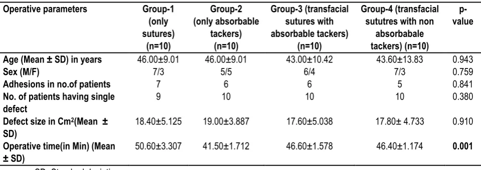
Table 1: Operative parameters.
SD- Standard deviation
Table 2: Pain VAS scores during follow up Pain
( Mean ± SD VAS Scores)
Group-1 (only sutures)
(n=10)
Group-2 (only absorbable
tackers) (n=10)
Group-3 (Transfacial sutures
with absorbable tackers) (n=10)
Group-4
(Transfacial sututres with non-absorbabale tackers)(n=10)
p-value
At day-1 2.40±0.62
(Moderate)
1.30±0.68 (Mild) 2.50±0.53 (Moderate)
2.70±0.48 (Moderate) 0.001
At 1st week 2.40±0.738
(Moderate)
1.30±0.483 (Mild) 1.90±0.316 (Mild) 2.50±0.516 (Moderate)
0.002
At 4th week 1.60±0.516
(mild)
0.50±0.527 (mild) 1.20±0.422 (mild) 1.80±0.632 (mild) 0.001
At 2nd month 0.70±0.483
(mild)
0.10±0.316 (mild) 0.50±0.527 (mild) 1.00±0.471 (mild) 0.003
At 4th, 6th and 12th month No pain No pain No pain No pain 1.000
Table 3: Other parameters
Other parameters Group-1
(only sutures)
(n=10)
Group-2 (only absorbable
tackers) (n=10)
Group-3 (Transfacial sutures
with absorbable tackers) (n=10)
Group-4 (Transfacial sututres with non-absorbabale tackers)
(n=10)
p-value
Hospital stay (in Days) (Mean±SD)
3.70±0.675 1.80±0.632 3.50±0.527 3.701±0.483 0.001
Ambulation on day-1 9 10 9 9 0.782
Foreign body sensation(n) 7/10 2/10 8/10 10/10 0.001
Operative parameters Group-1
(only sutures)
(n=10)
Group-2 (only absorbable
tackers) (n=10)
Group-3 (transfacial sutures with absorbable tackers)
(n=10)
Group-4 (transfacial sututres with non
absorbabale tackers) (n=10)
p-value
Age (Mean ± SD) in years 46.00±9.01 46.00±9.01 43.00±10.42 43.60±13.83 0.943
Sex (M/F) 7/3 5/5 6/4 7/3 0.759
Adhesions in no.of patients 7 6 6 5 0.841
No. of patients having single defect
9 10 10 10 0.380
Defect size in Cm2(Mean ±
SD)
18.40±5.125 19.00±3.887 17.60±5.038 17.80± 4.733 0.910
Operative time(in Min) (Mean ± SD)
Table 4: Complications during their stay and follow up
Complications Group-1
(only sutures)
(n=10)
Group-2 (only absorbable
tackers) (n=10)
Group-3 (Transfacial sutures
with absorbable tackers) (n=10)
Group-4 (Transfacial sututres with non-absorbabale
tackers) (n=10)
Seromas(n) 1/10 0 0 0
Inferior epigastric bleeding during passage of transfacial suture
1/10 0 0 0
Recurrence, post-operative ileus, mesh infection, wound infection
0 0 0 0
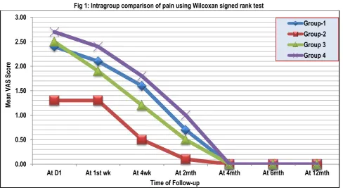
Fig 1: Intragroup comparison of pain using Wilcoxan signed rank test
RESULTS
Preoperative Parameters
The patients’ age ranged from 32 years to 78 years among the groups with more males (25 patients) as compared to females (15 patients). Primary ventral hernias were almost equally distributed among males and females (M18:F15) while incisional hernias were present in much higher frequency in females (12:3 80% females).
Operative Parameters
Almost similar trend was maintained after group wise distribution with no statistically significant (p=0.841) difference between the types of adhesions, number of defects (p value = 0.380) and the defect size (p value = .910) in the groups.
In the study groups, a single standard 15x 15 cm size composite mesh was used in all the patients.
Operative time distribution in groups: The mean duration of operation was 46.28±3.84 min. the operating time in study group 1 is 50.60±3.30 min. mean operating time in study group 2 is 41.50±1.76 min. mean operating time in study group 3 is 46.60±1.57 min and the mean operating time in study group 4 is 46.40±1.17 min. the difference was attributed to different techniques of mesh fixation, and more time was required for fixation of mesh with transfascial sutures as compared to fixation with tacks. On comparing the operative time in the study groups the p value (0.001) is highly significant.
All the patients in the groups study were completed laparoscopically and did not require conversion to open surgery. Post-Operative Pain
Postoperative pain was assessed using a visual analogue scale (vas). patients were explained that pain may presented by a straight line of 10 cm length, the extremes of which corresponds to no pain (0) at one cm and worst imaginable pain at the other end (10)
▪ Mild pain (1-3)
▪ Moderate pain (4-6)
▪ Severe (7-10)
Patients were asked to rate their pain depending upon severity:
• on the evening of the surgery 1st post op day,
• second one at 1st week (suture removal with clinical
examination on opd basis)
• then at 4th week, (clinical examination on opd basis)
• 2nd, 4th, 6th and 12th month (telephonic survey)
Post-Operative Pain Distribution in Groups from Day-1 to 2nd
Month: It was observed that from day 1 to 2nd month of their
follow up the p-value of mean pain scores showed significant difference in between the groups (p-value .001).
The p-value is significant:
• On comparison of mesh fixation technique with only sutures(Group-1) versus only tackers (Group-2)
0.00 0.50 1.00 1.50 2.00 2.50 3.00
At D1 At 1st wk At 4wk At 2mth At 4mth At 6mth At 12mth
M
ea
n V
AS
S
co
re
Time of Follow-up
• On comparing only tackers (Group-2) versus four cornered transfacial sutures with tackers (Group-3 and Group-4).
• On comparing four cornered transfacial sutures with absorbable tackers (Group-3) versus four cornered transfacial sutures with metallic tackers (Group-4).
• The p-value is not significant on comparison of only transfacial sutures (Group-1) versus four cornered transfacial sutures with absorbable/ non-absorbable tackers (Group-3 and Group-4) (p-value of 0.661 and 0.189 respectively). Post-Operative Pain Distribution in Groups At 4th, 6th and 12th
Month: the difference in the study groups was statistically not significant (p value 1.000)
Seroma: Seroma formation and persistence was not statistically significant in the study groups (p value 1.0). Only 1 patient developed seroma in post-operative period which resolved spontaneously.
Other Complications: 1 patient in group-1 had inferior epigastric bleeding during passage of transfacial suture there by increasing the operating time significantly to 58min.
None of the patient in the study groups developed hematoma, paralytic ileus, mesh infection, wound infection and recurrence within this follow up duration.
Hospital Stay Distribution in Groups: The mean hospital stay for the total study group was 3.18±0.98 days. The difference in hospital stay among the groups was statistically significant (p-value .001). Patients in whom mesh fixation was done with only tackers got discharged early compared to other techniques of mesh fixation.
Foreign Body Sensation in the Groups: Group 2 had
significantly less number of cases with F.B. sensation than in group-1, group-3 and group-4.
Ambulation in the Groups: Almost all the patients were ambulated on 1st postoperative day. The difference in ambulation
between the groups was not statistically significant (p value.782) Cost Analysis: On comparing the cost of the procedures in between the groups the p-value is significant. The cost of the procedure is more than 2.5 times in groups 2, 3, 4 than in group 1 where only transfacial sutures were used. On comparing the cost analysis of group 2, group-3 and group-4 the p-value is not significant.
DISCUSSION
Although laparoscopic incisional and ventral hernia repair has gained popularity, there are many technical issues, which need to be resolved. The issues of access to the abdominal cavity mesh overlap and mesh size have more or less been resolved. Further, issues like the ideal mesh to be used, the fixation technique and the necessity for closure of the defect before mesh fixation are areas of ongoing debate.
In our study we used different techniques of mesh fixation. In study Group 1 mesh fixation was done by transfascial sutures only, In Group 2 mesh fixation was done by tackers only, In Group 3 mesh fixation was done by absorbable tackers and four cornered transfascial sutures, In Group 4 mesh fixation was done by non-absorbable tackers and four cornered transfascial sutures. OPERATIVE TIME
Operative time has been one of the important determinants of assessing the effectiveness of the procedure. It has been observed that the operative time depends on a host of factors like
patient selection, visceral adhesions, contents of the hernia sac, time taken to create penumoperitoneum, type of mesh used and fixation technique and learning curve.
It was observed that the operative time was directly dependent on the presence of adhesions. The adhesiolysis of the adhesions accounted for increased operative time. The study groups were comparable in terms of visceral adhesions and contents of the hernial sac. In the present study it was also observed that the operative time was also depended on the feasibility and ease of intra operative handling of mesh.
The mean duration of operation was 46.28±3.84 min. On comparing the operating time in the study groups the p value is.001. This is statistically highly significant. The difference in operative time can be attributed to the reason that long time required for applying transfascial sutures as compared to applying tacks. It has been found that average time required for tying one suture was 1 min 58 seconds. The operative time was significantly low for mesh fixation technique with only tackers followed by four cornered transfacial sutures with absorbable/non-absorbable tackers then followed by only transfacial sutures.
HOSPITAL STAY
The difference in hospital stay among the groups was statistically significant (p-value .001). Patients in whom mesh fixation was done with only tackers got discharged early compared to other techniques of mesh fixation.
Laparoscopic repair can be carried out as a day care procedure, but it is the policy of our center to keep all patients, operated under general anesthesia for at least over-night for observation in the hospital. This is why; the patients could be discharged earliest on the postoperative day one only. The patients who were discharged late were because of patient's preference to stay in the hospital or having severe pain requiring injectable pain killers. POST-OPERATIVE PAIN
Laparoscopic incisional/ventral hernia repair has been termed as a painful laparoscopic procedure when compared to other minimal access surgical procedures in the immediate postoperative recovery phase. The incidence of chronic pain following laparoscopic incisional hernia repair has been reported to be about 1-3% in literature [72]. However, the overall pain scores may
not be higher and it may not affect the early ambulation and discharge from the hospital although the requirement of postoperative analgesia may be higher.
It was observed that from day 1 to 2nd monthof follow up low pain
scores were reported in patients where mesh was fixed with only tackers followed by transfacial sutures with absorbable tackers, followed by transfacial sutures with non-absorbable tackers and then by only sutures fixation.
At 4th, 6th and 12th months The difference in pain in the study
groups was statistically not significant (p value 1.000).
All patients experienced mild to moderate pain till 2nd month of
follow up. In long term duration neither of the patients in study groups had experienced pain after 2nd month.
COMPLICATIONS
seromas may be because of compression bandage applied post-operatively and seromas that formed were small and resolved by themselves without requiring aspiration.
Mesh Infection: None of the patients in our study had any mesh infection. This is due to strict asepsis followed during the procedure.
Recurrence: In present study none of the patient has reported with recurrence. This could be attributed to the fact that while performing mesh repair utmost care was taken to ensure that the mesh covered the hernia defect with atleast 3-5 cms of overlap on all sides. Care was also taken to ensure that mesh was properly anchored to the abdominal wall with the help of transfascial sutures on all four corners and tacks or transfsacial suture in between. On the completion of the surgery a careful survey of the entire abdominal wall was made to ensure that the mesh was placed properly.
Post-Operative Ileus: None of the patient in the study groups had developed post-operative ileus.
In our experience on follow up none of the patients in study groups developed any other complications on long term follow up like sub-acute intestinal obstruction, acute intestinal obstruction, mesh infection, bowel incarceration, intra-abdominal collections. Foreign Body Sensation: Patients in whom mesh was fixed using only tackers experienced less foreign body sensation than in other techniques.
COST ANALYSIS
On comparing the cost of the procedure, it is 2.5 times more in groups where tackers were used than in group where only transfacial sutures were used.
CONCLUSION
Thus it is concluded that laparoscopic repair should be the preferred method of repair of ventral hernia as it is associated with a shorter hospital stay, decreased post-operative pain, better cosmetic results decreased complication rate like recurrence, hematoma and seroma formation and decreased infection rate. With regard to the mesh fixation technique the suture fixation method is a cost-effective alternative to tacker fixation in laparoscopic incisional and ventral hernia repair.
Mesh fixation with tackers is easier and faster than transfascial suture fixation. Absorbable tackers usage showed low incidence of pain and foreign body sensation in the early postoperative period, but on long term follow up no such dependence was noted on either sutures or tackers.
The procedures are equally effective regarding the recurrence rates, complications, hospital stay and chronic pain. Though the procedures are equally effective in reducing the complications, hospital stay and early recurrence rate, still further studies are required to know the long term complications like chronic pain and late recurrence.
SUMMARY
▪ The inclusion criteria for selection of patients were patients having paraumblical hernia, incisional hernia, divarication of recti and epigastric hernia.
▪ The exclusion criteria of the patients was patients having densely scarred abdomen, acute abdomen with strangulated and infacted bowel, incarcerated hernia and children less than 18 years of age
▪ The age distribution, gender distribution, contents of hernia, number of defects, defect size and type and size of mesh were all comparable between the four groups.
▪ The mean duration of operation was 46.28±3.84 min. The operating time was significantly low in only tackers group and highest in only transfacial group. Whereas operating time was comparable in transfacial sutures and absorbable group and transfacial sutures and non-absorbable group.
▪ The difference in hospital stay and ambulation among the groups was not statistically significant.
▪ All patients experienced mild to moderate pain till 2nd month
of follow up. In patients where mesh fixation done by only tackers experienced significantly very low (mild) pain scores from day 1. In patients where mesh fixation was done using transfacial sutures with or without tacks had experienced moderate intensity of pain from day 1 to 2nd month of follow
up and in patients where absorbable tackers were used experienced low pain scores than in patients where metallic tackers were used. However In long term duration neither of the patients in study groups had experienced pain after 2nd
month.
▪ 1 patient developed seroma which resolved spontaneously without any intervention.
▪ None of the patient developed hematoma, post-operative ileus, mesh infection, wound infection and recurrence in the post-operative period.
▪ In patients where only tackers were used had significantly less number of cases with foreign body sensation than in groups where sutures were used.
▪ The cost of the procedure is more than 2.5 times in groups where tackers were used than in group where only transfacial sutures were used.
REFERENCES
1. Kingsnorth A, Bennet DM. Hernia, umbilicus, abdominal wall. Bailey and Love’s short practices of Surgery. 25th Edition Arnold Publication 2008;51:968-87.
2. Regnard JF, Hay JM, Rea S, Fingerhut A, Flamant Y, Maillard JN. Ventral incisional hernias: Incidence, date of recurrence, localization and risk factors. Ital J Surg Sci. 1988;18:259–65 3. Cassar K and Munro A. Surgical treatment of incisional hernia. J. Surg 2002;89:534-45.
4. DeMaria EJ, Moss JM, Sugerman HJ. Laparoscopic intraperitoneal ePTFE prosthetic patch repair of ventral hernia. Prospective comparison to open prefascial polypropylene mesh repair. Surgical Endoscopy 2000; 14(4):326-29.
5. Millikan KW. Incisional hernia repair. Surg Clin North Am 2003;83:1223-34.
6. Luijendijk RW, Hop WC, van den Tol MP, de Lange DC, Braaksma MM, IJzermans JN, Boelhouwer RU et al. A comparison of suture repair with mesh repair of incisional hernia. The New England Journal of Medicine Aug 2000;343(6):392-98. 7. Bucknell TE, Cox PJ, ellis H. Burst abdomen and incisional hernia : a prospective study of 1129 major laparotomies. BMJ 1982;284: 931-3
9. Bencini L, Sanchez LJ, Boffi B, Farsi M, Scatizzi M, Moretti R. Incisional hernia repair: Retrospective comparison of laparoscopic and open techniques. Surg Endosc 2003;17:1546-51.
10. McGreevy JM, Goodney PP, Birkmeyer CM, Finlayson SR, Laycock WS, Birkmeyer JD. A prospective study comparing the complication rates between laparoscopic and open ventral hernia repairs. Surg Endosc 2003;17:1778-80.
11. LeBlanc KA. Laparoscopic incisional hernia repair: Are trans-fascial sutures necessary? A review of the literature. Surg Endosc 2007;21:508-13.
12. Chowbey PK, Sharma A, Khullar R Mann V, Baijal M, Vashistha A. Laparoscopic ventral hernia repair. J Laparoendosc Adv Surg. Tech A 2000;10:79-84.
13. Chelala E, Thoma M, Tatete B, Lemye AC, Dessily M, Alle JL. The suturing concept for laparoscopic mesh fixation in ventral and incisional hernia repair: Mid-term analysis of 400 cases. SurgEndosc 2007;21: 391-95.
14. Carbajo MA, Martin del Olmo JC, Blanco JI, de la Cuesta C, Toledano M, Martin F et al. Laparoscopic treatment versus open surgery in the solution of major incisional and abdominal wall hernias with mesh. Surg Endosc 1999;13:250-52.
15. Olmi S, Magnone S, Erba L, Bertolini A, Croce E. Results of laparoscopic versus open abdominal and incisional hernia repair. JSLS 2005; 9: 189-95
16. Misra MC, Bansal VK, Kulkarni MP, Pawar DK. Comparison of laparoscopic and open repair of incisional and primary ventral hernia: results of a prospective randomized study. Surg Endosc 2006;20:1839-45.
17. Barbaros U, Asoglu O, Seven R, Erbil Y, Dinccag A, Deveci U et al. The comparison of laparoscopic and open ventral hernia repairs: a prospective randomized study. Hernia. 2007; 11:51-56.
18. Poreca MM, Mehta SG, Thanthvalia A, Udani D. Comparative study of laparoscopic versus Open ventral hernia repair. The internet Journal of Surgery 2009;22(2):1-7.
19. Stoppa RE. The treatment of complicated groin and incisional hernias. World J. of Surg 1989;13:545-54.
20. Bendavid R. The need for mesh. In Bendavid R (ed.); Prosthesis and abdominal wall hernias. Austin : R.G. Landes Company; 1994:207-23.
21. Kirshtein B, Lantsberg L, Avinoach E, Bayme M, Mizrahi S Laparoscopic repair of large incisional hernias. Surg Endosc 2002;16: 1717-19.
22. Carbajo MA, Martin del Olmo JC, Blanco JI, Toledano M, de la Cuesta C, Ferreras C et al. Laparoscopic approach to incisional hernia. Surg Endosc 2003;17:118-22.
[
Source of Support: Nil. Conflict of Interest: None Declared.
Copyright: © the author(s) and publisher. IJMRP is an official publication of Ibn Sina Academy of Medieval Medicine & Sciences, registered in 2001 under Indian Trusts Act, 1882. This is an open access article distributed under the terms of the Creative Commons Attribution Non-commercial License, which permits unrestricted non-commercial use, distribution, and reproduction in any medium, provided the original work is properly cited.