Special issue • 2020 • vol.1 • 11-22
MULTIMODALITY IMAGING OF BREAST TRAUMA:
DIAGNOSIS AND MANAGEMENT
Gatta G.
1, Ancona A.
2, Falco G.
3, Serra N.
1, Vallebona L.
1, Iannaccone T.
4, Bertozzi S.
5, Viola L.
1,
Di Grezia G.
61Radiology Department, University of Campania Luigi Vanvitelli Naples IT 2Radiology Department, Santa Maria Private Hospital, Bari IT 3Breast Surgery, Santa Maria Nuova Hospital-IRCCS Reggio Emilia IT
4Medicine Department, University of Salerno, Baronissi (SA) IT 5Breast Unit, University Hospital of Udine, Udine IT
6Radiology Department, “G Criscuoli” Hospital, Sant’Angelo dei Lombardi, Av IT
Email: [email protected]
Abstract
Purpose:
Breast traumas include road accident, assaults, falls or other minor events; the diagnosis is often delayed, despite several medical imaging techniques because of the priority to most relevant organs and anatomical districts involved in traumatic events. Aim of the study was to evaluate the imaging in breast traumas, the differential diagnosis and correlation with breast cancer and the medical or surgical management of each case described.
Methods: We performed a retrospective study over a period between February 2010 to June 2017. All patients diagnosed with breast injuries were included in the study. Breast clinical examination, mammography, ultrasonography and, in selected cases, Magnetic Resonance (MRI) and ultrasound-guided fine needle aspiration cytology were used for the diagnosis. Logistic regression models were used to estimate unadjusted and adjusted odds ratio and corresponding 95% confidence interval.
Results: Over the study period, 55 patients had a breast trauma; there were 12 cases of major event and 43 minor traumas. 2 males and 53 females have been included. Mean age was 47yo (range 27-71 yo). In 11 cases, surgical therapy was necessary, in 3 cases medical therapy and in 2 follow-up have been performed. In 10 cases, also a breast cancer has been diagnosed, but no association between breast trauma and the risk of breast cancer was observed (odds ratio: 0.85; IC 95%: 0.41-1.75; P: 0.665).
Conclusion: Breast traumas are underestimated if a major event occurred; imaging diagnosis of breast trauma requires often a multimodality approach and can be difficult expecially if a breast cancer also occurs. A dedicated breast radiologist is necessary for a correct differential diagnosis and to palinify a terapeutical approach and follow-up.
Introduction
The number and clinical consequences of breast traumatic accidents have not the same social relevance of breast cancer (incidence rate of 90-110/100,000 in Europe and US [1], 20% of cancer deaths in women [2] and this may be a consequence of the fact that breast surgery is easy to perform. The signs and symptoms of accidental trauma can mimic those of breast cancer especially if the patient has no memory of the traumatic event.
A major trauma (e.g. road accident) or minor (assaults, falls or other) can cause the formation of a hematoma, followed by central necrosis and liquefaction of the adjacent fat [3,4].
Unless a massive blood extravagation, requiring surgical drainage, occurs, this can lead to the development of a fibrotic or cystic tissues that will almost entirely calcify within 2 or 3 years. Scars associated with skin retractions can be also noticed [5].
In the literature, one case of pneumothorax in a 57 years old woman was reported. This was characterized also by multiple rib fractures, haemothorax and a large pneumothorax, which had passed the superficial fascia of the anterior chest wall reaching the virtual space between large and small pectoral and affecting the homolateral mammary gland [6,7].
Two main points should be taken into account when evaluating a breast trauma: the difficulty in differentiating recent outcomes of trauma (4-204 weeks) from signs/symptoms of neoplasia in the absence of specific symptomatology [8-10]; secondly, the possible correlation between breast trauma and cancer [11-13].
Aim of the study was to evaluate the imaging in breast traumas, the differential diagnosis and correlation with breast cancer and the medical or surgical management of each case described.
Methods
Study population
The population target of this retrospective cohort study were recruited from 1st February 2010 to30th June 2017 in our University Hospital and includes a total of 500 consecutive patients.
Only patients older than 23 years old were included. Exclusion criteria were: presence of breast cancer with maximum diameter > 2 cm (T2, T3, T4), patients
with prostheses, two or more first degree relatives with breast cancer.
Demographic characteristics collected for both cases and controls were: sex, age, previous or current estro-progestinic replacement therapy (average duration 3 years), history of trauma and breast trauma characteristics.
We evaluated the assessment of the dynamics of the traumatic event, months after trauma and the severity of the trauma (major – road accident; minor: assaults, falls or other).
All patients underwent breast examination and ultrasonography. Patients older than 40 underwent mammography in addition.
27% of patients (123/500) underwent ultrasound-guided fine needle aspiration cytology.
5% of patients (25/500) had also a Magnetic Resonance Imaging examination.
The study was designed in compliance with the Helsinki declaration.
All patients gave the permission for their images to be used.
Imaging
Clinical breast examination, mammography and ultrasound were performed by a breast radiologist. Mammography was performed with Instrumentarium Diamond Mammography (GE Healthcare, Wisconsin, USA). Two projections were performed (cranial-caudal and mediolateral oblique) and, in selected cases, also projections in targeted compression.
If necessary, additional screenings were performed in targeted compression and / or with direct radiographic magnification.
minutes after administration of intravenous contrast. It has been used Gadobutrol (Gadovist, Bayer Schering Pharma AG, Berlin, Germany) as a paramagnetic contrast agent with bolus injection of 0.1 mmol / kg of body weight.
Ultrasound-guided fine needle aspiration cytology was performed using the capillary aspiration technique. After obtaining informed consent, the patients were placed in a comfortable position and lesions were aspirated with 21-gauge needle, the needle being rotated during the sample. Local anaesthesia was not required.
The taken sample was swiped on a slide, fixed and analysed by an experienced pathologist at the Department of Pathology of our hospital.
Statistical analysis
Logistic regression was used to estimate odds ratios (ORs), their corresponding 95% confidence interval (CIs) and associated P values. Known or suspected risk factors for breast cancer (trauma, age, estrogen progestin therapy) were evaluated as potential confounding factors by multivariate analysis.
All P-values were two-sided and those less than 0.05 were considered statistically significant.
Results
Five hundred patients were examined in our study (mean age: 46 years; SD 11; median 47 years) (Table 1).
102 out of 500 consecutive patients (20.4%) were positive for breast cancer (Mean age 46.6 y.o., SD 7.78, range: 23-71 years).
99 out of 102 consecutive patients with breast cancer (97%) were female, 10 of them (10%) had history of trauma, 22 of them (21.6%) took estroprogestinic replacement therapy (average duration 3 years).
398 out of 500 consecutive patients (79.6%) were negative for breast cancer (Mean age 46.5 y.o., SD 9. 89, range: 24-72 years).
392 out of 398 consecutive patients without breast cancer (98.2%) were female, 45 of them (11.3%) had history of trauma, 68 of them (17%) took estroprogestinic replacement therapy (average duration 3 years).
The time interval between the trauma and the enrollment into the study was 32 months (SD 15; range 1-60 months; median value 29 months). 25% of patients with trauma (n=14) had a major trauma (car accident), 74% had a minor trauma following fall (n
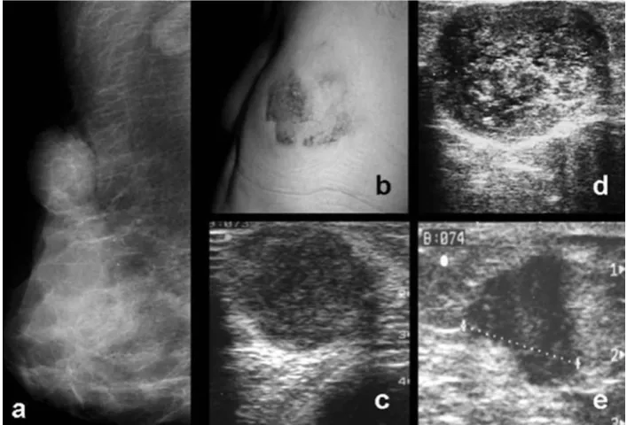
19), attack (n.18) or other (n.4). 71% per cent (n=39) of patients with a history of trauma had no sign of malignant disease at medical examination, nor mammography or ultrasonography. In two patients a hematoma was diagnosed; in one case it was treated with medical therapy (Fig. 1), in one case with surgery. A case of liponecrosis (Fig.2) and a case of fibrosis (Fig 3) were diagnosed. In two patients a post-traumatic Mondor syndrome on the site of a trauma was present (Fig.4) and treated with medical therapy.
10 patients (18%) had a mammarian carcinoma in the same quadrant of previous trauma, surgically treated (Fig.5). 9 out of 10 patients with breast cancer and breast trauma were female, 1 was male. The mean age of those patients was 48.9 years (SD 9.12; range: 40-70); 3 of them took estroprogestinic replacement therapy. 1 patient had a major trauma (road accident), 9 a minor trauma (5 attacks, 3 falls, 1 other cause). The time interval between the trauma and the enrollement in the study was 31.8 months (SD 12.7; range: 12-55).
These patients (9/500) underwent breast examination, mammography, ultrasonography and fine needle aspiration cytology. 3 patients had also Magnetic Resonance Imaging examination.
The result demonstrates that, in our experience, patients with a history of breast trauma have a chance of getting breast cancer almost identical to that of the general population (OR: 0.85, 95% C.I.: 0.41-1.75; p-value: 0.66) (Table 2a) .
Adjustment for age and for estroprogestinic replacement therapy did not change the Odds Ratios (OR: 0.84, 95% C.I 0.41-1.73, p-value: 0.64) (Table 2b).
Discussion
Breast trauma and its consequences, despite rare when compared to the thoracic and abdominal ones, have great clinical relevance because of the differential diagnosis with tumour lesions in the preclinical phase (parenchymal distortions and fibrous scars without typical lipo-necrotic calcifications). (Fig. 3) [14,15].
The findings of the present study show that patients who experienced breast trauma present instrumental negativity.
Furthermore, most of the occasional “minor”
Every breast trauma may potentially cause intraparenchymal haemorrhage followed by oedema, ischemia and subsequent colliquative necrosis of adipocytes characterized by the formation of vacuoles filled with necrotic lipidic material and surrounded by fibroblasts, foam cells and plurinucleate giant cells. This is later replaced by fibrous tissue. This picture of aseptic inflammation is
called “aseptic fat necrosis” and is directly related
to trauma, as reported in the literature since 1920 (Fig. 2). A nodulation may possibly be associated with a clinical picture characterized by ecchymosis, erythema, inflammation, pain, skin thickening or retraction. This can involve the entire organ or only a part of it which often correspond to the supero-external quadrants or the superficial retro areolar region. At mammographic evaluation the most common result of a trauma is represented by dystrophic calcifications with clear centre opacities [16]
.
In many cases it is however possible to find the thickening and deformity of the skin and subcutaneous tissue (16%), presence of focal (13% ) or spiculated masses with sharp margins (4%) and clusters of microcalcifications with suspicion of malignancy (4%). When a ultrasonography is performed, it is typical to notice a response of iso / hypoechoic solid nodules with posterior acoustic barrier or intracystic masses hyperechogenicity of subcutaneous tissue (27%), anechoic cysts with or without intralesional echoes with reinforced rear wall (17%), cystic mural nodules (4%) .
These findings may suggest a strong suspect of malignant lesion [17].
MRI is less accurate if performed within 6 months after the traumatic event. This because of the presence of a post-inflammatory granulation tissue which limits the differential diagnosis (risk of False Positive). When performed after at least six months from the traumatic event, the formation of a hypovascular scar with central necrosis makes the diagnosis by MRI more accurate.
The absence of specific symptomatology does not always allow the exclusion of malignancy. In doubtful cases, fine needle aspiration for cytology (sensitivity 87% specificity 99%) or core needle biopsy (False Negative estimated among 1.2-1.5%) are suggested [18-20].
Breast traumas may cause the immediate formation
of a hematoma, a contusion, as well as a post-traumatic Mondor's disease (superficial phlebitis). The latter has an incidence of 0.5-0.8% in symptomatic patients, both men and women [21]. In our experience, the two patients with a history of
traumatic accident and subsequent Mondor’s
disorder at trauma site were easily diagnosed during the breast control (visit, bilateral mammography and Doppler ultrasonography). Because of the risk; albeit rare, of association with breast cancer, we addressed the patients to a close follow-up with instrumental control at six months (CAT BIRADS 3) [22-24] (Fig 4).
The finding of 9 cases of breast cancer detected in patients with a history of traumatic event in the same quadrant of trauma, led us to hypothesize the existence of a correlation between trauma and onset of breast cancer. In our case-control study no correlation between recent trauma and onset of breast cancer was demonstrated (OR: 0.85, 95% Confidence interval: 0.41-1.75; P-value: 0.66) (Fig. 5). Breast traumas are underestimated if a major event occurred; imaging diagnosis of breast trauma requires often a multimodality approach and can be difficult expecially if a breast cancer also occurs [25-27].
A dedicated breast radiologist is necessary for a correct differential diagnosis and to plan a therapeutical approach and follow-up.
References
1. Cuzick J. Epidemiology of breast cancer
– selected highlights. The breast 2003; 12(6): 405-411
2. Robbins and Cotran Pathologic basis of disease. 8th ed. Philadelphia: Elsevier Inc. 2010
3. Gatta G, Pinto A, Romano S, Scaglione M, Volterrani L Clinical, mammographic and ultrasonographic features of blunt breast trauma. Eur j Radiol 2006 Sep;59(3);327-30
4. Di Grezia G, Romano T, De Francesco F, Somma F, Rea G, Grassi R, Gatta G –
Breast ultrasound in the management of gynecomastia in Peutz-Jeghers Syndrome: a case report. Journal of Medical Case Report 2014 ; (18) 8:440 5. Williams HJ, Hejmadi RK, England DW,
trauma: a pictorial review. The Breast 2002; 11:107-115
6. Walker D, Deakin CD, Smith G. Traumatic pneumomammothorax: Case report. Emerg. Med. J. 2002; 19:466-467
7. Vestito A, Mangieri FF, Gatta G, Moschetta M, Turi B, Ancona A Breast carcinoma in elderly women. Our experience. G Chir 2011 Oct;32(10):411-6 8. Pullyblank AM, Davies JD, Basten J,
Rayter Z. Fat necrosis of the female breast-Hadfield re-visited. Breast 2001; 10: 388-91
9. Tan PH, Lai LM, Carrington EV, Opaluwa AS, Ravikumar KH, Chetty N, Kaplan V, Kelley CJ, Babu ED. Fat necrosis of the breast – A review The breast 2006; 15: 313-318
10. Capobianco G, Crivelli P, Piredda N, Maiore M, Cherchi PL, Dessole M, Virdis G, Dessole S, Meloni GB. Hysterosalpingography in infertility investigation protocol: is it still useful? Clin Exp Obstet Gynecol. 2015;42(4):448-51
11. Aqel .M., Howard A., Collier D.St.J., Fat necrosis of the breast: a cytological and clinical study, The Breast 2001; 10:342-345
12. Ho K.C.K., McKessar M., Giuffre B.M., Review of fat necrosis cases in patients recalled from a breast screening program, The Breast 2008; 17 :209 13. Rigby JE, Morris JA, Stewart M, Gatrell
AC Can physical trauma cause breast cancer? Eur J Cancer Prevention 2002; 11:307-311
14. Di Grezia G, Gatta G, Brunese L, Falco G State of the art in Integrated Breast Imaging BioMe Research International Volume 2019, Article ID 7596059, 2 pages 15. Amadu AM, Marras V, Crivelli P, Soro D, Conti M, Meloni GB. Isolated breast metastasis 4 years after nephrectomy. Breast J. 2018 Jan;24(1):85-87
16. Fancellu A, Sanna V, Sedda ML, Delrio D, Cottu P, Spanu A, Giuliani G, Conti M, Piras R, Crivelli P, Porcu A. Benefits of Organized Mammographic Screening
Programs in Women Aged 50 to 69 years: A Surgical Perspective. Clin Breast Cancer. 2019 Oct;19(5): e637-e642
17. Amadu AM, Soro D, Marras V, Satta G, Crivelli P, Conti M, Meloni GB. Primary breast chondrosarcoma: Imaging and pathological findings. Eur J Radiol Open. 2017 Nov 5;4:138-140
18. Ancona A, Capodieci M, Galiano A Vacuum assisted biopsy diagnosis of atypical ductal hyperplasia and patient management. Radiol Med 2011 Mar; 116(2):276-91
19. Crivelli P, Ledda RE, Parascandolo N, Fara A, Soro D, Conti M. A new challenge for Radiologists: Radiomics in Breast Cancer Biomed Res Int. 2018 Oct 8;2018:6120703
20. A Poggiu, M Tamponi, F Sanna, R Bona, F Pinna, P Crivelli, D Gabriele, R Montella, E Sanna, M Conti, P Marini. A radiomics analysis process using free software platforms: Our experience In Sassari. Physica Medica 56, 252-253
21. Conant EF, Wilkes AN, Mendelson EB, Feig SA. Superficial thrombophlebitis of
the breast (Mondor’s disease):
mammography findings. AJR 1993; 160:1201-1203
22. Yanik B, Conkbayir I, Oner O, hekimoglu
B. Imaging findings in Mondor’s disease.
J. Clin. Ultrasound 2003; 31:103-7
23. Fietta P, Manganelli P. Mondor’s
disease. Spectrum of the clinical and pathological features. Minerva Med. 2002; 93:453-6
24. Kinoshita T, Yashiro N, Ihara N, Yokogawa M. MR findings of seat belt injury in the breast. J. Comput. Assist. Tomogr. 2002;26: 1054-6
25. Di Grezia G., Prisco V., Iannaccone T et al. Personality disorders and temperamental traits in patients with breast disease: Preliminary results Minerva Psichiatrica 2016; 57(3): 85-92 26. Gatta G., Di Grezia G., Ancona A et
breast biopsy European Journal of Inflammation 2013, 11(3):825-835
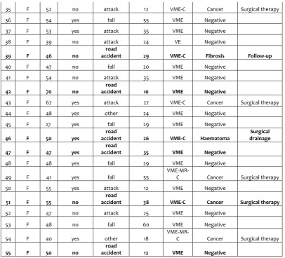
Table 1. Summary of findings from the review of patients with trauma (n: 55).
Legend - EP: estrogen progestin therapy; MONTHS: months since injury; VME: visit, mammography, echography; MR: Magnetic resonance; C: Fine needle aspiration
No. SEX AGE EP THERAPY TRAUMA MONTHS IMAGING DIAGNOSIS THERAPY
1 F 71 no Fall 19 VME-C Fat necrosis Follow-up
2 F 70 no attack 15 VME Negative
3 F 60 no
road
accident 30 VME Negative
4 F 47 no attack 27 VME Negative
5 F 50 no Fall 54 VME Negative
6 F 58 no
road
accident 50 VME Negative
7 F 38 no Fall 35 VE Negative
8 M 44 no Fall 29 VME
Mondor's
disease Medical therapy
9 F 43 no attack 48 VME-C Negative
10 F 28 no
road
accident 18 VE Negative
11 F 29 no Fall 26 VE Negative
12 F 49 no Fall 45 VME-C Cancer Surgical therapy
13 F 48 no
road
accident 1 VME Haematoma Medical therapy
14 F 40 no attack 35 VME-C Cancer Surgical therapy
15 F 43 no attack 25 VME-C Cancer Surgical therapy
16 F 40 no fall 36 VME Negative
17 F 37 no fall 47 VE-C Negative
18 F 35 no other 35 VE Negative
19 F 34 no attack 48 VE Negative
20 F 38 no
road
accident 15 VE
Mondor's
disease Medical therapy
21 F 38 yes attack 23 VE Negative
22 F 41 no attack 26 VME-C Negative
23 F 37 no
road
accident 48 VE Negative
24 F 42 no attack 29 VME Negative
25 F 46 no fall 14 VME Negative
26 F 55 no fall 38 VME Negative
27 M 49 no attack 36
VME-MR-C Cancer Surgical therapy
28 F 50 no fall 27 VME-C Cancer Surgical therapy
29 F 39 no other 28 VE Negative
30 F 36 no fall 23 VE Negative
31 F 27 no
road
accident 29 VE Negative
32 F 56 no fall 25 VME Negative
33 F 56 no
road
accident 23 VME Negative
35 F 52 no attack 12 VME-C Cancer Surgical therapy
36 F 54 yes fall 55 VME Negative
37 F 53 yes attack 35 VME Negative
38 F 39 no attack 24 VE Negative
39 F 46 no
road
accident 29 VME-C Fibrosis Follow-up
40 F 47 no fall 20 VME Negative
41 F 54 no attack 35 VME Negative
42 F 70 no
road
accident 10 VME Negative
43 F 67 yes attack 27 VME-C Cancer Surgical therapy
44 F 48 yes other 24 VME Negative
45 F 27 yes fall 29 VME Negative
46 F 50 yes
road
accident 26 VME-C Haematoma
Surgical drainage
47 F 47 yes
road
accident 35 VME Negative
48 F 48 yes fall 29 VME Negative
49 F 41 yes fall 55
VME-MR-C Cancer Surgical therapy
50 F 55 yes attack 12 VME Negative
51 F 55 no
road
accident 38 VME-C Cancer Surgical therapy
52 F 47 no attack 25 VME Negative
53 F 48 no fall 60 VME Negative
54 F 40 yes other 18
VME-MR-C Cancer Surgical therapy
55 F 50 no
road
accident 12 VME Negative
Table 2. Results of retrospective cohort study with pairing the relationship between breast cancer and breast trauma
a) Logistic regression and b) Multiple logistic regression (adjusted for age and Estrogen Progestin therapy) - Odds ratio (OR) with
b) 95% confidence interval (95% C.I.) and respective p-values. Legend: EP: estrogen progestin intake
breast cancer odds
ratio 95%CI
P-value
a) Logistic regression trauma 0,85
0.41-1.75 0.66
b) Multiple logistic regression
trauma 0.84
0.41-1.75 0.64
age 1.00
0.98-1.02 0.97
EP 1.34
Figure 1. Unresolved outcomes of breast traumas: post-traumatic hematoma difficult to differentiate from mucinous carcinoma and medullary carcinoma.
Case n.13 –A forty-eight years old female, with a history of car accident une month earlier. a) Mammography in oblique projection: oval opacity with sharp and regular margins.
b) Clinical picture of soft tissue hematoma
c) Ultrasound findings of hematoma: oval hypoecogenity lacuna with disomogeneous aspect due to hyperechogenic spots and not well defined margins
d) Ultrasound finding of mucinous carcinoma: oval hypoecogenity lacuna with clear hyperechogenic spots with clear and regular margins
e) Ultrasound findings of medullary carcinoma: oval hypoecogenity lacuna with disomogeneous aspect and sharp and irregular margins.
Figure 2. Unresolved outcomes of breast traumas: fat necrosis
Case n. 1_ Seventy-one years old femal, with a history of fall 19 months earlier a) Mammography in CC left projection: Multiple calcified liponecrotic post-traumatic cysts
Figure 3. Outcomes of major trauma: Fibro-post-traumatic scar
Case n. 39 _Forty-six years old female, with a history of car accident 29 months before.
Left mammography in CC (a) and MLO (b) projection: vaguely star shaped blurred opacity with dendritic striae, difficult to differentiate from infiltrating lobular carcinoma.
Figure 4. Mondor’s post-traumatic superficial thrombophlebitis
a) Case n.20_ Thirty-eight woman, with a history of car accident 15 months earlier b) Case n.8 – Forty-four years old male, with a history of fall 29 months earlier.
a, b) Clinical image of Mondor’s post-traumatic superficial thrombophlebitis at trauma site.
210x121mm (150 x 150 DPI)
Figure 5. Ductal infiltrating carcinoma with focal areas of post-liponecrosis in absence of calcification Case n. 43 Seventy-years old female, with a history of attack 27 months earlier
Right mammography, CC a) and LL b) projection showing clear and voluminous opacity, with star shape and dendritic striae, short and disomogeneous c) Ultrasound findings: circle shaped mass with hypoecogenic aspect